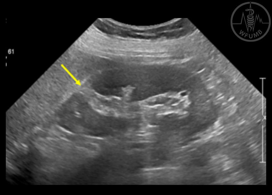
Fig 17.1a
Junctional parenchymal defect in a 61-year-old woman. Echogenic cortical defect (arrow) in the upper polar area of the kidney which is continuous with the renal sinus echo
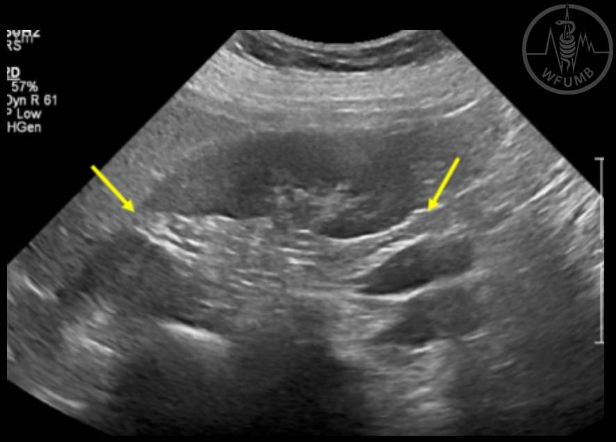
Fig 17.1b
Junctional parenchymal defects in a 61-year-old woman. Echogenic cortical defects (arrows) in the upper and lower polar areas of the kidney which is continuous with the renal sinus echo
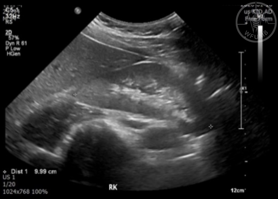
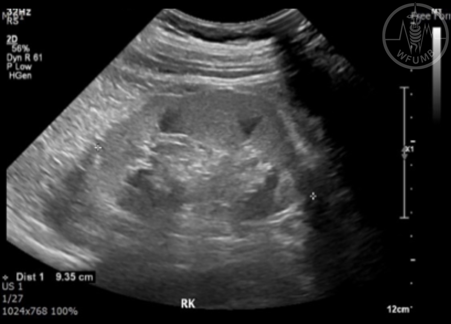
Fig 17.2a
35-year-old man with IgA nephropathy. Grey-scale US of the right kidney shows diffusely increased echogenicity of the renal parenchyma and obliterated CMD
Fig 17.2b
35-year-old man with IgA nephropathy. Grey-scale US of the left kidney shows diffusely increased echogenicity of the renal parenchyma and obliterated CMD
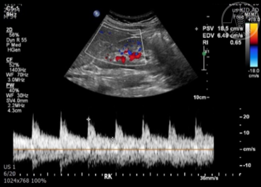
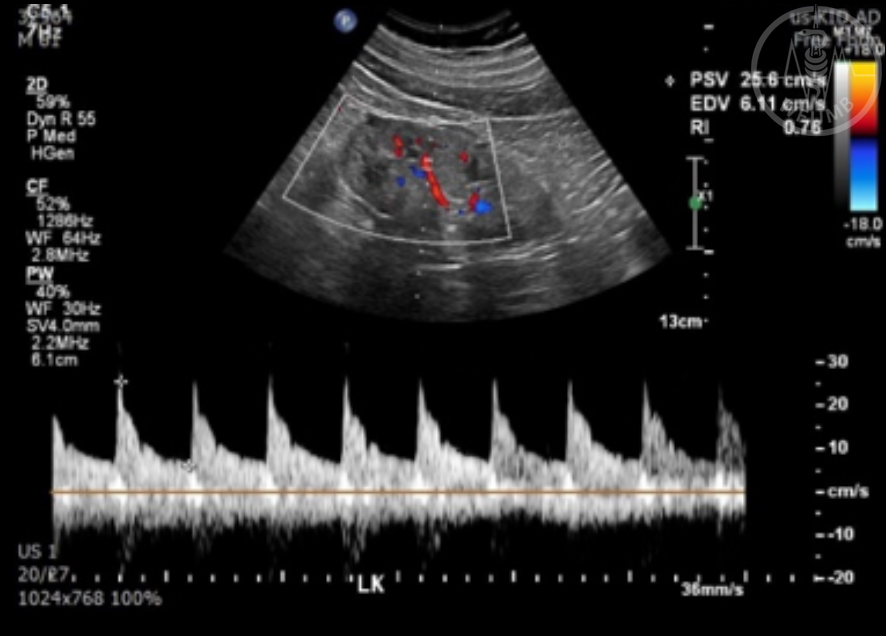
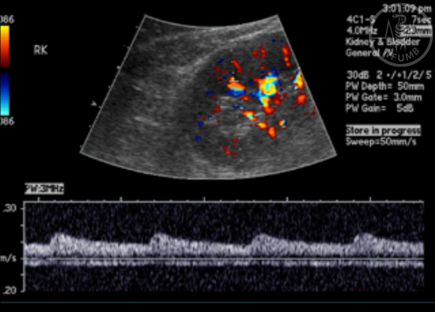
Fig 17.2c
35-year-old man with IgA nephropathy. SDUS image of the right kidney shows normal Doppler spectral patterns and normal RI (right 0.65; left 0.63 - not shown)
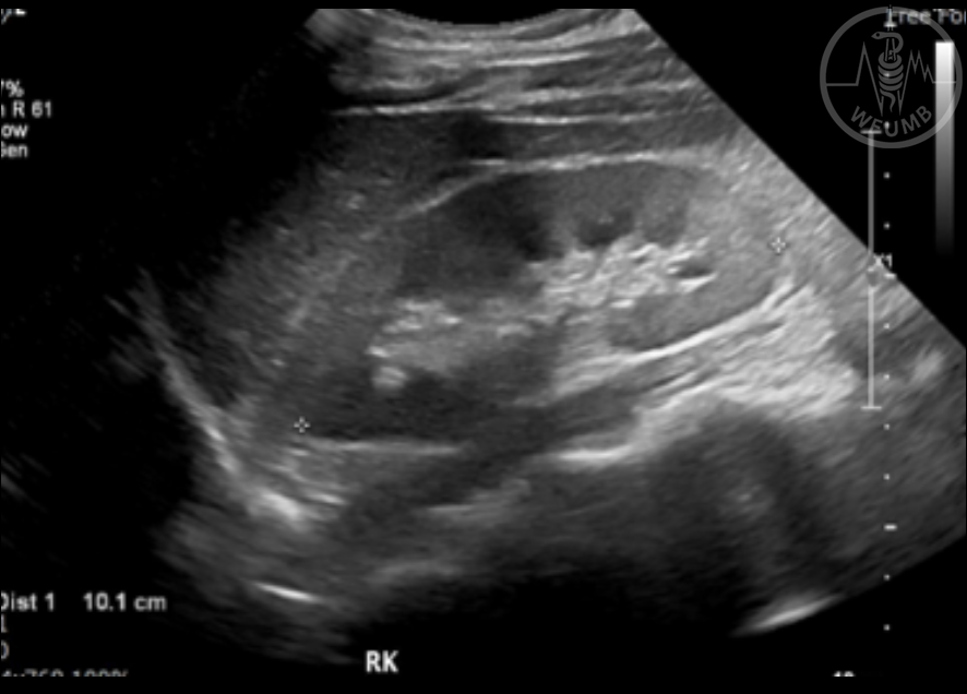
Fig 17.3a
Tubulointerstitial nephritis in a 62-year-old man. Grey-scale US image of the right kidney shows diffusely increased echogenicity of the renal cortex and slightly accentuated CMD
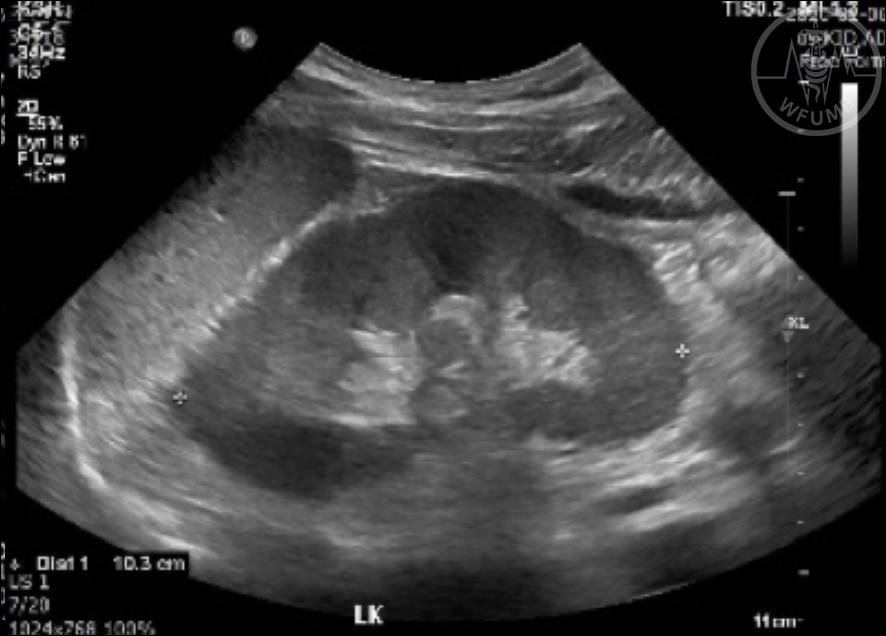
Fig 17.3b
Tubulointerstitial nephritis in a 62-year-old man. Grey-scale US image of the left kidney shows diffusely increased echogenicity of the renal cortex and slightly accentuated CMD
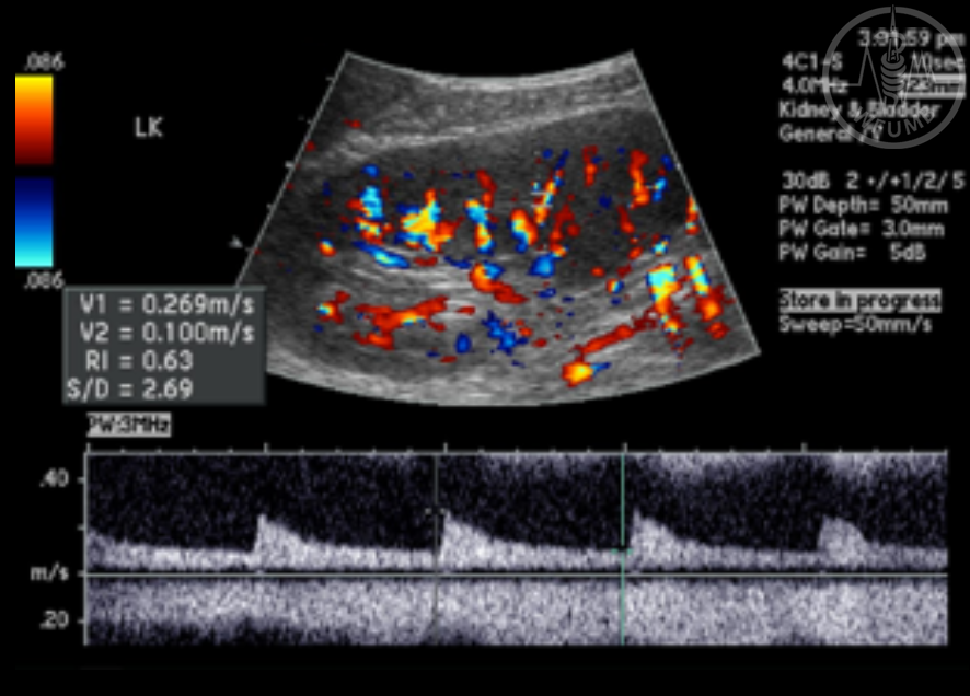
Fig 17.3c
Tubulointerstitial nephritis in a 62-year-old man.SDUS image of the right kidney shows elevated Doppler resistive index (right 0.76; left 0.78 - not shown)
Fig 17.4a
47-year-old man with diabetic nephropathy. Grey-scale US image of the right kidney shows normal size and shape of the kidney. Note renal parenchyma is slightly thick and CMD is slightly poor
Fig 17.4b
47-year-old man with diabetic nephropathy. Grey-scale US image of the kidney shows normal size and shape of the kidney. Note renal parenchyma is slightly thick and CMD is slightly poor
Fig 17.4c
47-year-old man with diabetic nephropathy. SDUS shows high resistance pattern of the Doppler spectra and elevated RI (right 0.80; left 0.77 - not shown)
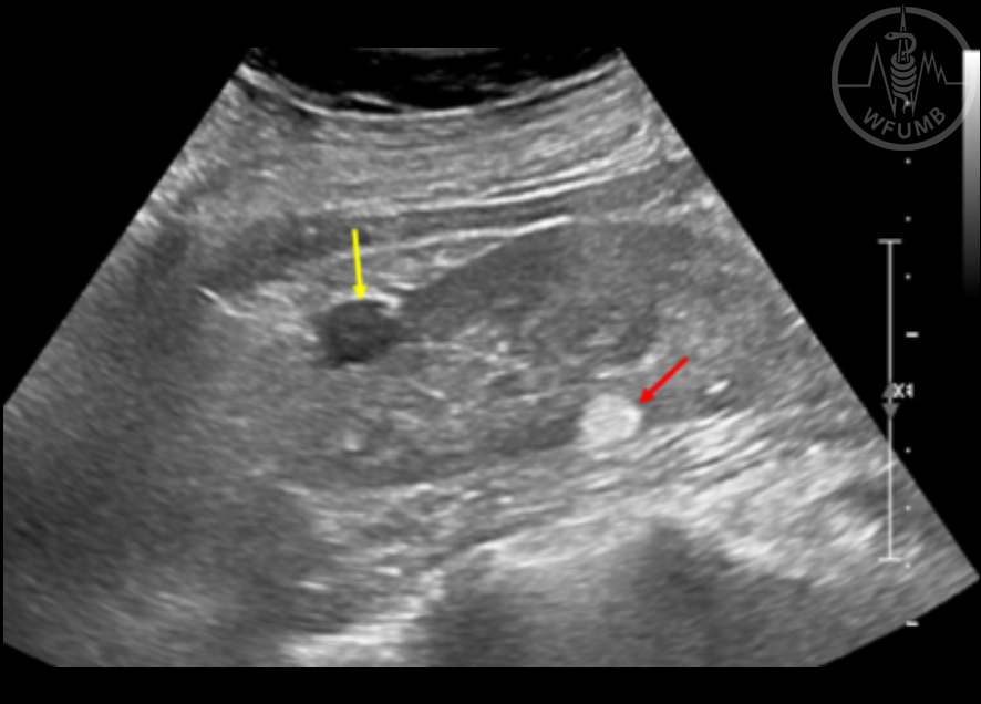
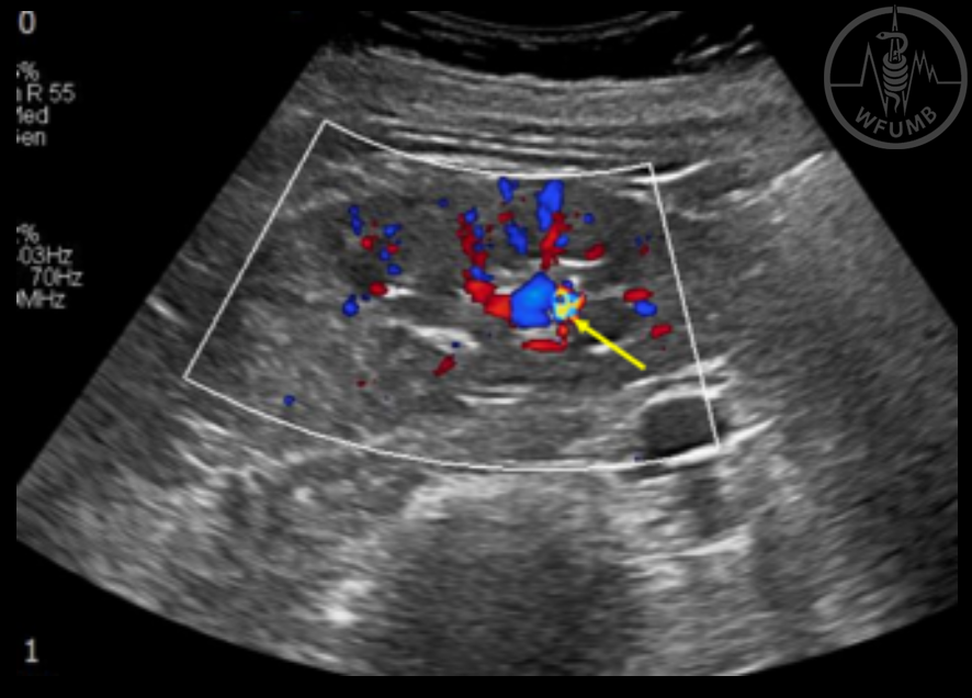
Fig 17.5
Small cortical cyst and angiomyolipoma in the same kidney in a 62-year-old woman. Longitudinal US image of the right kidney shows a small anechoic renal cyst (yellow arrow) in the anterior aspect and a hyperechoic angiomyolipoma (red arrow) in the posterior aspect of the right kidney
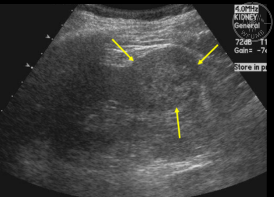
Fig 17.6a
Two most common subtypes of renal cell carcinomas, clear cell type and papillary type. Clear cell RCC in a 49-year-old man showing heterogeneous appearance on B-mode US (arrows)
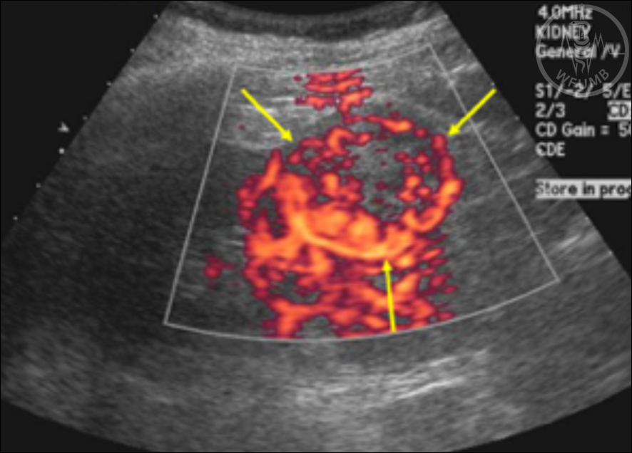
Fig 17.6b
Two most common subtypes of renal cell carcinomas, clear cell type and papillary type. Clear cell RCC in a 49-year-old man showing and hypervascular appearance in CDUS (arrows)
Fig 17.6c
Two most common subtypes of renal cell carcinomas, clear cell type and papillary type. Papillary renal cell carcinoma in an 83-year-old man showing homogenous appearance in B-mode UD (arrows)
Fig 17.6d
Two most common subtypes of renal cell carcinomas, clear cell type and papillary type. Papillary renal cell carcinoma in an 83-year-old man showing hypovascular appearance in CDUS (arrows)
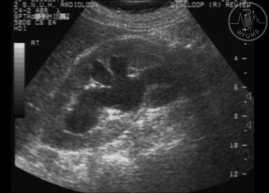
Fig 17.7a
Hydronephrosis due to ureteral obstruction by uterine cervical carcinoma in a 69-year-old woman. US of the right kidney in longitudinal plane shows dilated renal pelvic and branching calyces of the right kidney
Fig 17.7b
Hydronephrosis due to ureteral obstruction by uterine cervical carcinoma in a 69-year-old woman. Doppler US of the intrarenal artery of the right kidney shows slightly elevated Doppler RI of 0.74
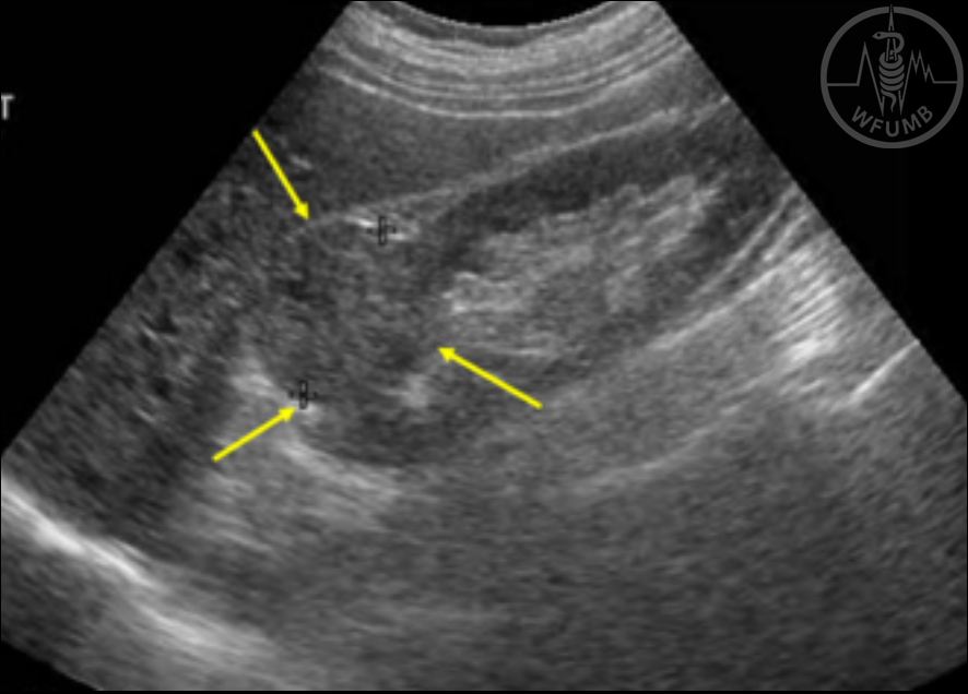
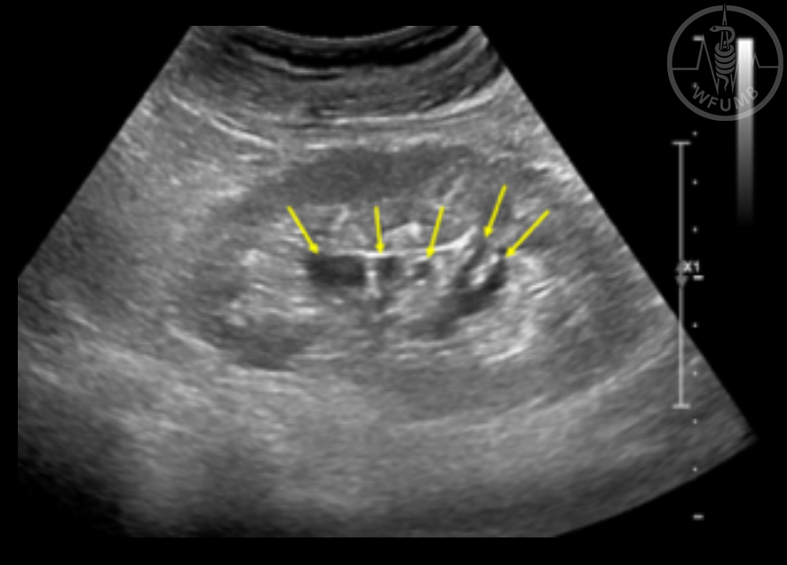
Fig 17.8a
Parapelvic cysts mimicking hydronephrosis in an 81-year-old man. Grey-scale US image of the left kidney in longitudinal plane shows multiple cystic lesions of variable shape (arrows) in the central renal sinus area. Note that those cystic lesions are not connected to each other
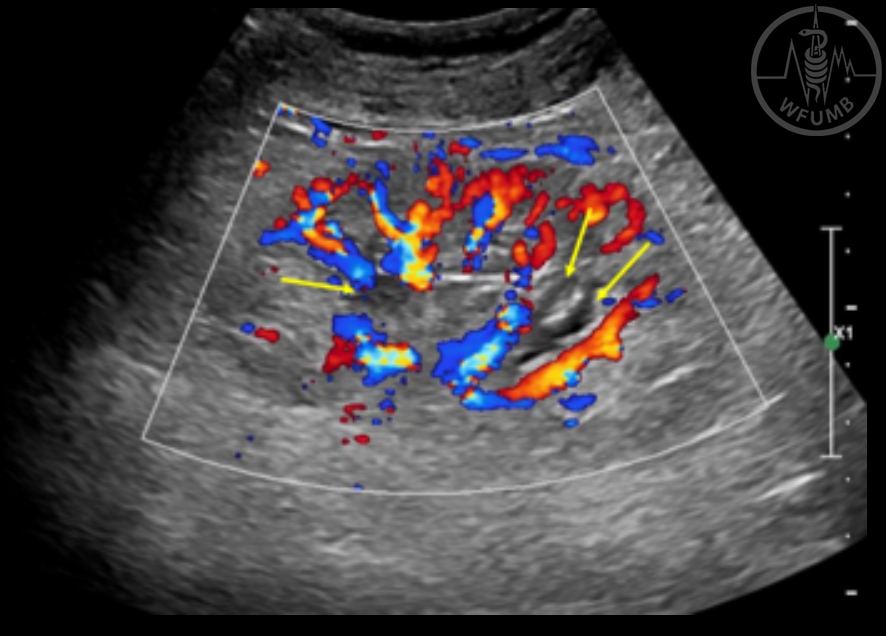
Fig 17.8b
Parapelvic cysts mimicking hydronephrosis in an 81-year-old man. CDUS image shows that these lesions (arrows) are not vessels
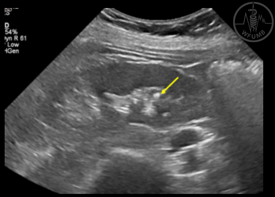
Fig 17.9a
Twinkling artifact in a 61-year-old woman with a small stone in a tip of the calyx. Grey-scale US image of the right kidney shows a small echogenic lesion (arrow) in the lower pole area. The lesion does not have definite posterior sonic shadowing
Fig 17.9b
Twinkling artifact in a 61-year-old woman with a small stone in a tip of the calyx.
CDUS shows strong twinkling artifact (arrows) that is helpful in detecting small stones
Fig 17.10a
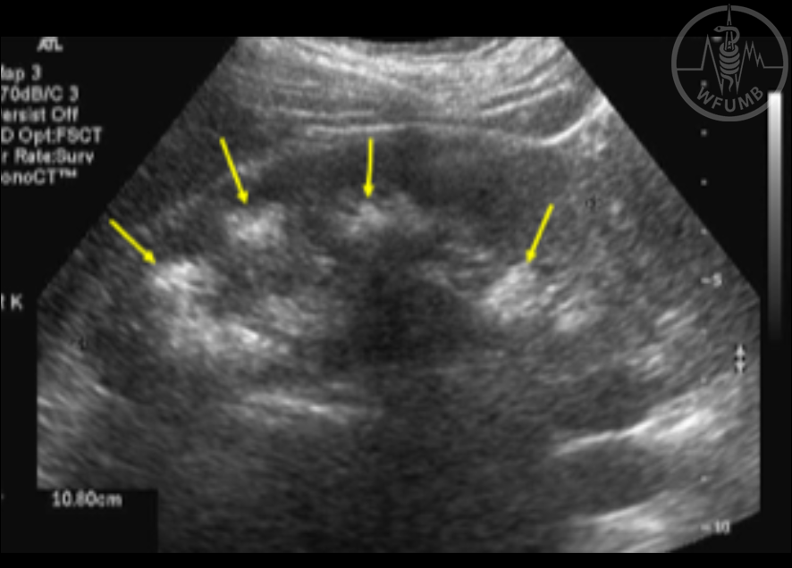
Medullary nephrocalcinosis due to renal tubular acidosis in a 45-year-old woman.
Grey-scale US image of the right kidney in longitudinal plane shows hyperechoic renal medulla (arrows) due to medullary nephrocalcinosis
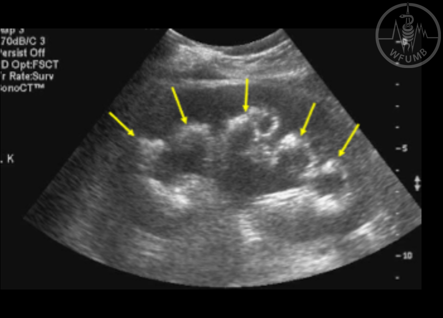
Fig 17.10b
Medullary nephrocalcinosis due to renal tubular acidosis in a 45-year-old woman.
Grey-scale US image the left kidneys in longitudinal plane shows hyperechoic renal medulla (arrows) due to medullary nephrocalcinosis. Mild hydronephrosis of the left kidney due to ureter stone
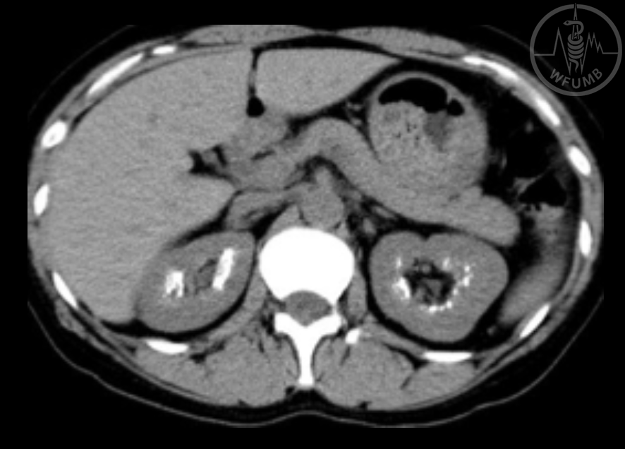
Fig 17.10c
Medullary nephrocalcinosis due to renal tubular acidosis in a 45-year-old woman.
Non-enhanced CT of the kidney shows fine calcification sin the renal medullary regions, both kidneys
Fig 17.11a
Unilateral renal artery stenosis in a 70-year-old hypertensive man. SDUS of the right kidney shows slow and weak pulse (pulsus tardus and parvus) pattern
Fig 17.11b
Unilateral renal artery stenosis in a 70-year-old hypertensive man. SDUS of the left kidney shows normal Doppler spectral pattern with sharp systolic peak
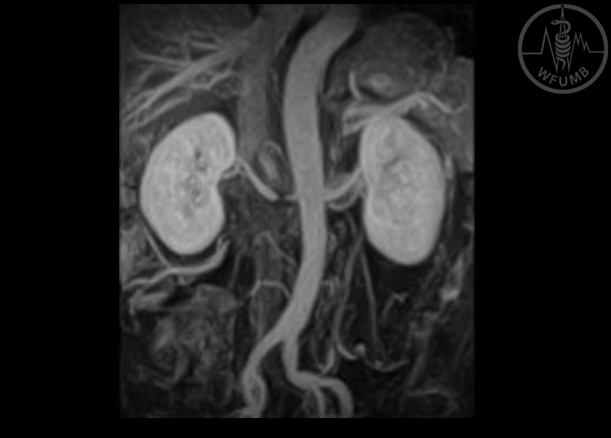
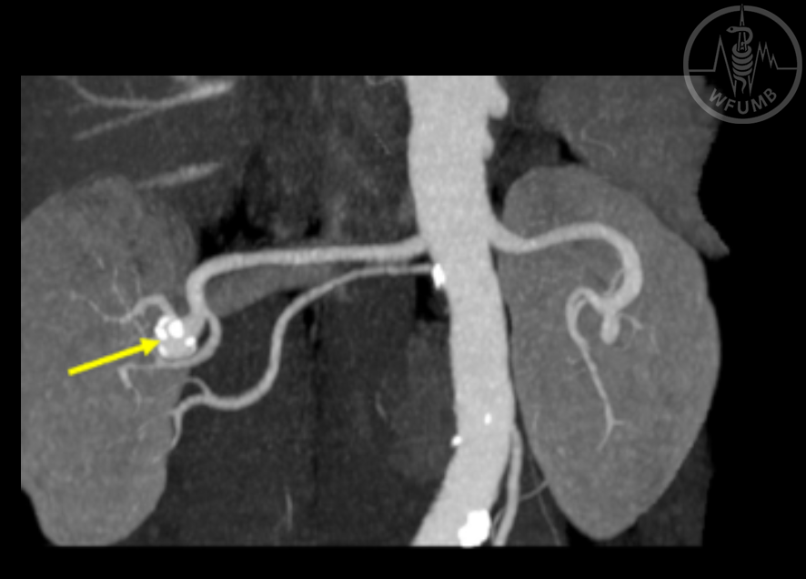
Fig 17.11c
Unilateral renal artery stenosis in a 70-year-old hypertensive man. MR angiography shows a stenotic lesion (arrow) in the right renal artery
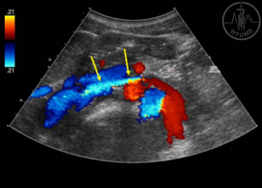
Fig 17.12a
19-year-old man with gross hematuria caused by nutcracker syndrome. Grey-scale US image in transverse plane shows left renal vein (LRV) compressed between the aorta and superior mesenteric artery (SMA) (arrow)
Fig 17.12b
19-year-old man with gross hematuria caused by nutcracker syndrome. CDUS shows jetting of bright-colored high-velocity blood flow (arrows)
Fig 17.12c
19-year-old man with gross hematuria caused by nutcracker syndrome. Peak flow velocity of LRV at aortomesenteric portion was 112 cm/sec
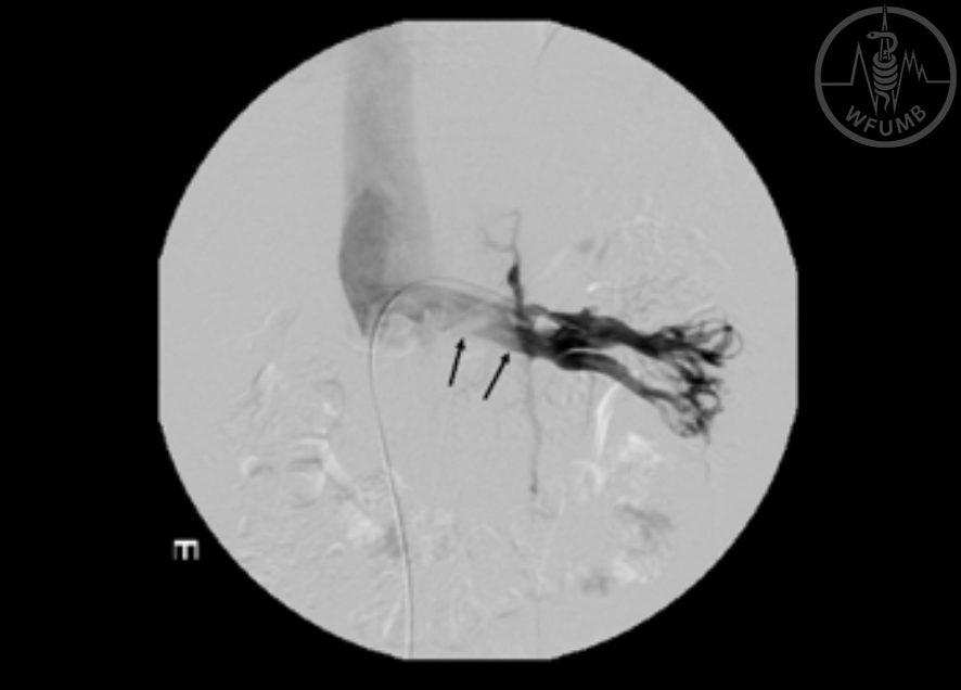
Fig 17.12d
19-year-old man with gross hematuria caused by nutcracker syndrome. Left renal venography shows compression of LRV (arrows) and pressure gradient across the compressed aortomesenteric LRV was 4mmHg
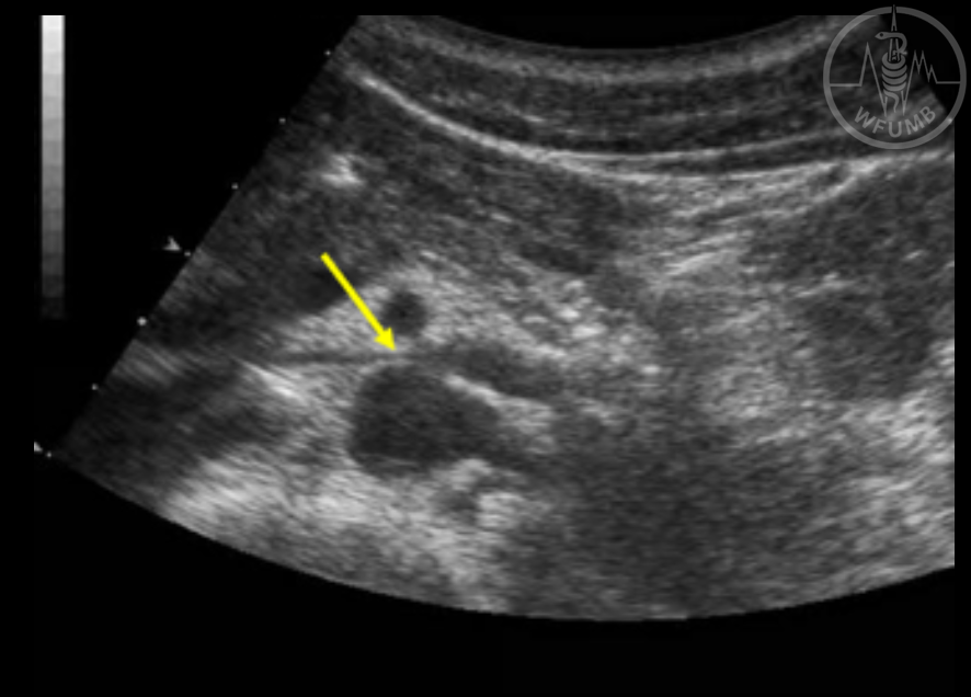
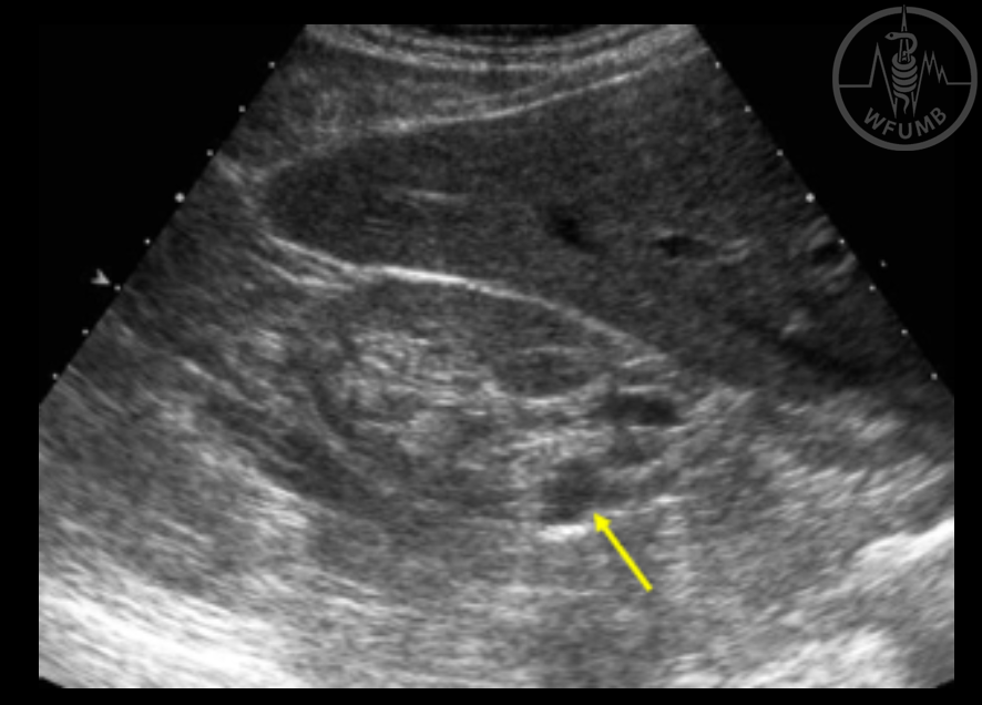
Fig 17.13a
Calcified renal artery aneurysm in a 63-year-old man. Grey-scale US image of the right kidney shows a small round anechoic lesion (arrow) in the posterior aspect of the right kidney
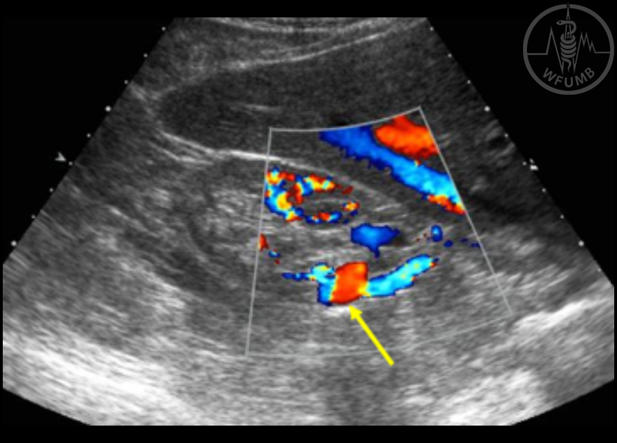
Fig 17.13b
Calcified renal artery aneurysm in a 63-year-old man. CDUS shows that the lesion is filled with flow signals of various colors (arrows) indicating that the lesion is an aneurysm
Fig 17.13c
Calcified renal artery aneurysm in a 63-year-old man. CT angiography shows a calcified aneurysm in the right kidney
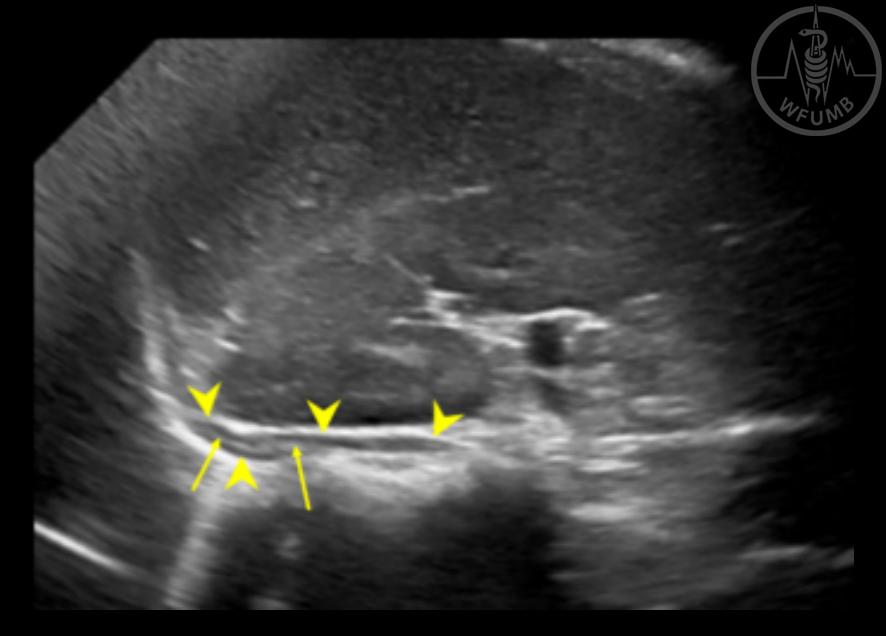
Fig 17.14a
Normal US images of the adrenal gland in a 59-year-old woman. Longitudinal (US image of the right adrenal gland shows an elongated structure with hypoechoic peripheral cortex (arrowheads) and hyperechoic central medulla (arrows)
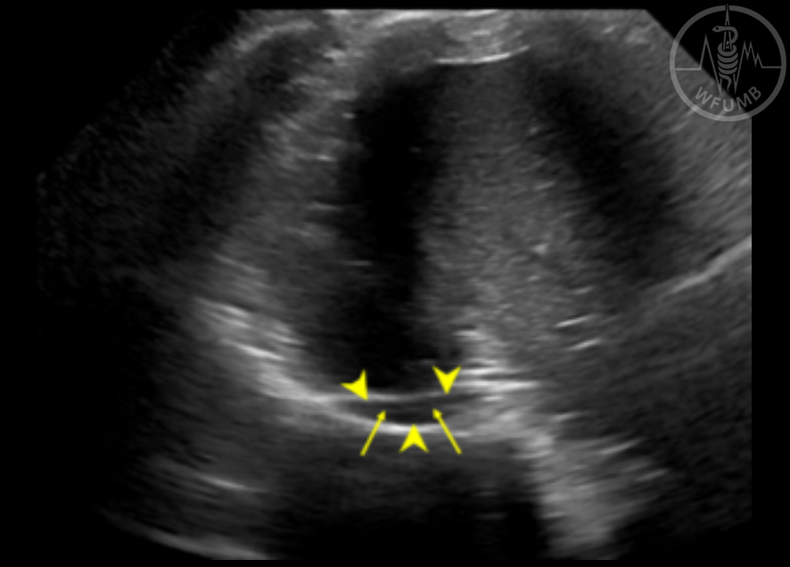
Fig 17.14b
Normal US images of the adrenal gland in a 59-year-old woman. Transverse US image of the right adrenal gland shows an elongated structure with hypoechoic peripheral cortex (arrowheads) and hyperechoic central medulla (arrows)
Fig 17.15a
Adrenal myelolipoma in a 41-year-old man. Longitudinal US image of the right kidney shows a round mass of heterogeneous hyperechogenicity (arrows)
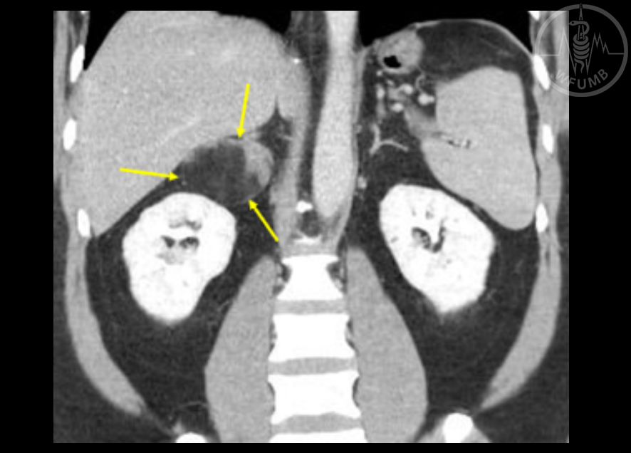
Fig 17.15b
Adrenal myelolipoma in a 41-year-old man. Contrast-enhanced CT in coronal plane shows a right adrenal mass with fatty and non-fatty components (arrows)