WFUMB Course Book
Abdominal Vessels and Portal Vein System Chaper 11 Media Library
Close window and return to Chapter 11
Chapter Images
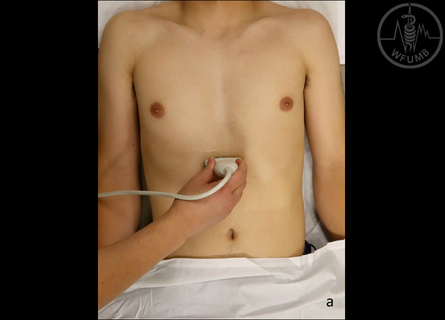
Fig 11.1a Patient in supine position, arms relaxed, along the body, transverse probe in epigastric location
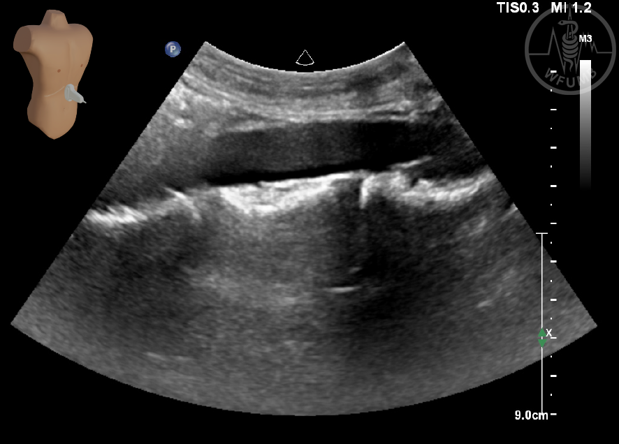
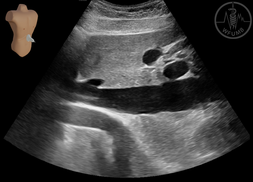
Fig 11.1b Typical anatomical structures in a transverse section in the epigastrium
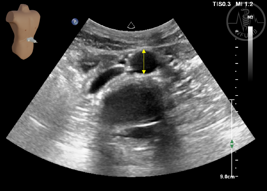
Fig 11.2a Measurement of the aorta diameter in transverse plane
Fig 11.2b Measurement of the aorta wall thickness in longitudinal plane
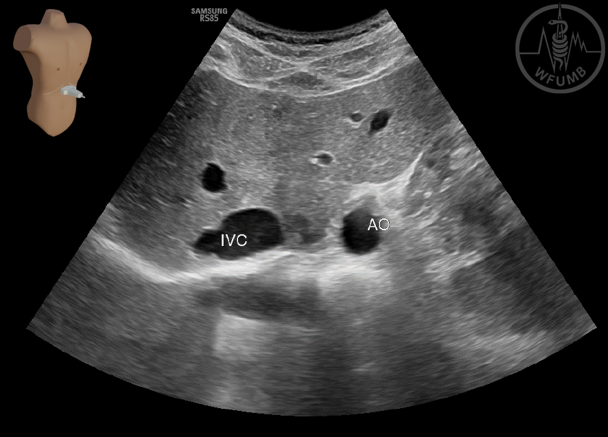
Fig 11.3a IVC in transverse plane
Fig 11.3b IVC in longitudinal plane
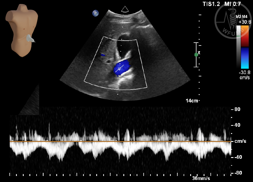
Fig 11.3c Normal flow in IVC
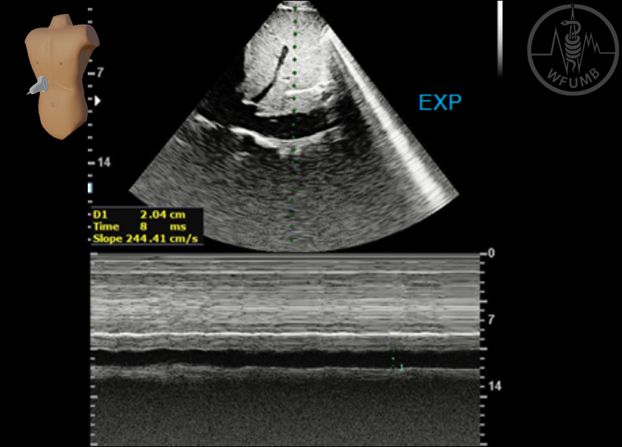
Fig 11.4a IVC collapsibility index – expiration phase patent vein
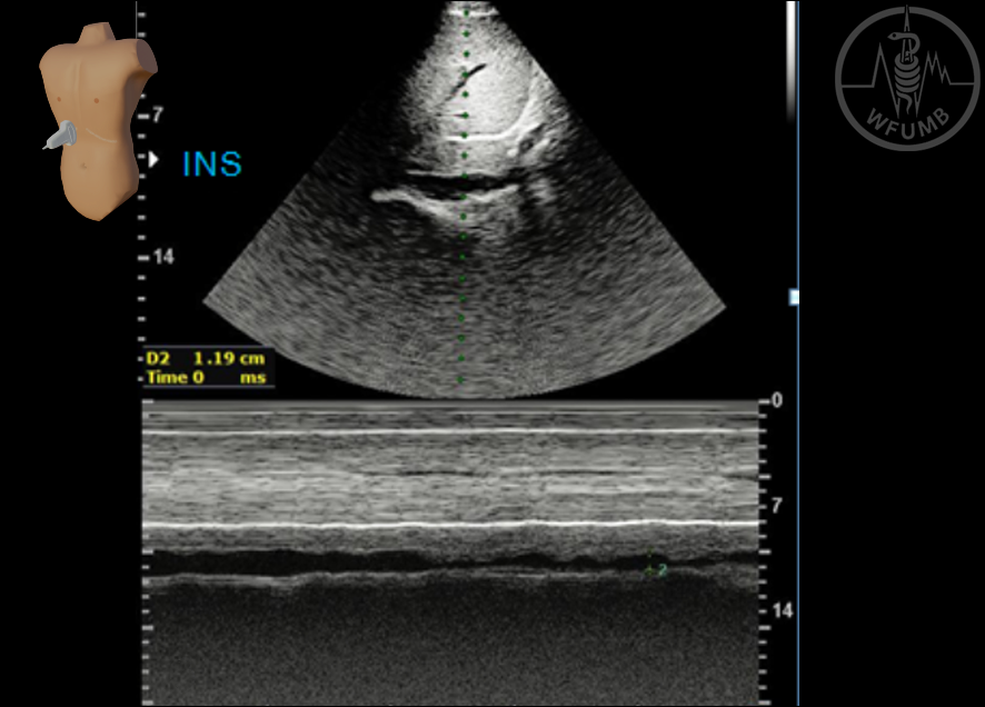
Fig 11.4b IVC collapsibility index – inspiration phase collapsing vein
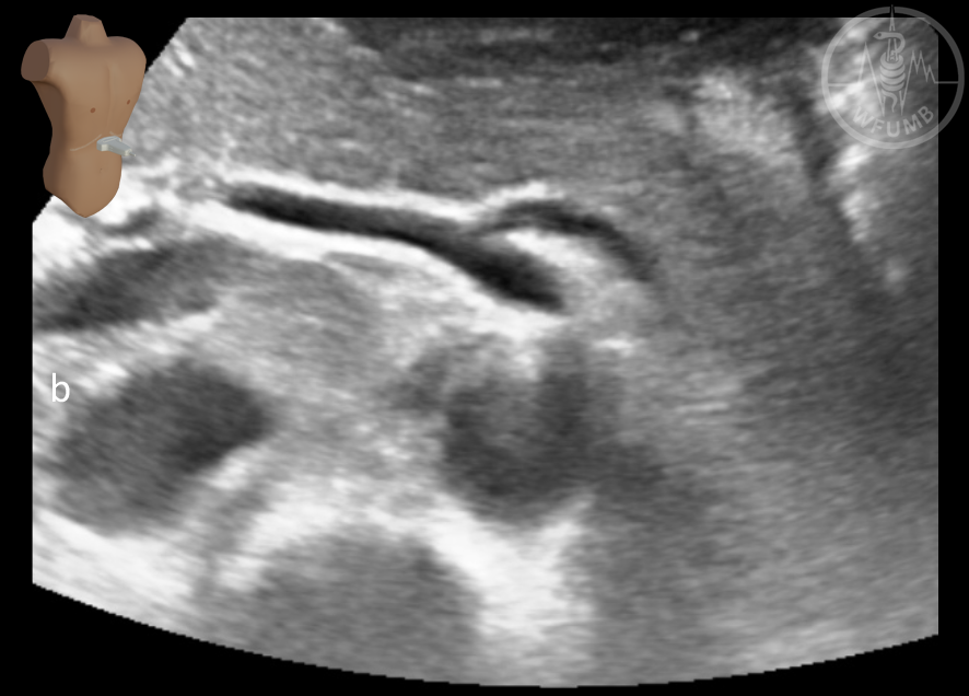
Fig 11.5a Celiac trunk (CT) and superior mesenteric artery (SMA) in longitudinal plane
Fig 11.5b Celiac trunk (CT) - seagul sign of celiac bifurcation
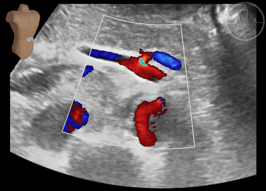
Fig 11.5c Celiac trunk (CT)– seagull sign with Color Doppler
Fig 11.6 Pulsed wave Doppler of celiac trunk
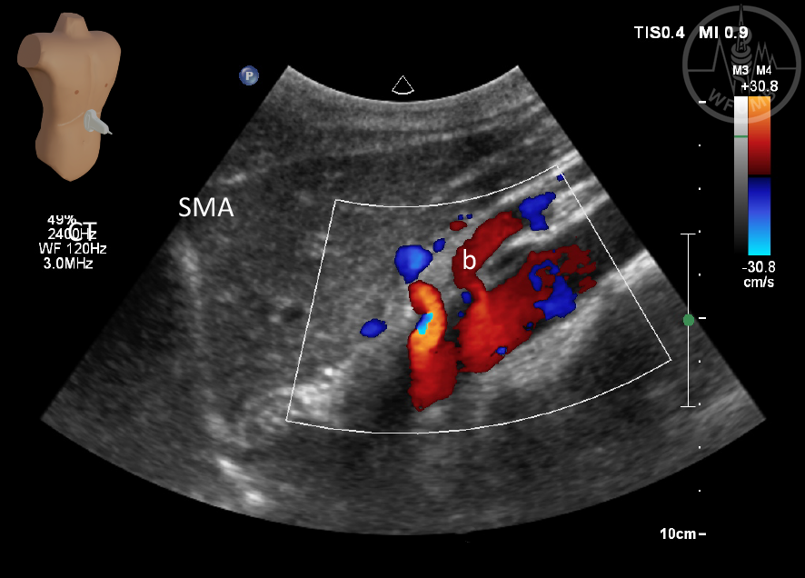
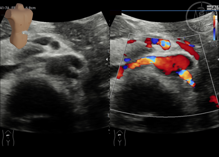
Fig 11.7a SMA and SMV in a transverse plane
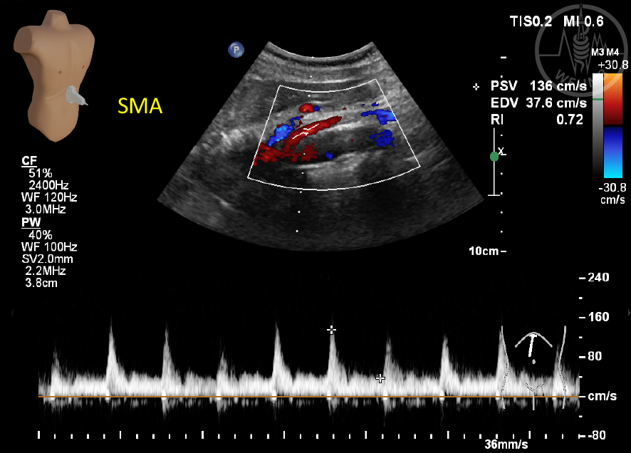
Fig 11.7b Flow in the SMA in a longitudinal plane
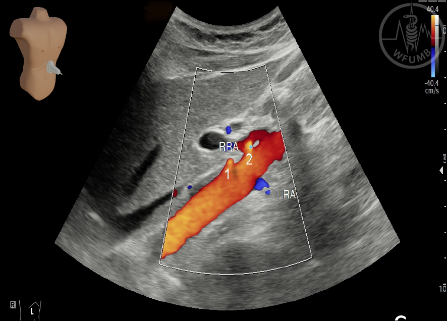
Fig 11.8a Renal arteries. Typical both arteries in transverse section
Fig 11.8b Renal arteries. Probe position for investigation of right artery on transverse oblique section
Fig 11.8c Renal arteries. Both right renal arteries and left renal on coronal section
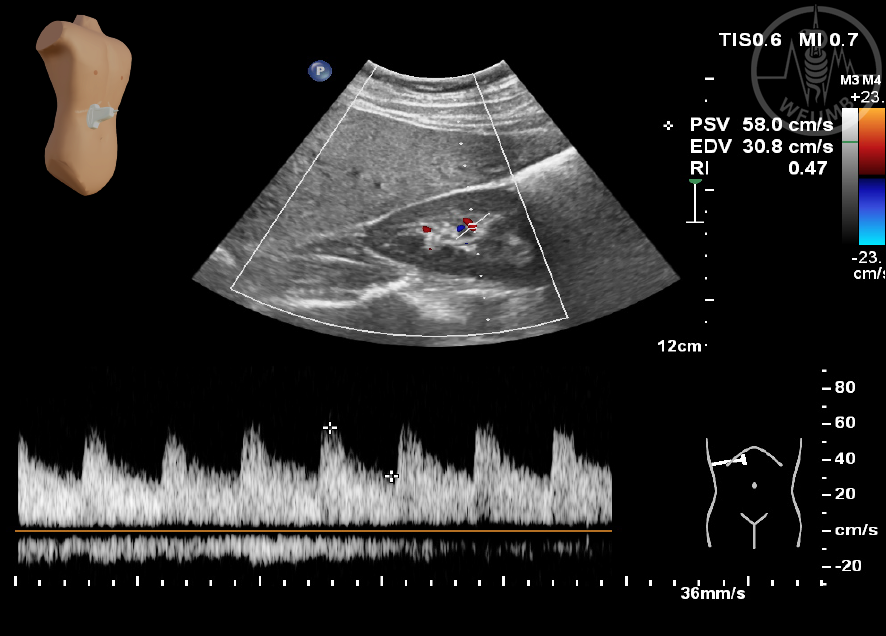
Fig 11.8d Renal arteries. Intrarenal artery flow waveforms on pulsed wave Doppler
Fig 11.9a Portal vein in longitudinal plane, flow color red – towards probe
Fig 11.9b Flow in portal vein on Pulsed Wave Doppler
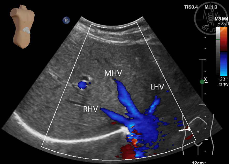
Fig 11.10 Hepatic vein confluence (Color Doppler), color blue – flow away from the probe, right, middle and left hepatic veins (RHV, MHV, LHV)
Chapter Videos
This website uses cookies to improve your experience. By using this website you agree to our
Data Protection Policy
.
Read more
Accept all
Translate »