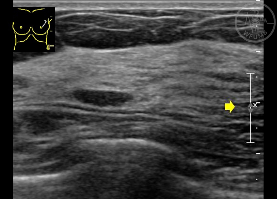
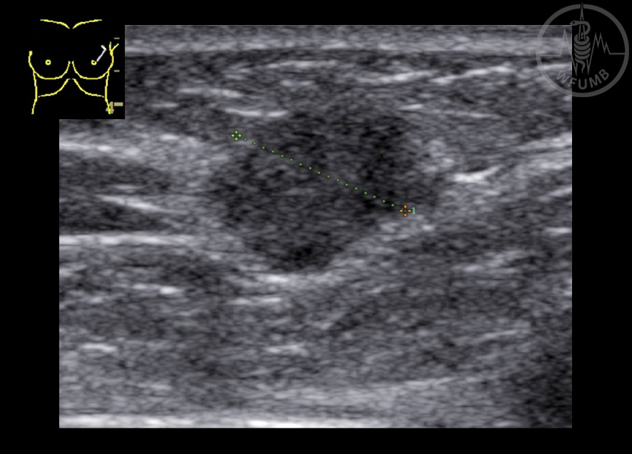
Fig 20.3.1
Image demonstrates the focal zone (arrow) in the plane of the mass
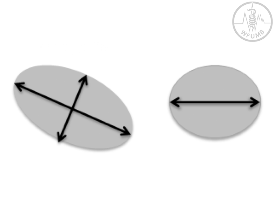
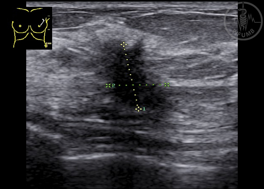
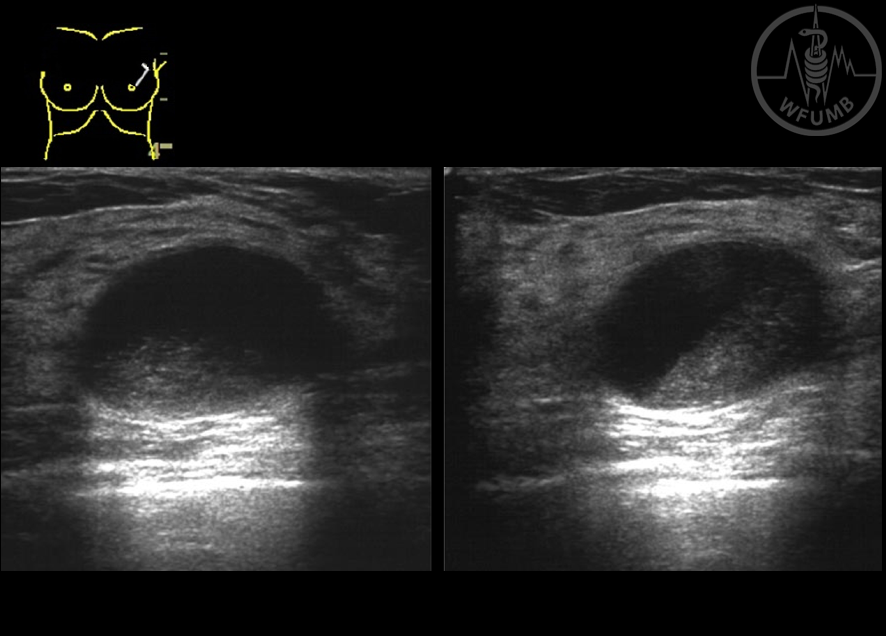
Fig 20.3.2
Correct way to measure the longest axis and its perpendicular axis (left). On the right we see the orthogonal axis
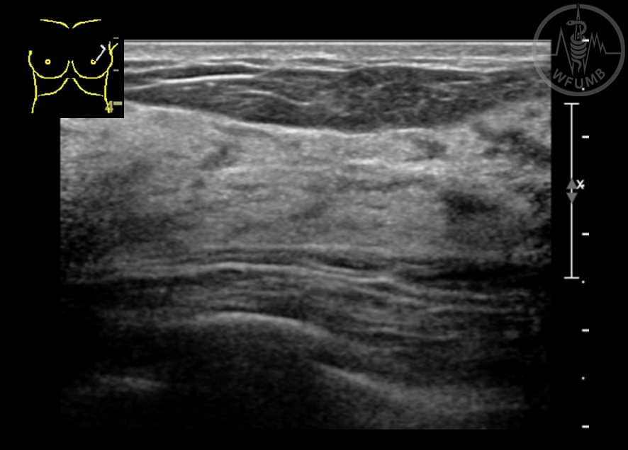
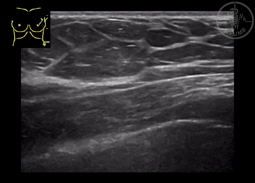
Fig 20.3.3a
Normal breast anatomy: notice the different layers of the breast in the ultrasound image
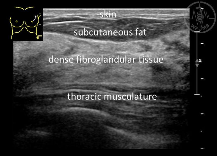
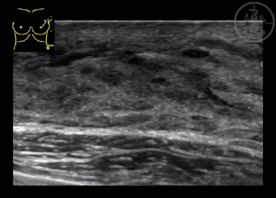
Fig 20.3.3b
Normal breast anatomy: notice the different layers of the breast in the ultrasound image
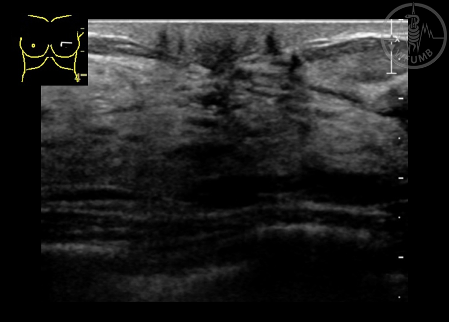
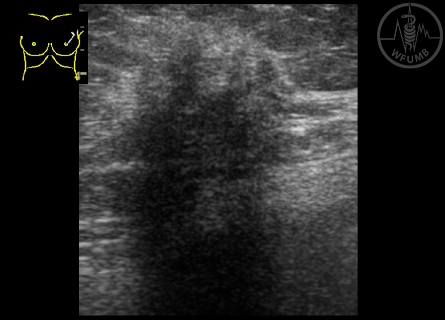
Fig 20.3.3c
Normal breast anatomy. Retroareolar region with the characteristic acoustic shadow
Fig 20.3.3d
Breast composition: Fatty infiltration in a 70 year old lady
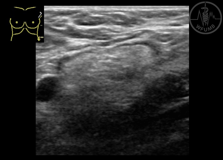
Fig 20.3.3e
Breast composition: Fibroglandular structure in a 20 year old female
Fig 20.3.4a
Normal axillary lymph node, with thin hypoechoic peripheral cortical (arrow)
Fig 20.3.4b
Pathological lymph node, with complete obliteration of the hilum (biopsy confirmed metastatic carcinoma)
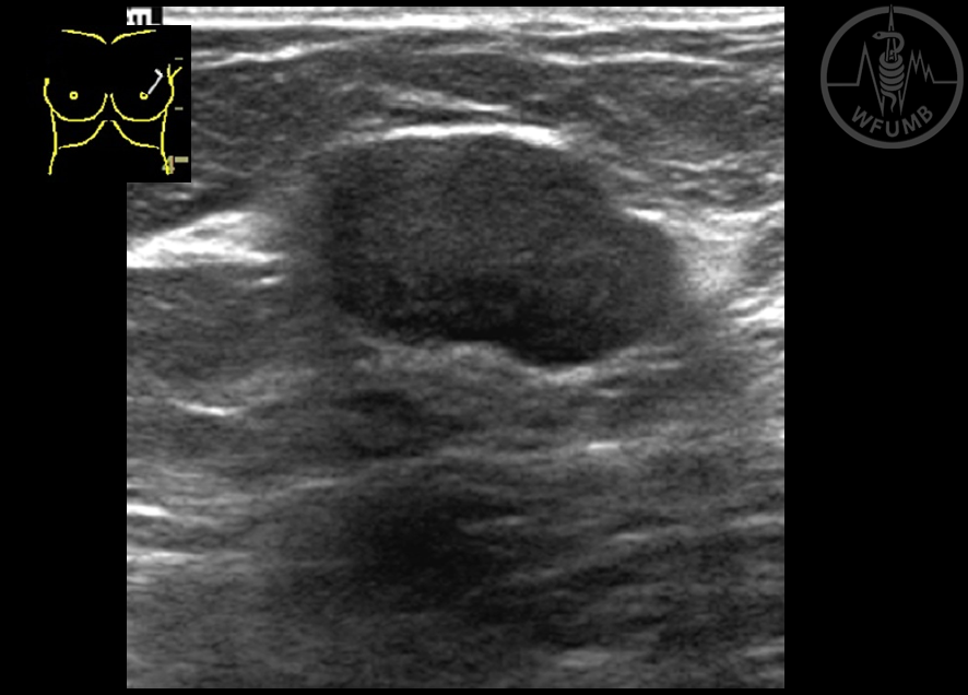
Fig 20.3.5a
Lexicon. Shape: Oval x Round x Irregular. Hypoechoic oval and circumscribed mass
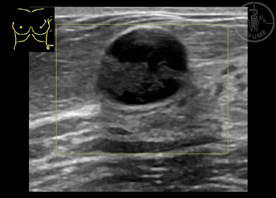
Fig 20.3.5b
Lexicon. Shape: Oval x Round x Irregular. A complex (solid and cystic) round and circumscribed nodule
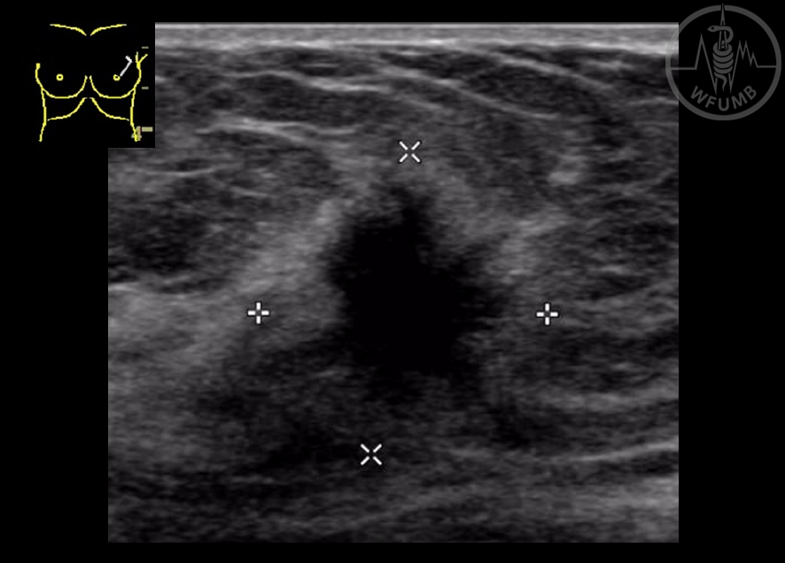
Fig 20.3.5c
Lexicon. Shape: Oval x Round x Irregular. An irregular hypoechoic mass with spiculated margins (histopathology: infiltrating ductal carcinoma at percutaneous biopsy)
Fig 20.3.6a
Lexicon: Orientation: Parallel x Not Parallel. Oval circumscribed mass with parallel orientation
Fig 20.3.6b
Lexicon: Orientation: Parallel x Not Parallel. Irregular spiculated mass with no parallel orientation
Fig 20.3.8
Lexicon. Margin: Not circumscribed - Indistinct margin lesion with significant posterior shadowing
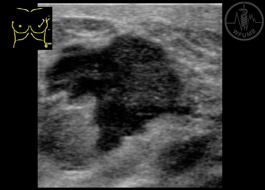
Fig 20.3.9
Lexicon. Margin: Not circumscribed - angular. Complex (solid and cystic) mass with angular margin
Fig 20.3.10
Lexicon. Margin: Not circumscribed - microlobulated. A pattern that is often difficult to identify
Fig 20.3.11
Lexicon. Margin: Not circumscribed - spiculated. Hypoechoic mass with irregular shape and spiculated margin. US guided core biopsy confirmed malignant result
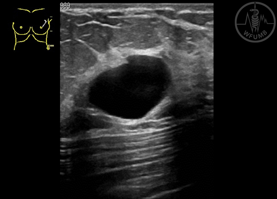
Fig 20.3.12
Lexicon. Echo pattern - anechoic: Simple cyst with posterior enhancement. A classic BI-RADS 2 finding
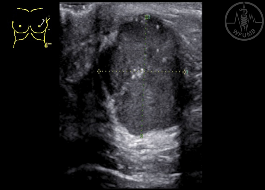
Fig 20.3.13
Lexicon. Echo pattern - hypoechoic: Image shows a hypoechoic no parallel (vertical) circumscribed mass
Fig 20.3.14
Lexicon. Echo pattern - hyperechoic: Image shows a hyperechoic mass (arrow) with no suspicious features
Fig 20.3.15
Lexicon. Echo pattern - complex: Image shows a complex (solid and cystic) mass
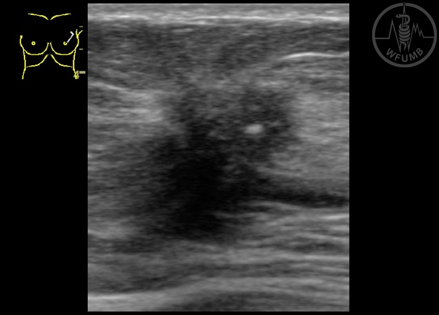
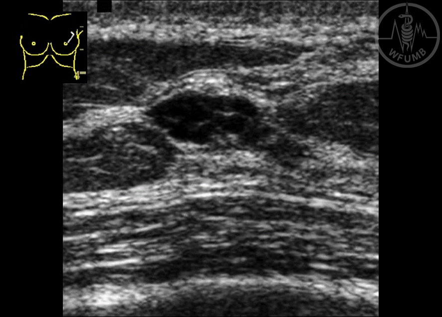
Fig 20.3.16a
B-Mode US: suspicious lump, no parallel, with microlobulated margins and small calcifications inside
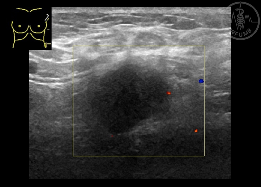
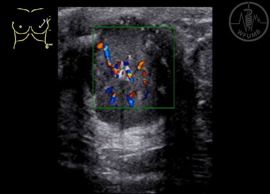
Fig 20.3.16b
Suspicious lump. Color Doppler can prove the solid nature of the lesion
Fig 20.3.17a
Special cases. Clustered microcysts
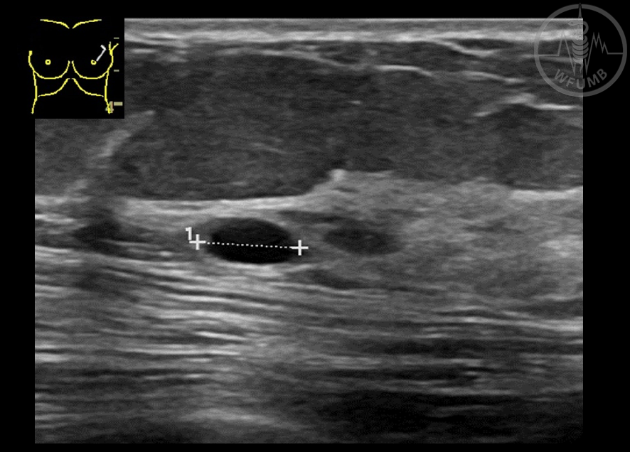
Fig 20.3.17b
Special cases. Complicated cyst (circumscribed cyst with debris inside that moved with the patient’s movement). When isolated these findings can be classified in category BI-RADS 3. If multiple and bilateral, benign assessment can be chosen (BI-RADS 2)