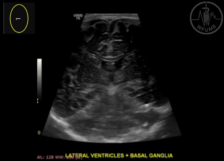
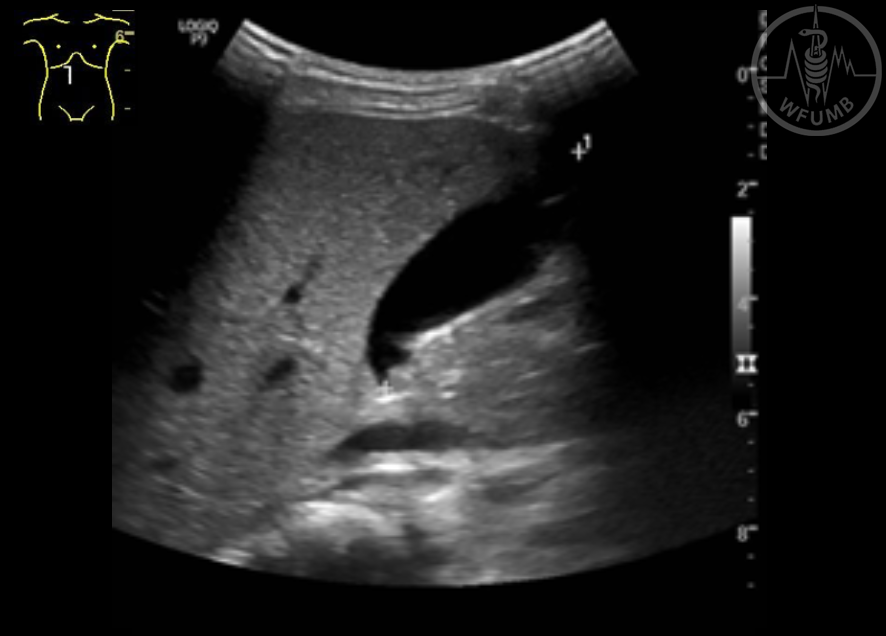
Fig 22.1
Coronal view, in gray scale showing lateral ventricles, third ventricle, basal ganglia and some of the tentorium
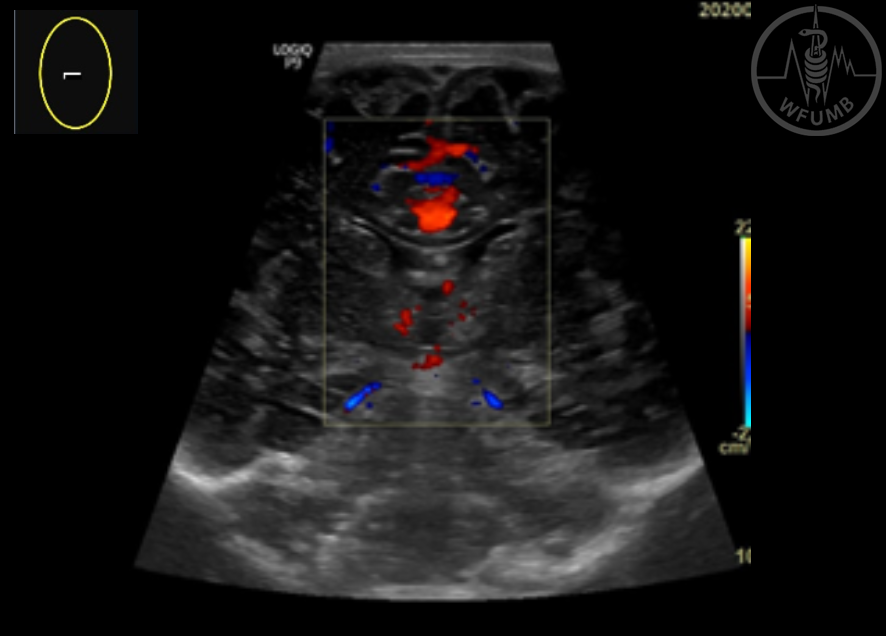
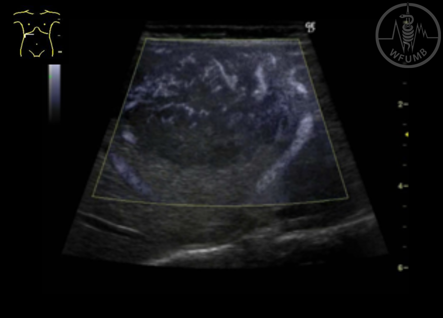
Fig 22.2
Coronal view, in gray scale and color Doppler showing lateral ventricles, third ventricle, basal ganglia and some of the tentorium
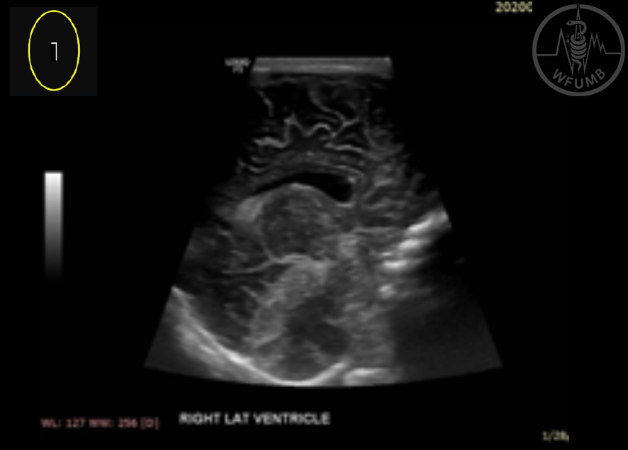
Fig 22.3
Sagittal view, gray scale showing lateral ventricle, some of the corpus callosum, cerebellum and parietal brain
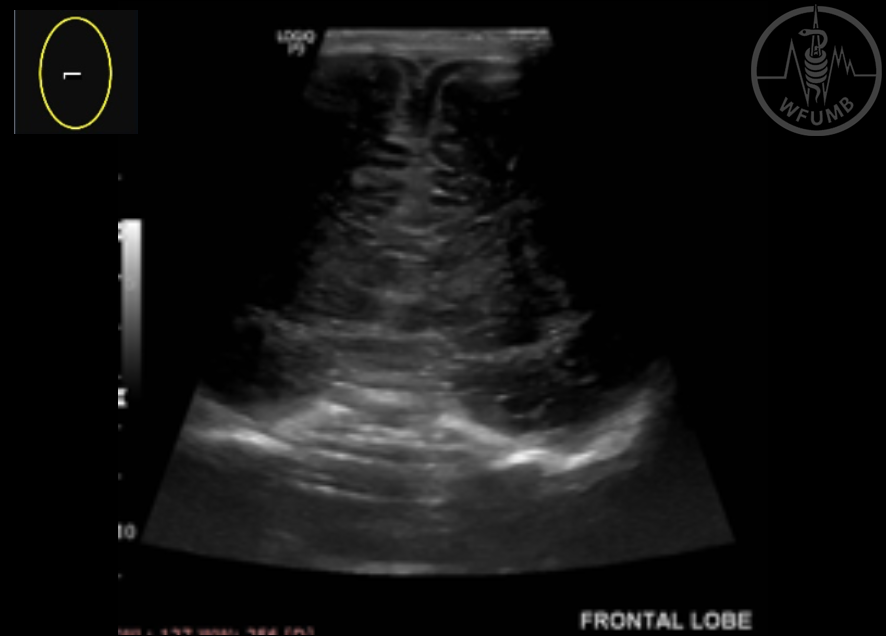
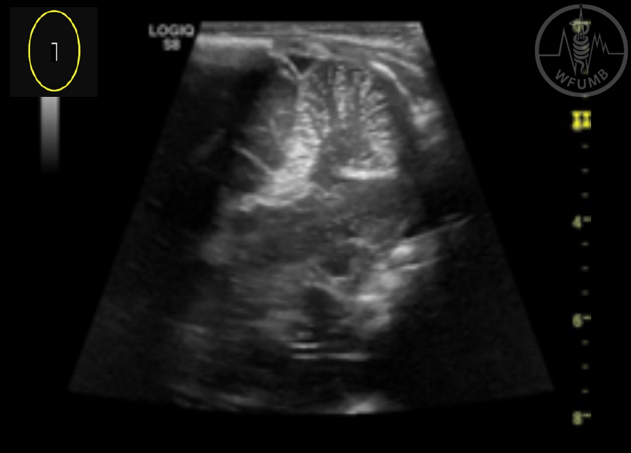
Fig 22.4
Coronal view, gray scale US showing frontal brain and some of the anterior commissure
Fig 22.5
Sagittal view, gray scale US showing frontal brain and some of the anterior commissure
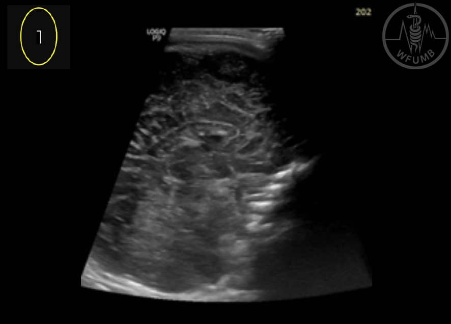
Fig 22.6
Coronal view, gray scale US showing temporal brain and cerebellum and some of the ventricular system
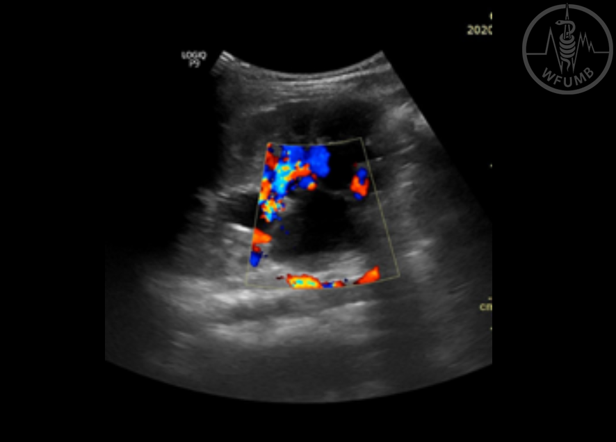
Fig 22.7
Coronal view, gray scale and Doppler showing temporal brain and cerebellum and some of ventricular system
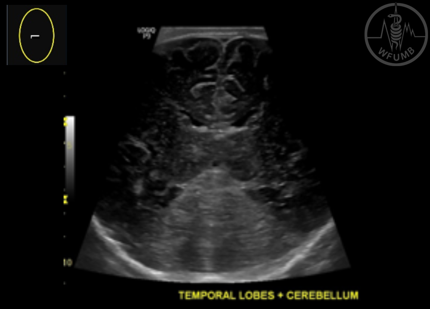
Fig 22.8
Gray scale US showing brain and cerebellum
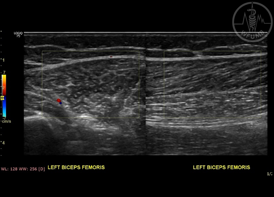
Fig 22.9
Grayscale US, sagittal view showing the echotexture of the normal muscle and subcutaneous tissue
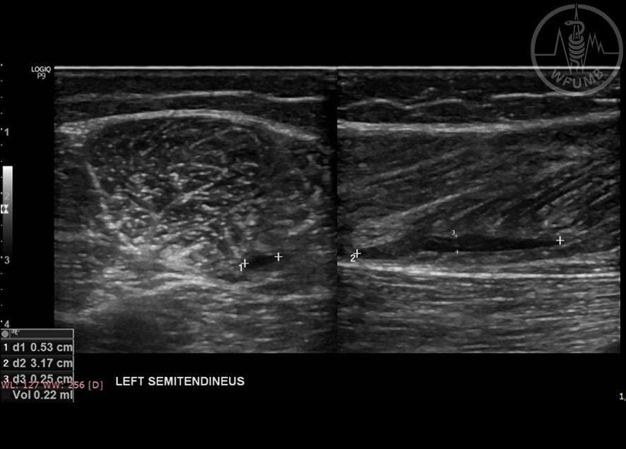
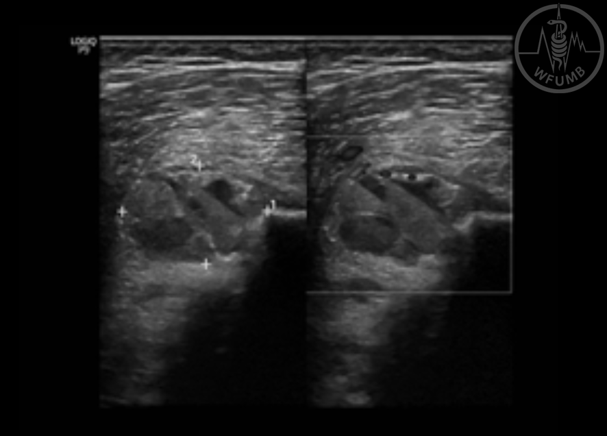
Fig 22.10
Axial and coronal view, gray scale showing some fluid collection underneath left semitendinosus muscle insertion
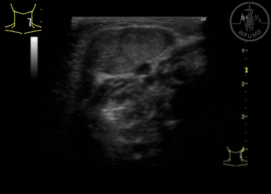
Fig 22.11
Fibromatosis colli in 1 month old baby girl
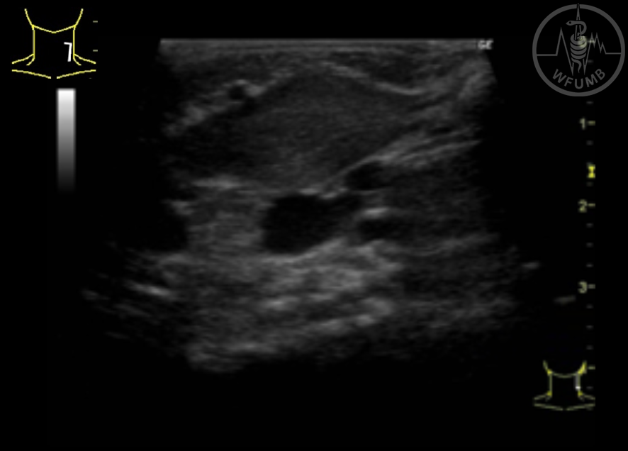
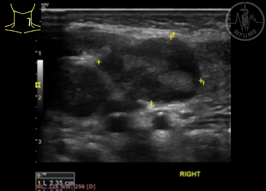
Fig 22.12
Fibromatosis colli in 1 month old baby girl
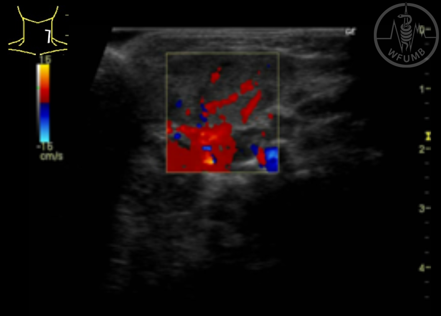
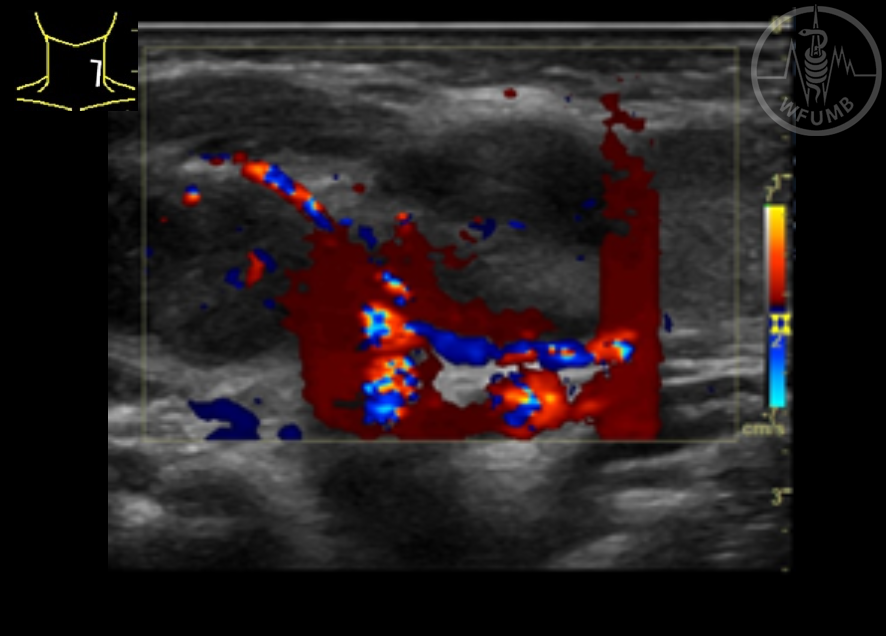
Fig 22.13
Gray scale US and color Doppler scan showing fibromatosis colli in an 1 month old baby girl
Fig 22.14
Axial view, gray scale US showing inflammatory lymph nodes in the neck
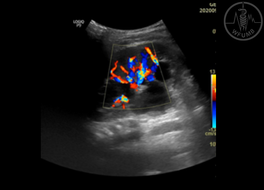
Fig 22.15
Axial view, gray scale and color Doppler showing inflammatory lymph nodes in the neck
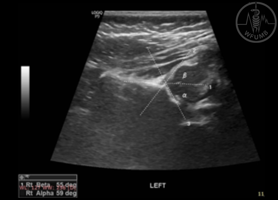
Fig 22.16
Coronal view, gray scale showing osseus and musculotendinous structure of the hip
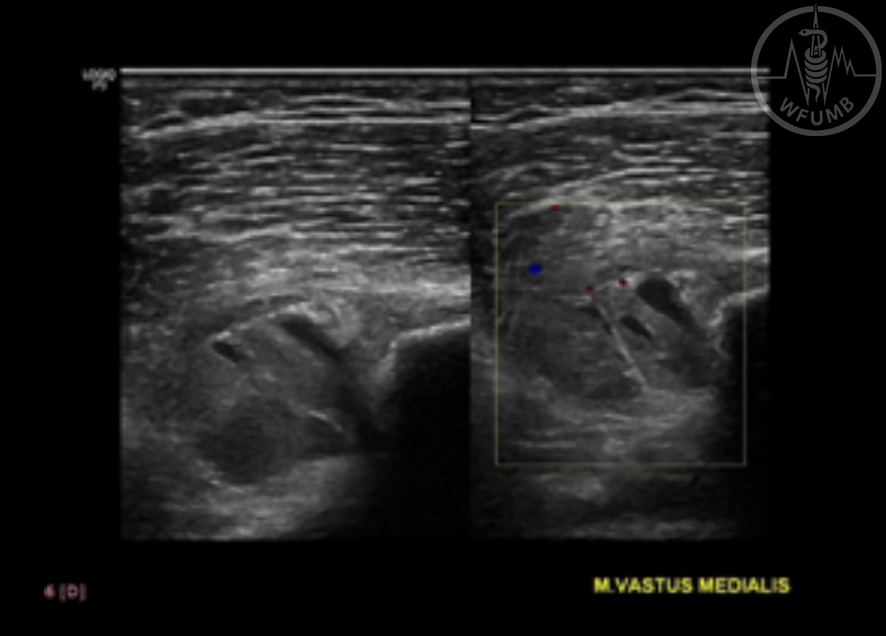
Fig 22.17
Axial view, gray scale and color Doppler showing a heterogeneous lesion nearby vastus medialis muscle, which resulted in MRI a osseous sarcoma
Fig 22.18
Axial view, gray scale and color Doppler showing fluid collection in the mass
Fig 22.19
Sagittal view, gray scale and power Doppler showing the aorta and the superior mesenteric artery
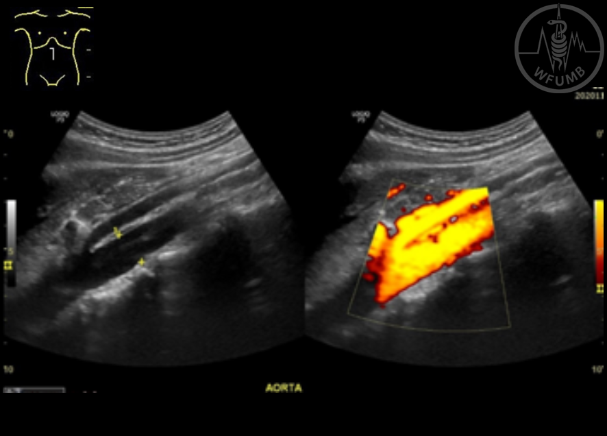
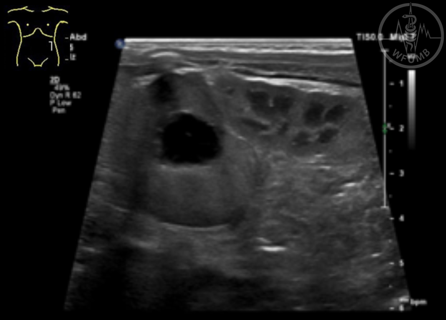
Fig 22.20
Axial and sagittal view, gray scale showing a umbilical hernia
Fig 22.21
Coronal view, gray scale US showing the liver and right kidney
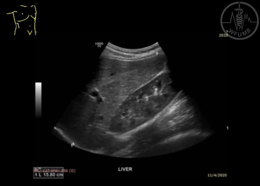
Fig 22.22
Coronal view, gray scale US showing a normal gallbladder
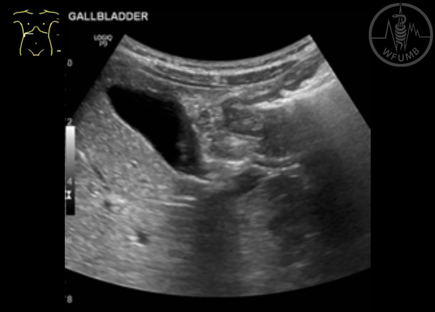
Fig 22.23
Sagittal view, gray scale US showing a normal gallbladder
Fig 22.24
Ultrasonographic images of hepatic hepatoblastoma
Fig 22.25
Ultrasonographic images of hepatic hepatoblastoma
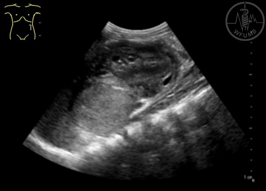
Fig 22.26
Ultrasonographic image of neuroblastoma
Fig 22.27
Ultrasonographic image of a neuroblastoma
Fig 22.28
Ultrasonographic image of Wilms tumor
Fig 22.29
Ultrasonographic image of Wilms tumor
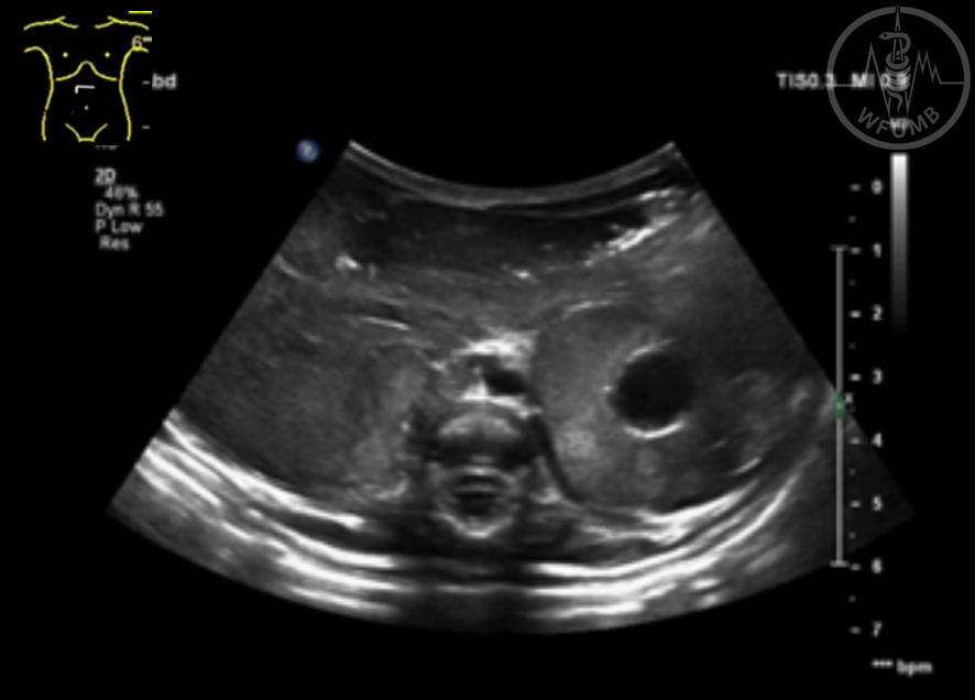
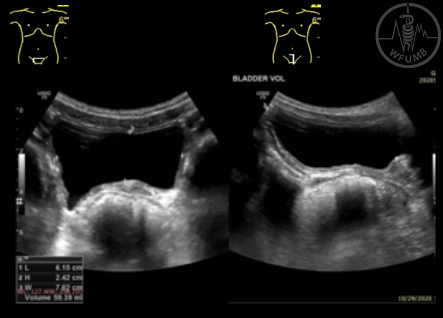
Fig 22.30
Axial and sagittal view, gray scale US showing normal bladder
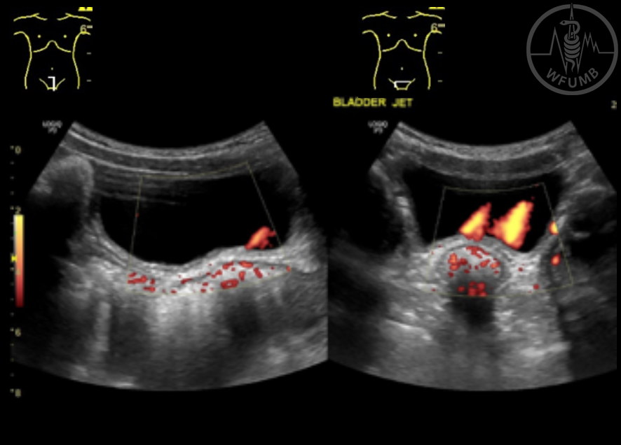
Fig 22.31
Sagital and axial view, power Doppler showing bladder ``jet``
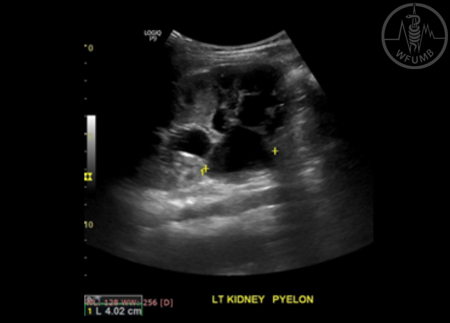
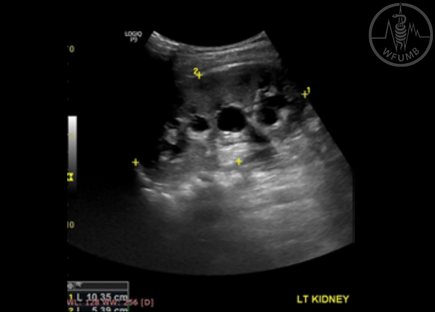
Fig 22.32
Coronal view, gray scale US showing left kidney ureteropelvic junction obstruction pre void
Fig 22.33
Coronal view, color Doppler showing left kidney ureteropelvic junction obstruction pre void
Fig 22.34
Coronal view, gray scale US showing left kidney ureteropelvic junction obstruction post void
Fig 22.35
Coronal view, color Doppler US showing left kidney ureteropelvic junction obstruction post void