Fig 29.1
Schematic image of lower extremity deep veins
Fig 29.2
High frequency linear transducer
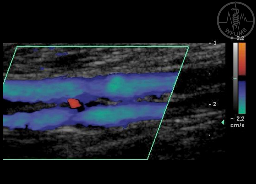
Fig 29.3
Distal leg. Low velocity flow in posterior tibial veins well depicted adjusting PRF to low settings
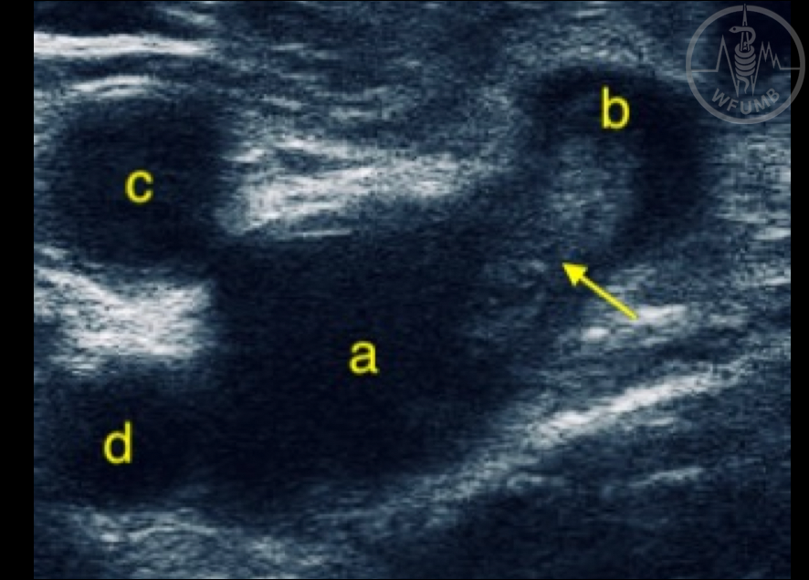
Fig 29.4
Transversal plane in crural region: a: common femoral vein; b - great saphenous mouth with thrombus; c - superficial femoral artery; d - deep femoral artery; Arrow - thrombus
Fig 29.5a
Transversal plane in crural region, no compression
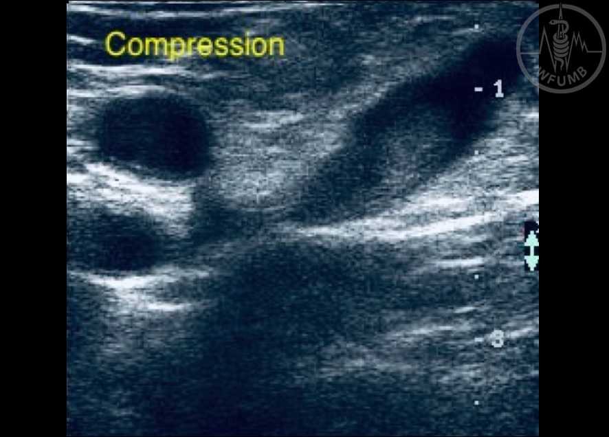
Fig 29.5b
Transversal plane in crural region. Right positive compression maneuver in great saphenous with thrombus. Common femoral with normal wall contact
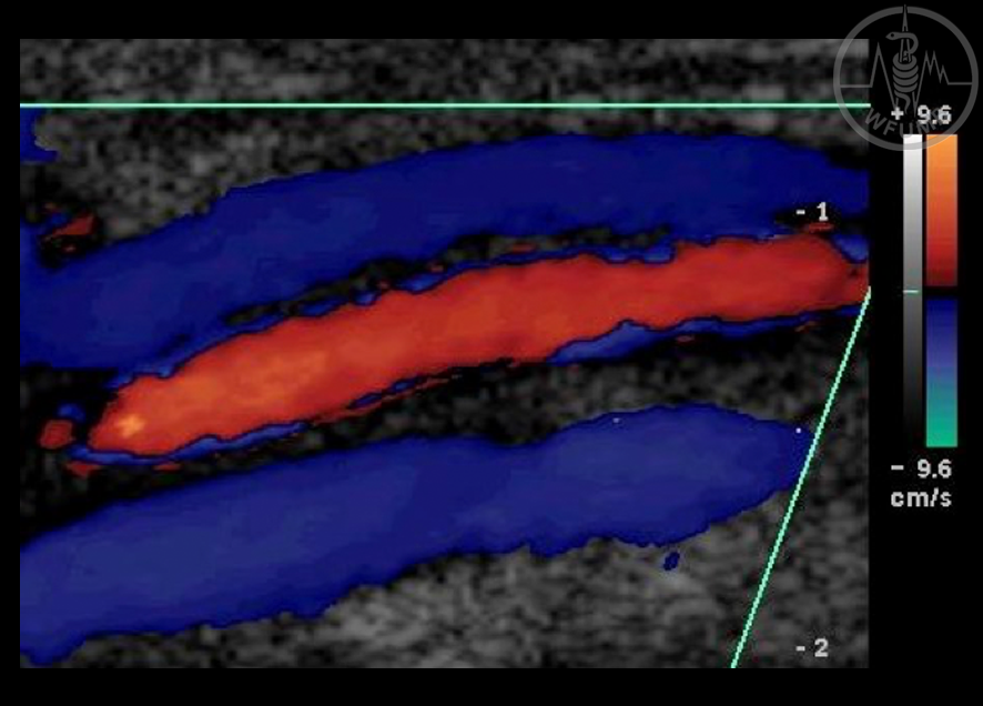
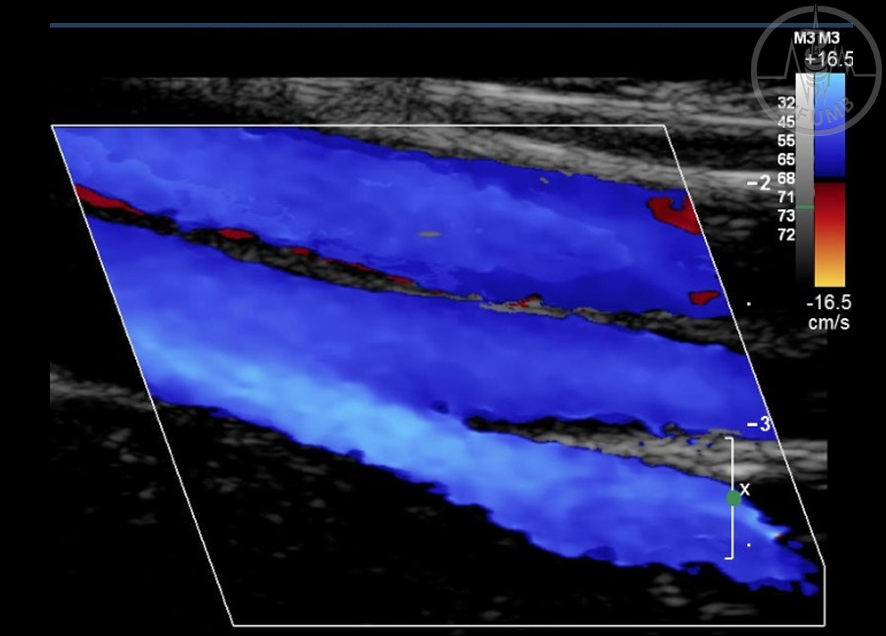
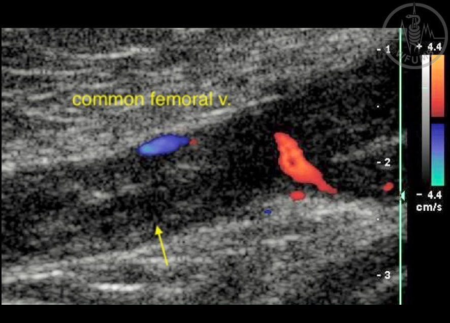
Fig 29.6
Power Doppler longitudinal image of femoral v. and deep femoral v. confluence to common femoral vein. Because non-directional Power Doppler was used, the superficial femoral artery on top is coded with the same color
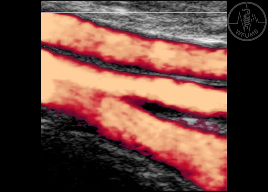
Fig 29.7
Longitudinal plane of posterior tibial veins. Color Doppler shows 2 veins coded in blue and a tiny communicant vein coded in red, bringing flow from lower to upper vessel
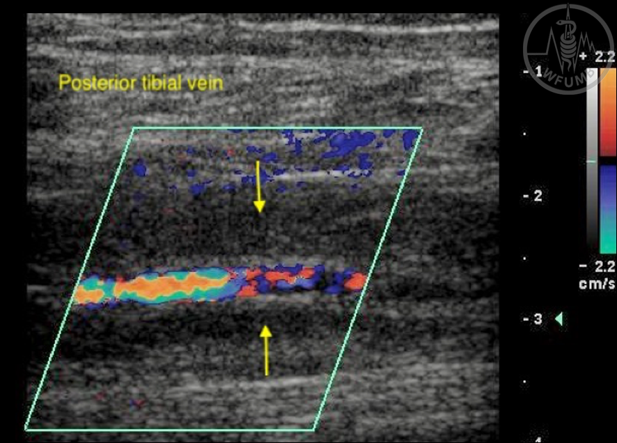
Fig 29.8
Posterior tibial v. thrombosis (arrows) Low amplitude echoes (hypoechoic) within both vessels. No color signal with the lowest PRF of the system. Aliasing in the posterior tibial artery due the adjusted low PRF (pulse repetition frequency)
Fig 29.9
Normal longitudinal CD image of femoral v. and deep femoral v. confluence to common femoral vein
Fig 29.10
Normal spectral analysis shows phasic and oscillating flow associated with respiratory movements
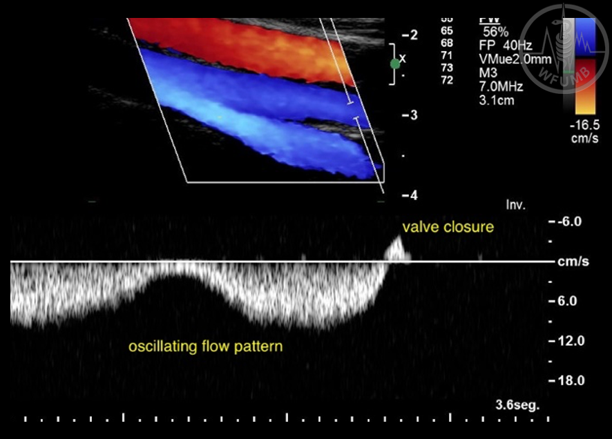
Fig 29.11
In the case of proximal thrombosis of measurement site, the flow distal to the thrombus will be continuous with loss of its oscillating pattern
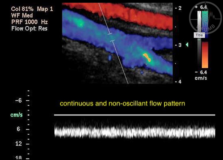
Fig 29.12
In the case of partial thrombosis, the flow of the residual lumen is continuous and accelerated
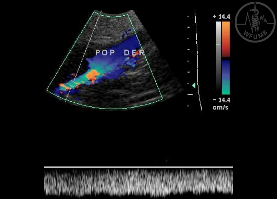
Fig 29.13
Normal longitudinal CD of popliteal vein. Total filling with no echoic images within the vessel
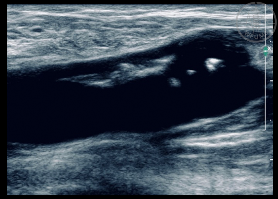
Fig 29.14
Longitudinal B mode image with popliteal incipient thrombosis affecting the valve. The vein is dilated. Painful compression
Fig 29.15
Longitudinal image of common femoral vein thrombosis. After unsuccessful thrombolytic treatment the vessel is dilated, has echogenic material within the vein and the Color box (ROI, region of interest) shows no flow signal
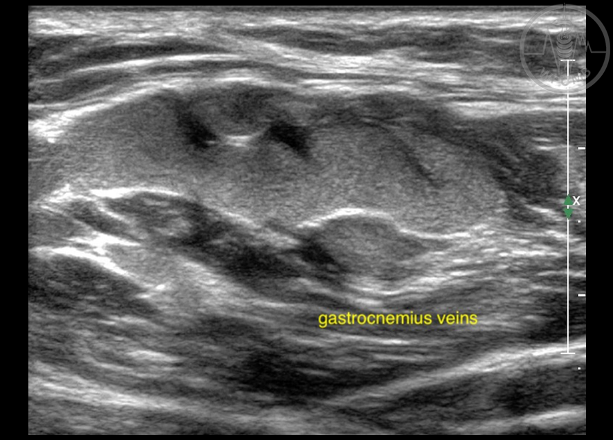
Fig 29.16
Incipient gastrocnemius veins thrombosis. The site, extension and characteristics of thrombosis must be reported
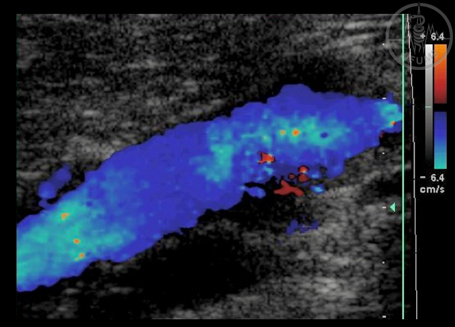
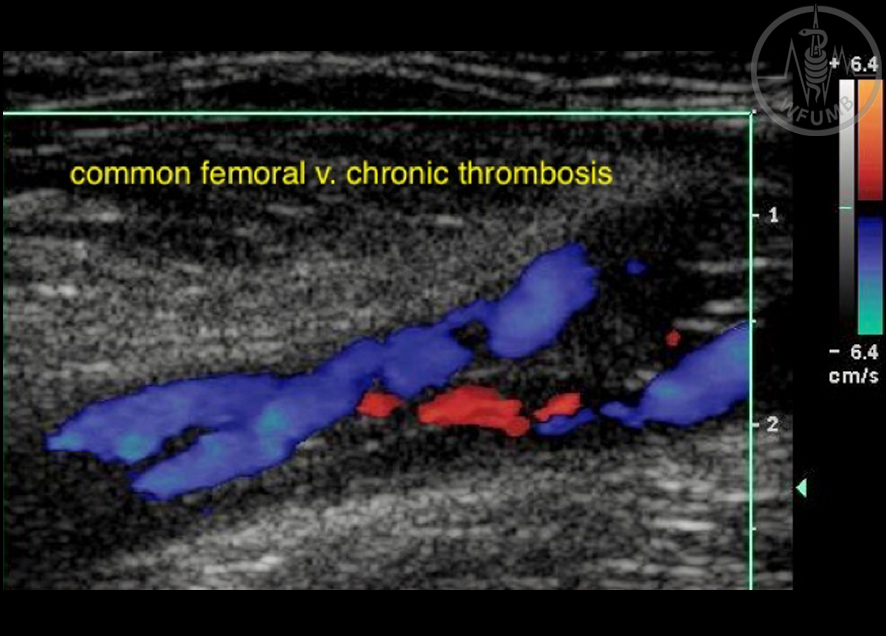
Fig 29.17
Common femoral v. chronic thrombosis. Hypoechoic B mode image with scattered hyperechoic dots. Vessel diameter is normal. Insignificant pass of flow coded in blue and retrograde flow in red due the obstruction caused by the thrombus
Fig 29.18
Common femoral vein with chronic post-thrombotic changes. Partial obstruction of the vessel and clot remnants with well-defined edges
Fig 29.19
Common femoral vein with chronic post-thrombotic changes. Partial recanalization is seen with CD that shows flow circulating between the clot remnants adhered to the vein walls
Fig 29.20
Common femoral vein with chronic post-thrombotic changes. “Thrombotic scar”