Echoes Issue No. 35 [ March 2024 ]

Welcome to Issue 35 of ECHOES

It is hard to believe that we are already 3 months into 2024 and WFUMB has used this traditionally quieter part of the year to continue improving its website. Our Centres of Education have actively been providing much needed education in under resourced regions and in this edition we showcase the POCUS courses in Fiji and the trial of clinical assistance provided to the small Pacific nation of Vanuatu. The Publication & Safety committees have been undertaking the time consuming task of updating guidelines and recommendations and these will be published over the coming months.

Recent correspondence from our Federations has a recurring theme in regards to education.

Although individual countries have their own requirements in regard to the performance of clinical ultrasound, there is a common necessity to find effective teaching methods.

It has been suggested by our representative from MASU, Teresa Abbattista, that sessions be included at conferences dedicated to the different methods of, and experience of teaching ultrasound. Teaching techniques will vary depending on the needs of the ‘students’ involved. From total beginners to experienced practitioners seeking to advance their scanning skills, there could be many ways that teaching can be delivered. As an example the teaching of obstetrics to obstetricians will be significantly different to the teaching of obstetrics in a PoCUS situation. Recognising that there are differences will assist in planning teaching methodologies. We would appreciate hearing from those interested in this topic so that we can progress the discussion further.

Please let us know what you would like to see in future editions of ECHOES and remember to spread the word by forwarding this newsletter to your colleagues.

Sue Westerway,  Publications Committee


Cambodia Project - Peter Coombs

Maternal morbidity and mortality during childbirth represent critical issues faced by many low- and middle-income countries, particularly compounded in rural and remote regions. Cambodia, a small country with a devastating history, grapples with these challenges. The 2h Project, founded by Kate and Kevin Taylor, is an Australian-based charity that has been supporting maternal healthcare in Cambodia since 2007. In 2017, the program began exploring ways ultrasound could be integrated into its midwifery training (Safer Arrivals). In 2021, the 2h Project Ultrasound Midwife Training program was launched with support from the World Federation for Ultrasound in Medicine and Biology (WFUMB). This is the story.

In Cambodia, women living in outlying provinces encounter significant risks during pregnancy, childbirth, and the postpartum period. These maternal health issues not only claim mothers' lives but also disrupt families and communities. Local health centres (day hospitals) in Cambodia serve as the key pillar for providing antenatal care and uncomplicated deliveries, with midwives at the forefront. Currently, Cambodian women inconsistently seek antenatal care, leading to delayed deliveries and limited transfer opportunities. Australian sonographers in partnership with the 2h Project envisioned that ultrasound be part of a solution.

The aims of the ultrasound program were to train and support midwives in remote health centres with handheld ultrasound devices to provide basic obstetric ultrasound services. This would have obvious diagnostic benefits: Is the baby breech? Is there enough amniotic fluid? Any placenta previa? Early identification would prompt earlier intervention within at the larger central hospital, some distance away. The hope was that ultrasound would also more broadly support antenatal care. Mothers being able to visualize their unborn would be more likely to travel to the health centres for more regular antenatal care. Training midwives would provide opportunistic education, role extension, and empowerment, further enhancing the care they provide.

In 2022, Australian Volunteer Sonographers Peter Coombs (previous WFUMB Ethiopian Ultrasound Project) and Angel Lee set up a 'gofundme' to raise money for handheld ultrasound devices. Six Butterfly Ultrasound systems were procured, including the device donated by WFUMB and a device donated by City Imaging (Obstetricians Dr. Andrew Edwards, Dr. Sofie Piessens). This provided enough devices for pilot projects in the provinces of Pailin (Thai border) and Ratanakiri (Vietnamese border). Butterfly also supports the project ongoing through their Global Healthcare Initiative.

This vision was that the project would be sustainable. All the health centres have wifi, enabling all examinations from the individual health centres to be sent to the secure online platform to be reviewed and audited when Angel/ Peter returned to Australia. This meant that the project could have ongoing remote support and opportunities for audit. Digital communication enabled ongoing feedback and encouragement to midwives. Refresher webinars were provided in the early months after this first implementation visit.

Self-funded return visits for additional one-on-one training were organized through 2023: Angel and Joyce Chen in May, Peter and Celia Lim in November. (All are sonographers at Monash Health in Melbourne). These trips reinforced the basic image series and introduced biometry. At the end of November, after three visits, the midwives had performed over 2000 examinations that were available for remote support and audit.

This ultrasound project has been incredibly enriching for all involved. The midwives are phenomenal in their engagement and enthusiasm. They are thrilled to be provided this opportunity, and the empowerment they experience is evident. Peter, Angel, Joyce, and Celia all share stories of the grace and care shown by the local teams and reflect on many touching moments with patients. This is collaborative learning, with much learned from the midwives and people of Cambodia. For the 2h Project, this innovative pilot has been valuable in shedding light on the broader midwifery training that has been ongoing for many years.

But most importantly, it is the pregnant mothers who have the most to gain. Safer arrivals!

Prof Sudhir Vinayak Update


Since the WFUMB congress Sudhir has been making a good recovery, albeit at a slow pace. He has advised that he wishes  to step down from his position as President of WFUMB and hand over to the Vice President, Prof Ioan Sporea. 
He will continue to be part of the Executive Board  and has taken this decision only for health reasons and to enable him to focus on a return to good health and to play a part in WFUMB in the future.
 
We wish Sudhir a speedy recovery and a return to good health and resumption of his role as President in WFUMB.

WFUMB Webinars in the first quarter of 2024

EFSUMB Report

Two Schools to highlight in this report:

6th International Paediatric Advanced Ultrasound School, London, UK 27 – 28 June

The European Society of Paediatric Radiology (ESPR) and the European Federation of Societies in Medicine and Biology (EFSUMB) have come together, to present the 6th International Paediatric Advanced Ultrasound Course, to be held once again in London, at King’s College Hospital.

The venue is the beautiful Fetal Institute, with outstanding facilities, and with easy connection to central London by public transport.

The topics are mainly those related to Contrast Enhanced Ultrasound (CEUS) and their application to paediatric practice, but the introduction of more advanced imaging techniques such as liver elastography and fat quantification will be introduced for the first time.

The organisers in London have presented Contrast Enhanced Ultrasound meetings since 2014, and this is the first time a paediatric onsite face-to-face meeting has been scheduled since the pandemic. We look forward to meeting all the delegates and faculty in London.

The programme has been well-designed, in collaboration with ESPR and should appeal to all paediatric imaging specialists, but also contains enough information to interest the adult practitioner.

The 2-day conference is designed so that on the first day liver imaging is examined in detail, with leading experts in the field presenting clinical aspects and future research prospects. The delegates will be introduced to concept of CEUS, the position of the European countries in relation to the use of CEUS in paediatric patients, and how to set up a service if you do not already run one. Experienced experts will highlight common and uncommon findings of focal liver lesions in paediatric patients. Physicians and surgeons will join the faculty during the day, and present the clinical aspects alongside the imaging expertise offered.

On the second day, the now established “David Cosgrove Lecture” will be delivered by Dr Christian Greis, who has recently retired, as one of the leading experts in the development of CEUS. He will be looking at aspects of regulatory approval for the use of CEUS in paediatric practice.

On the second day, less common uses of CEUS will be presented with an emphasis on innovative research, renal disease, bowel, testicular and splenic abnormalities. The day will conclude looking at future prospects for the use of CEUS in paediatric patients in many unexplored areas.

The faculty lined up is unique, all are experts in their field and have been at the forefront of the development of new ultrasound technology for many years. It would be safe to say that the assembled faculty is probably the “best at the job” in the world. This presents a unique opportunity for delegates to listen, question and socialise with experts in the field.

To this effect, the faculty and delegates dinner will be on the middle night and is already included in the registration fee, which is being kept at a very reasonable level to attract delegates and to impart knowledge.

We invite you to London at the time of the year where the weather is kind, to come and listen to experts in the field and develop your own practice of advanced ultrasound techniques for children.

Refer to the website for further information and full registration.

We look forward to seeing you.

Euroson POCUS School Vienna University of Medicine, Austria and also online, on 6-7 September 2024, chaired by Dr. Mihai Iacob, EUVEKUS President


The POCUS Curriculum and Guidelines for Frontline Physicians in Daily Practice, is being organised by EUVEKUS /EADUS – The European Ultrasound Working Group in Primary and Ambulatory Healthcare
.

Its aim is to advance common approaches and POCUS skills for frontline physicians to improve early diagnosis. It will host over 120 delegates and a 12 member world-renowned faculty.  Our International Keynote Speakers are Prof Caroline Ewertsen - EFSUMB President and Prof. Shlomo Vinker, the President of WONCA  Europe. In addition to lectures there will be practical hands on workshops and several social gatherings. Ultrasound in primary care is a valuable tool to add to the traditional skills of history taking and clinical examination.

Our presentations will allow you to understand at a basic level the role and limitations of POCUS in patients with clinical problems frequently encountered in the practice of family doctors including gastroenterology, nephrology, respiratory disease and cardiology.

EFSUMB has established a task force group of general practitioners, pediatricians, and frontline physicians to establish a POCUS core curriculum for frontline physicians in primary care. The task force group launched a Delphi study in December 2023 to develop a list of POCUS examinations suitable for a core curriculum. Close to 100 primary care doctors from 25 European countries participated in the first Delphi round and the study has now progressed to the second round.  The study is expected to be completed by Summer 2024 and the results will be presented at the Euroson POCUSS School in Vienna in September 2024 and at the EUROSON 2024 conference in Napoli in November. 

WFUMB Guidelines on Multiparametric Ultrasound for Evaluation of Chronic Liver Disease: the development process

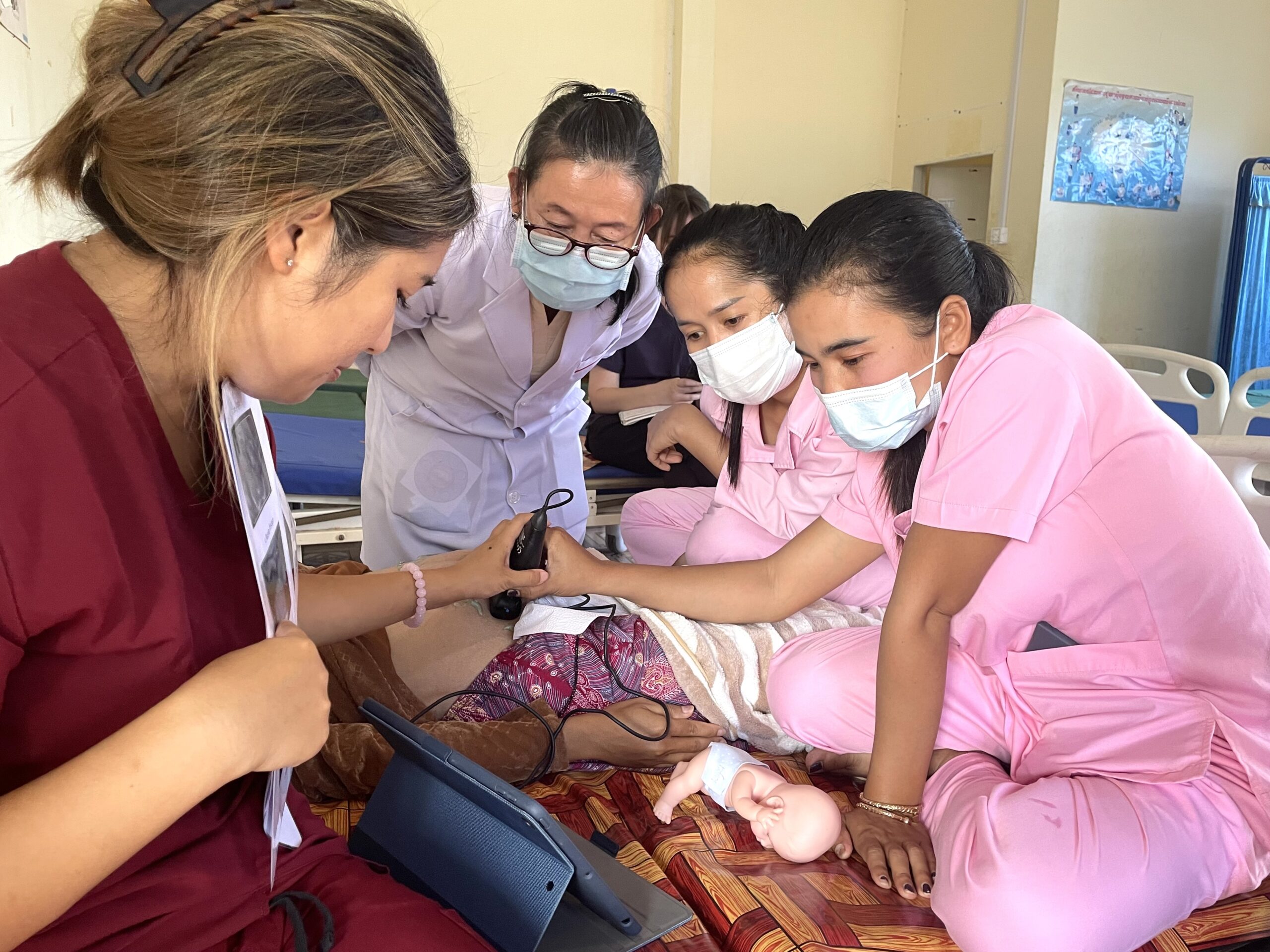
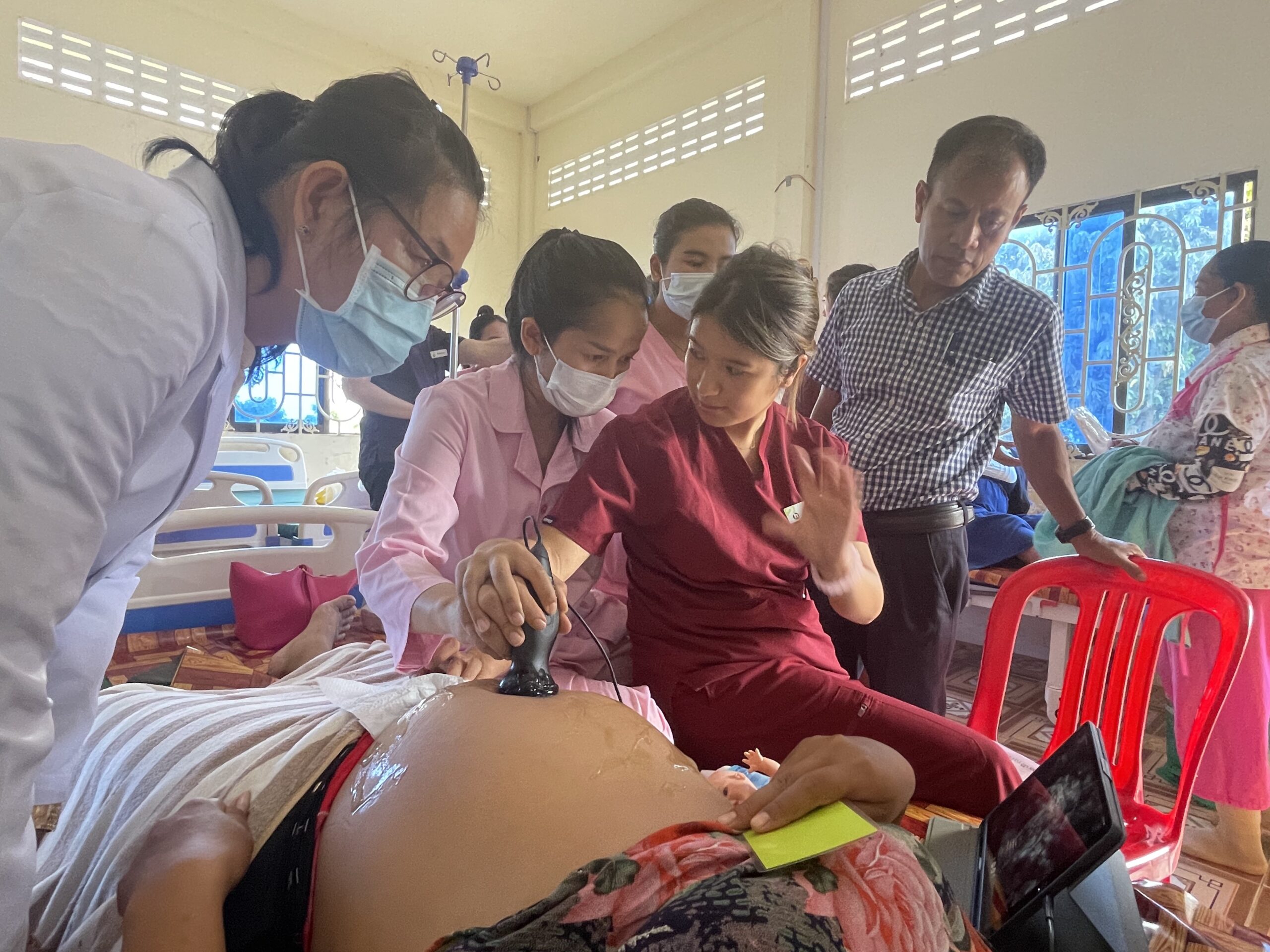
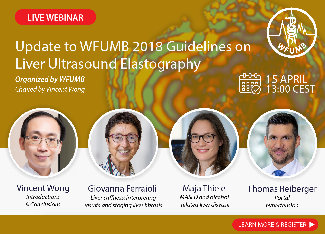
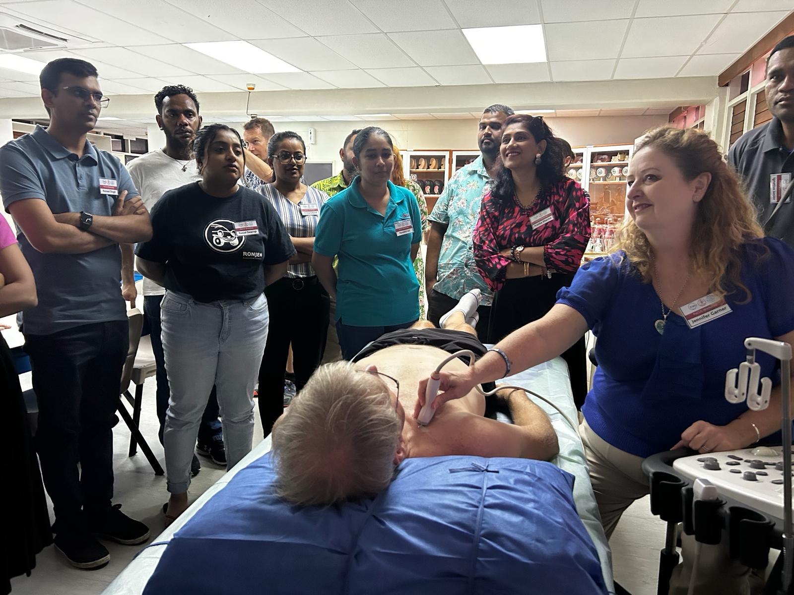
The two sets of “WFUMB Guideline/Guidance on Liver Multiparametric Ultrasound” (Part 1: Update to 2018 Guidelines on Liver Ultrasound Elastography; Part 2: Guidance on Liver Fat Quantification) are coming very soon.

The document is clinically oriented and is also a useful guide for the users. In part 1 it is explained how the liver stiffness values should be interpreted, and the usefulness of stiffness assessment for evaluating the outcome of patients with chronic liver disease is highlighted. Both in part1 and part2, the protocols for a reliable acquisition of the two quantitative parameters are detailed and the reasons that lead to the inclusion of each specific item in each protocol are explained. For the assessment of liver stiffness with elastography, each etiology of chronic liver disease is discussed in a dedicated section. There are also sections for the use of elastography in particular settings, such as in the pediatric population or in the evaluation of patients with portal hypertension. Where there is solid evidence, the recommendations were rated in a standardized manner, following the Oxford Centre for Evidence-Based Medicine released in 2009. For ultrasound fat quantification the recommendations were based on published studies and experts’ opinion but were not graded because the level of evidence was low at the time of publication of the document.

The following is the account of the process that led to the release of the document in a short period of time.

On May 2023, the WFUMB Executive Board asked me to lead a further update to the liver ultrasound elastography guidelines that were released in 2018 and published in the official journal of the WFUMB, i.e. Ultrasound in Medicine and Biology. This time, the goal was to also release a guidance on fat quantification with ultrasound, because steatotic liver disease has become the leading cause of chronic liver disease worldwide and the availability of noninvasive methods for fat quantification is therefore of great interest. The document was on “Multiparametric Ultrasound”, even though other parameters for the noninvasive assessment of chronic liver disease, such as viscosity among others, were not included because of insufficient literature and discordant results.

The WFUMB Executive Board relied on me for the choice of the members of the steering committee and of the experts’ panel. This was, of course, a huge responsibility and a big challenge.

For the development of guidelines on noninvasive techniques the “experts” should not merely be “expert users” of the specific technique(s). Rather, they should be leading experts who have substantially contributed with their research to the advancement in the topic(s) of the guidelines.

The criterion followed for choosing the experts (for all the authors of these guidelines) was a PubMed and Scopus search for authors of high-quality research studies on ultrasound liver elastography and ultrasound fat quantification. Among them, the panel of experts was selected also considering the different Ultrasound Federations that constitute the WFUMB. Therefore, different geographic areas of the world were considered.

In the selection process, the specific specialty of the expert was not (and I strongly believe that it should not be) a criterion that was followed.

Other criteria applied for choosing the experts were: no more than one expert per institution; no more than one expert per country (exceptionally two); no more than five experts per Ultrasound Federation (AFSUMB, ASUM, EFSUMB, FLAUS, MASU); at least one expert per Federation unless there was no expert identified - based on the proposed rule - to include.

The WFUMB publication committee agreed on these rules and on the list of experts selected utilizing these criteria.

The experts received the invite at the end of May 2023 and by the middle of June 2023 the panel was completed. The experts’ panel is composed of 17 members belonging to the different Ultrasound Federations adhering to the WFUMB.

The steering committee identified the areas of interest and established  a dedicated section for each one. The topic of each area was assigned to a group of experts with a leader who had the task to assign duties to each member, to receive the feedback from all of them, to collect the contributions and to organize a draft. The draft reported the evidence from the literature; based on them, the recommendations were made and graded when it was deemed appropriate based on the body of evidence available.

Two online meetings to discuss specific matters, also including the agreement on statements made in the “work-in-progress” drafts, were held on August and October 2023.

By the end of November 2023, the draft was ready and discussed in a hybrid meeting held on November 28 in Chicago, Illinois, USA, on the occasion of the RSNA meeting. The two parts of the manuscript, i.e. part 1 on liver elastography and part 2 on liver fat quantification, were shared in Google Drive and all authors had the possibility to comment and revise them. The recommendations that were made in part 1 of the document were formally voted in an online meeting scheduled on January 10.

The document was also sent for comments to all the members of the WFUMB publication committee.

Part 1 of the document was submitted to Ultrasound in Medicine and Biology in January 23, part 2 in January 28.

Overall, the development of the document took about six months and it proceeded at a speedy pace, even though this period also included the summer vacation. This achievement must be ascribed to the dedication and enthusiasm of the panel members, let alone their expertise and knowledge of the topics.  

These guidelines were not supported by any vendors in any manner, and this strengthens their content. Most of the experts have conflicts of interest that are clearly and honestly declared in the published articles and that, however, does not bias or diminish the scientific value of the document, because it is based on published studies that were rated using objective criteria, i.e. those of the Oxford Centre for Evidence-Based Medicine.

Giovanna Ferraioli, MD, FAIUM
Leader of the panel of experts for the development of the WFUMB Guideline/Guidance on Liver Multiparametric Ultrasound
Member of the WFUMB Publication Committee
Affiliation: Department of Clinical, Surgical, Diagnostic and Pediatric Sciences, University of Pavia, Pavia, Italy
A Webinar: Update to WFUMB 2018 Guidelines on Liver Ultrasound Elastography is taking place on 15th April 2024.


View the programme and register here >

FIJI COE

Clinical assistance in the Pacific

The WFUMB Centre of Education (CoE) in Fiji has an expansive reach across the Pacific Ocean. Although the CoE runs a minimum of three hands-on courses each year out of the Fiji National University in Suva, the vast distance between the Pacific nations limits access to ultrasound education for many users. Other limitations include hospitals releasing staff to attend courses and the substantial cost of airfares and accommodation.

To assist in overcoming these problems, a new concept has been trialled at the Port Villa Central Hospital in Vanuatu. In a WFUMB / RAD-AID collaboration, two self-funding volunteers spent four days working with the radiology staff as they scanned their routine clinical load. There are two ultrasound machines in the department and an average of 50 patients a day attend for obstetric, abdomen, thyroid, paediatric and pelvic scans amongst others. Simple worksheets were provided, scan protocols were suggested, as were machine settings, scanning techniques and report writing.

All of the staff that spent time with the visiting tutors were most appreciative of the training and are looking forward to return visits.

The huge number of patients reflects the increasing need for ultrasound services within the hospital. It was noted that there were machines in both maternity and the emergency department but with limited staff trained in performing ultrasound, these systems are being underutilised. This situation highlights the need for more education in the region.

With the success of this trial it is hoped to repeat the concept not only within the Pacific but in other locations around the world.

Sue Westerway
Co-Director WFUMB Fiji CoE
Jo McCann
WFUMB Council
RAD-AID Program Manager – South Pacific

Report on Fiji POCUS courses

The Fiji CoE conducted two PoCUS courses in February 2024 at the Fiji National University campus in Suva. Thirty four participants came from Fiji, Tuvalu, American Samoa & Papua New Guinea.

An Advanced 2 day course for medical doctors with intermediate ultrasound skills covered cardiac & lung. There were 12 attendees from emergency medicine, anaesthetics, internal medicine and public health.

This was followed by a 2 day Basic PoCUS course for doctors with minimal skills which covered the aorta, gallbladder and renal. 22 doctors participated, mainly from the Government Hospital in Suva.

There were 4 teaching stations for the hands on component, utilising 3 FNU machines, a WFUMB Butterfly and tablet, phantoms and attendees as models. The Butterfly was particularly useful for the Basic PoCUS with the larger cohort. 

A pre and post course quiz showed significant improvement in knowledge for all participants

The international faculty volunteered their time and included two emergency doctors, an applications specialist and two experienced tutor sonographers.

A quote from a participant:

‘I see how scanning could become intuitive, like driving or breathing'


ASUM Report

ASUM24 Ultrasound Excellence

The ASUM 24 Scientific Committee is pleased to invite presenters to submit abstracts for oral and poster presentations to be delivered at the 2024 conference from 1-3 November at the W Hotel, Brisbane.

The ASUM meeting is conducted annually and always attracts a multi-disciplinary group of healthcare professionals from Australia, New Zealand and the surrounding Asia and Oceania region to discuss and explore the latest trends, research findings, and innovative solutions within the field of diagnostic ultrasound.

The theme of ASUM 24 is, ‘Ultrasound Excellence: Forging Connections, Enhancing Collaboration, Amplifying Communication’. The conference format intends to target key areas of ultrasound specialisation, while bringing together the various disciplines to share their intelligence.

Please refer to the ASUM 24 Abstract Submission Guidelines. Abstract submissions are due 11:59 pm AEST Friday 31 May 2024. Abstracts will be reviewed and submitters will receive a notification regarding the outcome of their submission by 30 June 2024.

Access the Abstract Submission Portal here.

The ASUM Board and ASUM 24 Scientific Committee greatly appreciate you taking the time to submit your work for consideration.

ASUM New Zealand 2024

The ASUM New Zealand 2024 conference will be held in Auckland from 19 – 21 July and will provide a wonderful opportunity for delegates to meet face-to-face with friends, both old and new, to learn from leading voices across our diverse ultrasound community both in New Zealand and beyond. The program will cover various topics in obstetrics and gynaecology, POCUS and general ultrasound. Registration will open soon.

Event sponsor: Tātaki Auckland Unlimited

Scholarship: Sonographer Students

On behalf of the estate of the late Dr Delwyn Nicholls, ASUM launched a new scholarship in 2023 to support student sonographers in rural Australia.

The scholarship provides a $5,000 grant for successful applicants who are in their third or fourth year of study.

Congratulations to Sarah Williamson, who was awarded the inaugural Delwyn Nicholls Scholarship in 2023.

Applications for the 2024 scholarship are now open and close Friday, 16 August 2024. Find out more here.  


ASUM Outreach, Canon Medical and Tonga’s Ministry of Health partnership in 2023 delivered ultrasound training in Tonga.

In November 2023, the ASUM Outreach team, consisting of Peter Coombs, Jackie Spurway and Brett Ayres attended Vaiola Hospital for five days. The primary focus of this visit was to support the development of vascular ultrasound services in Tonga. Didactic presentations and hands on clinical training were planned, while also adapting to the needs of the department and patients, as required.

The week's activities not only imparted valuable knowledge and skills, but also fostered a culture of learning, encouragement, affirmation, and support among the Tongan sonographers. Reflecting on the quieter pace of clinical activities during this visit, there was a notable increase in teaching and training opportunities. The next visiting team is recommended to possess the capability to provide comprehensive training in all areas of ultrasound. Additionally, input from the local team regarding the repetition of specific aspects from the current visit should be sought.

We are looking forward to joining our friends in Tonga in 2024 to continue learning from each other.

AJUM

The Australasian Journal of Ultrasound in Medicine (AJUM) is a multi-disciplinary journal with an extensive international readership. The February 2024 edition of AJUM focuses on the latest research and applications in ultrasound medicine, highlighting original research such as artificial intelligence as a teaching tool for gynaecological ultrasound and the utility of POCUS in the emergency department for acute cholecystitis The issue also examines advances in the detection of sialoliths in paediatric patients and offers new perspectives on rheumatoid vasculitis through ultrasound imaging.

Explore the February issue at https://onlinelibrary.wiley.com/toc/22050140/current

If you have original research, systematic reviews or a “How to…..” article, please submit your manuscripts to the AJUM online. “How to…..” articles are one of our top downloaded articles and made freely available through our publisher Wiley for our national and international audience.

ASUM Drive

Get streaming access to a vast library of on-demand ultrasound content, covering a wide range of specialties. Enhance your skills and knowledge anytime, anywhere.

ASUM members simply need to log in to access the full library of on-demand content. Not a member of ASUM? You can purchase an annual subscription to ASUM Drive for only AUD 200/year at https://drive.asum.com.au/browse.

WFUMB AFSUMB Student Webinars

The WFUMB-AFSUMB Student Webinars are taking place across 2023 – 2024 on the second Saturday of the scheduled months. Each webinar consists of two, 1 hour lectures (see the full enlarged scheduled below) and will take place via Zoom connection in English language.
Time: 10:00am Central European Time (CET or CEST)

Register by clicking on the schedule below >

View the last three webinar presentations below >

Arab Health Dubai UAE 29 January - 1 February

Arab Health has become a significant event for the healthcare industry and the 49th edition was no exception. Hosted in Dubai, the event brought together a diverse participation of healthcare professionals, businesses, and organisations from around the world to showcase the latest innovations and advancements in medical technology, equipment, and services.
 
The WFUMB team met with a number of ultrasound companies at this year's Arab Health meeting . The meetings are a useful opportunity to understand the specific regions and areas  of interest of these companies.  We learnt from Samsung about their ultrasound learning programme -SONOSYNC - for use in remote areas. Sonoscape work closely with veterinarians and all are interested in sponsoring WFUMB's next congress. We met with Supersonic Imagine now up and running after the buyout from Hologic and keen to expand in China, Mexico and the Nordic countries. The next stage is to follow up and match these interests with WFUMB's activities.

A facelift for the WFUMB Web Site


To keep our online presence fresh and current the Publications Committee has been looking at ways to improve the WFUMB web site. Working alongside the WFUMB Web Admininstrator we've identified areas for better access, content and a more user friendly layout.

The new look web site with improved search facilities will be available online shortly - Watch this space!

Letter from wife of deceased past president M.Fukuda

Educational resources from WFUMB's partner, ISUOG


WFUMB recently received a letter of thanks for the gold medal from the wife of the deceased Professor Morimichi Fukuda. Below is the contents of the letter translated into English
.

January 12, 2024,

I received a commemorative medal and a letter today. I immediately made it an offering at the alter.
I am sure that my deceased husband would be grateful and pleased. I would like to express my sincere gratitude for your kindness and consideration. I would also like to express my sincere gratitude to everyone involved.
Sapporo is in full swing in winter. It is the whole place covered with completely white snow. Because your place would be also cold from now on, I pray that you will take good care of yourself.
Thank you very much.

Best Regards
Yoshiko Fukuda

ISUOG provides education for practitioners and trainees at all levels to enhance their skills and knowledge in ultrasound in obstetrics and gynecology with the ultimate aim of improving health outcomes for women across the world.

Our lectures and courses are accessible onsite, online, via live-streaming and On Demand. 
Explore the different ways you can learn with us below.

EUROSON 2024

35th congress of the European Federation of Ultrasound Societies in Medicine and Biology

Congress Centre “Stazione Marittima”, Naples, 9-11 November

In conjunction with SIUMB Educational Programme, 9-12 November

  • Complete programmes and registrations will be available from April 15
  • Call for Abstract will be open from March 15 until May 15.


Upcoming Congresses


A luminary interview with Francis Duck

Francis A Duck PhD DSc MBE

My interest in ultrasonics was sparked as a basic-grade medical physicist at University College Hospital (UCH) London. I had a BSc in physics and, in 1966, I had been appointed as a junior nuclear medicine physicist by the dynamic young head of the medical physics department, John Clifton. In the same cohort of new entrants with me, John had recruited Roland Blackwell as the physicist to look after the Nuclear Enterprises Diasonograph that had been installed for evaluation. John encouraged each new recruit to take an interest in the all aspects of modern medical physics, from the new computer-based radiation treatment planning system to the biomechanics of open-heart surgery. Roland, who would eventually become professor of medical physics and head of department, learned about ultrasonics on the job and shared his insights with me, including me in trouble-shooting, testing transducers and changing valves.

The selection of an experimental MSc project gave my own opening into ultrasonics. At that time, blood-flow detection using continuous-wave ultrasonic Doppler was causing interest, with Ray Gosling at Guy’s already demonstrating the use of audio frequency spectrum analysis for detailed investigation of flow in peripheral arteries. Peter Fish was starting to explore pulsed Doppler for imaging at King’s College Hospital. Dawood Parker, at UCH, was looking at an intra-arterial transit time ultrasonic probe for blood speed measurement. My project was to develop an intra-arterial Doppler system. It worked, although the project was only tested with a blood mimic. I was then recruited by the head of radiology at UCH, John Hodson, who was leaving London to become head of Radiology in the new medical school at Memorial University, St John’s, Newfoundland, Canada, and wanted to investigate Doppler techniques in a renal animal laboratory. Before duplex imaging and pulsed Doppler had been developed to reach deep arteries, our approach was to use the venous system as a conduit, introducing a miniature, angled Doppler transducer to lie alongside any deep artery of interest. By the time I left St John’s in 1974 I had developed the probe to create two orthogonal beams, the vector sum of their outputs having the potential for angle-independent velocity measurement, and a third central pulse-echo transducer with echo-tracking, to measure arterial diameter and its pulsation. In theatre, we using an angled intra-oesophageal probe to monitor blood flow in the thoracic aorta of an anaesthetised patient, a precursor of intra- oesophageal cardiac imaging. My work on vascular dynamics formed the basis of my PhD thesis (London 1977). 

I then spent the following 18 months in Jim Greenleaf and Steve Johnson’s prolific ultrasonic laboratory at the Mayo Clinic, exploring where ultrasonic imaging might be going. While our transmission tomography never achieved its apparent potential, work on harmonic generation gave a glimpse of the future, as did our 6-element synthetic aperture array, one image taking 1 hour to process on the CDC 3500 computer. There was greater clinical impact from the SRI mechanical real-time carotid scanner: pulsed Doppler was added after I left. 

Kit Hill then invited me to return to the UK and join him at the Institute of Cancer Research, Royal Marsden Hospital in Sutton. His team included several with whom I had the privilege to work over a period of many years, including Jeff Bamber, Gail ter Haar, Bob Chivers and Dariush Nassiri, Here I learned the fundamentals of ultrasonic scattering, and developed my first software for predicting pulsed ultrasonic beam structure. 

With a growing family, I needed to leave the uncertainty of grant-funded posts, and secured a position as a hospital-based medical physicist at the Royal United Hospital in Bath. Strictly I was assigned to nuclear medicine, but the appointment of Orhan Berktay, with an international reputation in non-linear ultrasonics, as Physics Professor at Bath University, initiated a creative relationship with his laboratory that lasted until my retirement, by which time I had been appointed as a visiting professor. Using the new pvdf membrane hydrophones, a world of non-linear acoustics opened up, requiring a much deeper understanding of the physics of ultrasonics and its interaction with tissue than had previously been recognized. Guided by Victor Humphrey, we explored harmonic imaging and developed a strategy to avoid acoustic saturation, which invalidated the linear estimation of in-situ exposure at high amplitudes.

Our method was endorsed by the IEC. The 1989 Ultrasonic Conference at Allerton, Illinois, run by Bill O’Brien, emphasized that non-linear acoustics had become a central issue in modern medical ultrasonics (Fig 1). Hazel Starritt’s beautiful experiments on acoustic streaming, caused by imaging pulses, and its non-linear enhancement, led to considerations of the importance of radiation force for safety. In turn, this led to a new way of defining acoustic dose-rate in general terms, as total energy deposition per unit mass, the unit being fundamental for both temperature rise and radiation force. 

Safety, and its underpinning requirement for high fidelity ultrasonic metrology, established a creative liaison with Roy Preston’s group at the National Physical Laboratory, including David Bacon, Adam Shaw and Bajram Zeqiri, and with Andrew Hurrell from Precision Acoustics. Our published surveys of acoustic output, together with those of Tony Whittingham and Kevin Martin in Newcastle, underpinned the development of the WFUMB Safety Guidelines. Work with national and international organizations including WFUMB, EFSUMB AIUM, IEC and ICRU gave me life-long friendships with colleagues from around the world, including particularly Stan Barnett, Marv Ziskin and Tom Szabo. Greater seniority in the UK, working with the Department of Heath on education and professional matters, let to the award of MBE for services to health care, and a hand-shake from Queen Elizabeth II. 

However, we never managed to enter the marketplace with the devices we developed. Closest was Mike Perkins’ portable milliwatt power balance.

About a dozen, manufactured in-house, were sold at cost. But David Hughes’ patented automated attenuation-correction failed, largely because shadows were clinically familiar and diagnostic. We reached a clinical evaluation of a compression-free automatic 3D breast scanner, but were unable to raise commercial interest. A project for radiation-force elastography with Jeff Bamber never reached in-vivo testing. Mark Tooley’s automated Doppler blood pressure system, remains under development. Perhaps we should have tried harder.

But it has been of considerable pleasure to see scientists who spent time in our laboratories in Bath then go on to forge their own successful careers in medical physics and ultrasonics. These include Barry Ward, Jackie Shipley, Ellie Martin and Prashant Verma. 

Since retirement, I have taken a particular interest in the historical origins of ultrasonics, leading to its later exploitation in medical diagnosis from the 1950s onwards. Of specific interest is the work of the French physicist Paul Langevin (1872-1946). From 1915 he led the WW1 development of underwater ultrasonics, predicting is use in medicine as early as 1918. His understanding that the piezoelectricity of quartz could be exploited for ultrasonic pulse-echo detection formed the basis for all subsequent laboratory and industrial developments. It explains, incidentally, why, when I first started working in ultrasonics in the late 1960s, the PZT ceramic transducers were still called ‘crystals’. Last year, an IEEE commemorative plaque was unveiled at ESPCI in Paris, where he worked and was the director. This plaque records Langevin’s role in the development of Sonar, and records that this ‘led to other applications including depth sounding and medical echography’. 

Looking back at more than half a century of medical ultrasonics, it was the echnical developments in the 1970s and 1980s that made the most impact on its clinical acceptance. This was when the change occurred from static, slow, imaging using single transducers to dynamic imaging using arrays. Once physiological information was integrated using pulsed Doppler, what followed was steady technical innovation together with dissemination, clinical training and acceptance, leading to the present dominant position of ultrasonic methods in modern medical imaging. 

gdpr-image
This website uses cookies to improve your experience. By using this website you agree to our Data Protection Policy.
Read more
Translate »