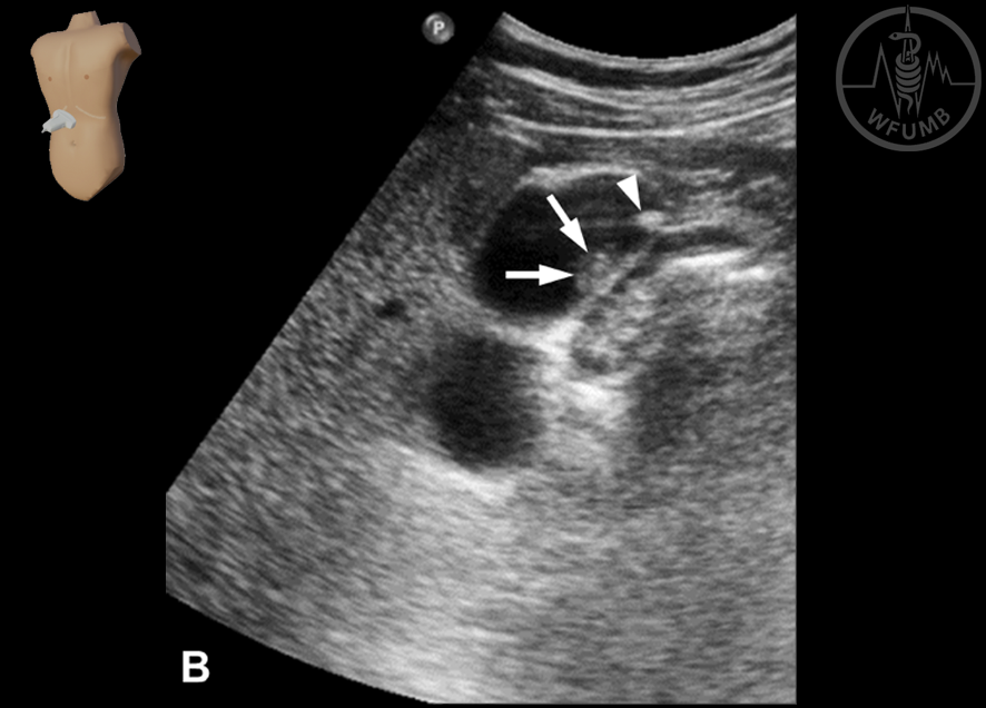
Fig 7.1a
Longitudinal scan of the gallbladder in the supine position. A tiny polyp
(echoic lesion attached to the gallbladder wall -
arrows) is seen
Fig 7.1b
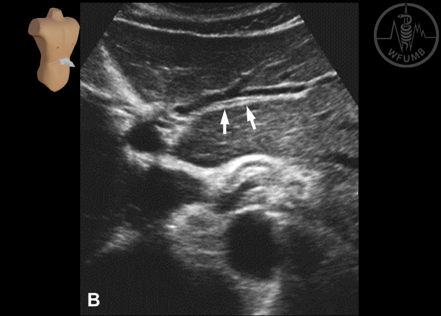
Longitudinal scan of the gallbladder in the right anterior oblique position (same case as Fig. 7.1a). With the postural change,
a tiny stone (arrowhead) in the neck moves into the fundus and becomes visible, while a tiny polyp (arrows) doesn’t move
Fig 7.2a
Longitudinal scan of the gallbladder in the fundamental imaging mode
Fig 7.2b
Longitudinal scans of the gallbladder in the and harmonic imaging (same case as Fig 7.2a). The fundal area of the gallbladder becomes clearer in the harmonic imaging by minimizing reverberation artifact, as compared with the fundamental imaging
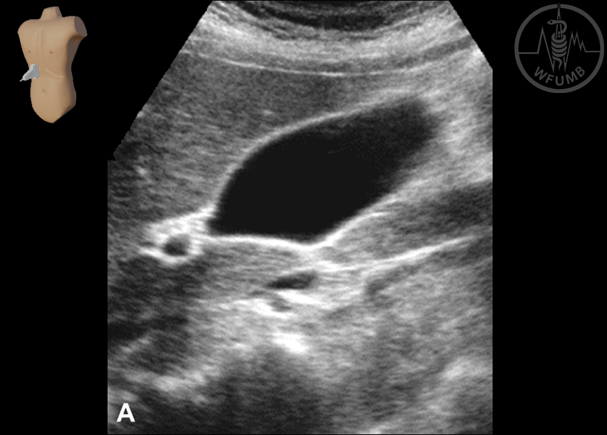
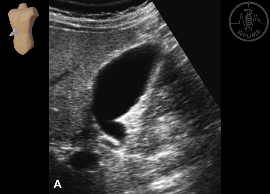
Fig 7.3a
Longitudinal scan of the normal gallbladder in fasting state. Note that the echo-free gallbladder appears pyriform or elliptical, becoming tapered and tortuous towards the neck
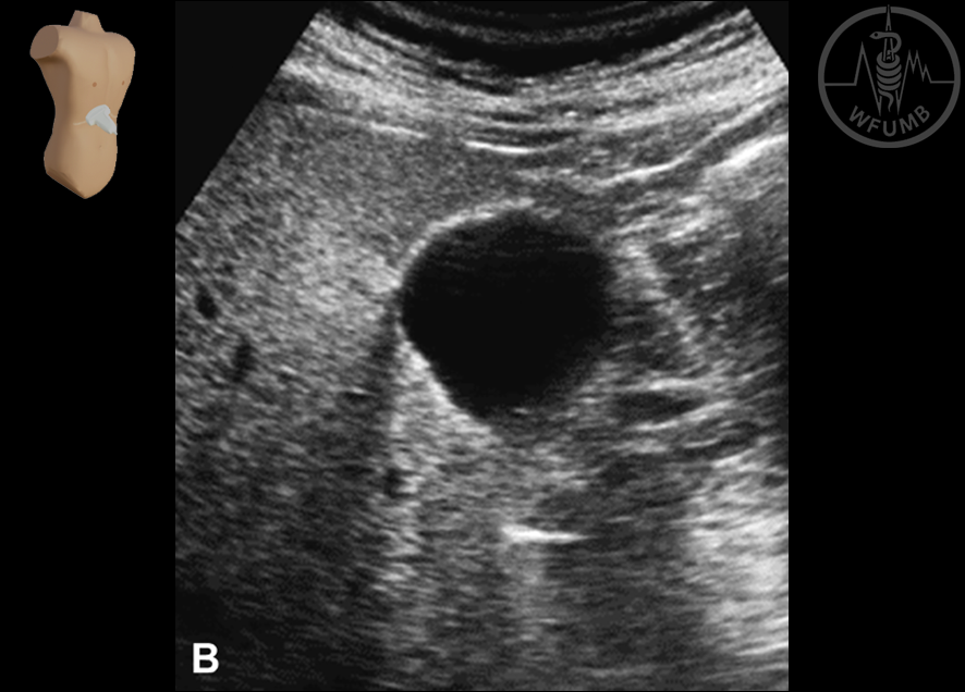
Fig 7.3b
Transverse scan of the normal gallbladder in
fasting state. Note that the echo-free gallbladder appears circular in the transverse scan
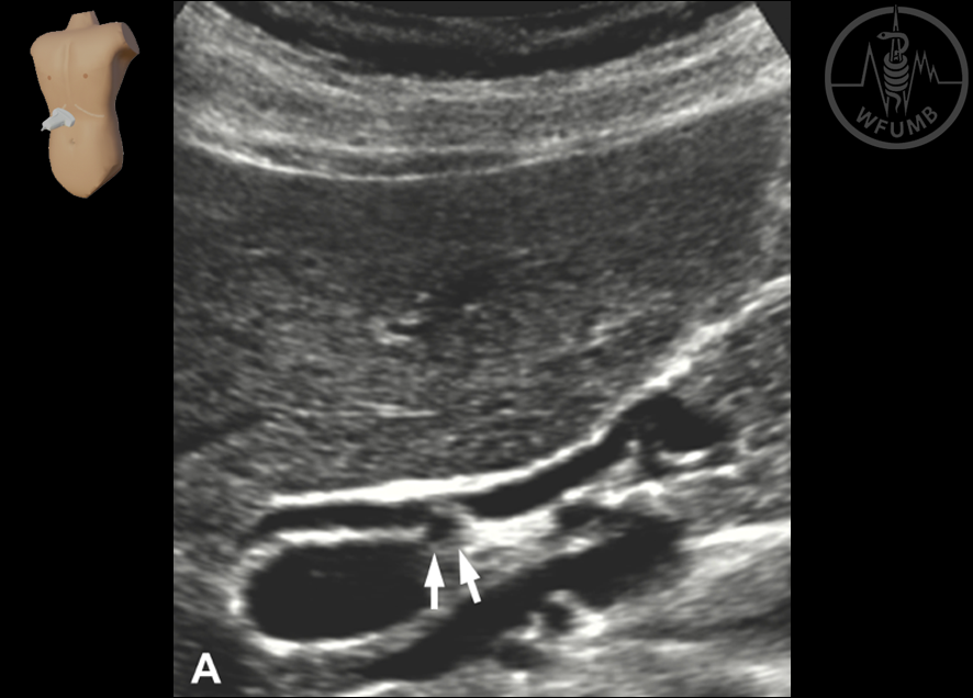
Fig 7.4a
Transverse scan of the normal intrahepatic bile ducts in the right hepatic lobe. Note that the intrahepatic bile ducts (arrows) are visualized as thin tubular structures with a diameter of less than the accompanying portal vein branches
Fig 7.4b
Transverse scan of the normal intrahepatic bile ducts in the left hepatic lobe. Note that the intrahepatic bile ducts (arrows) are visualized as thin tubular structures with a diameter
of less than the accompanying portal
vein branches
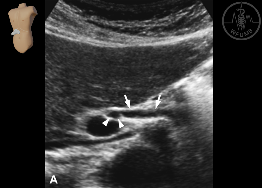
Fig 7.5a
Longitudinal scan of the normal proximal common duct (arrows). Note the right hepatic artery (arrowheads)
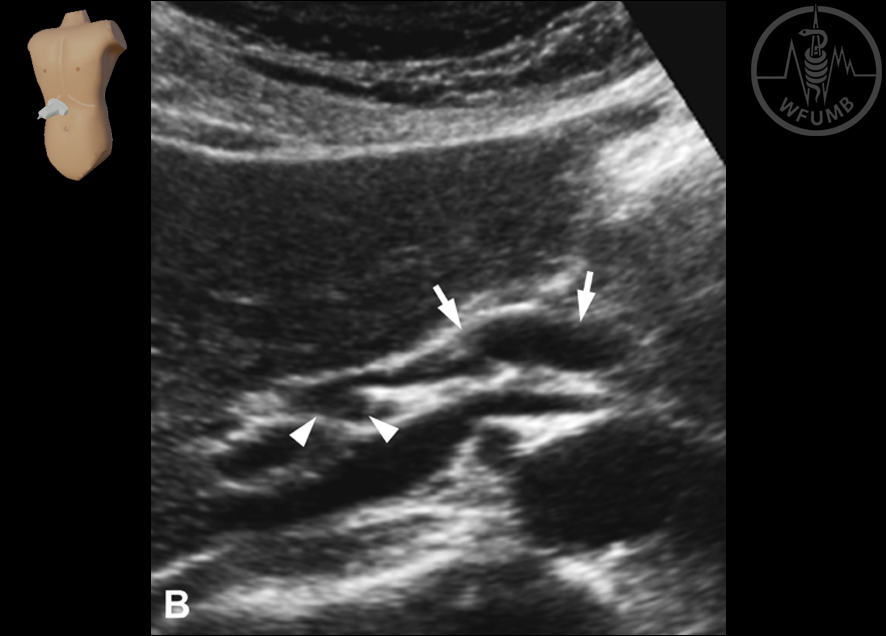
Fig 7.5b
Longitudinal scan of the normal distal common duct. The distal common duct (arrows) is slightly widened than the proximal common duct as it passes in the free edge of the hepatoduodenal ligament. Note the right hepatic artery (arrowheads)
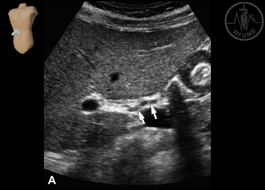
Fig 7.6a
Longitudinal scan of the common duct in B-mode ultrasound. The round right hepatic artery (arrows) crosses between the common duct and main portal vein
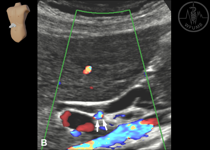
Fig 7.6b
Longitudinal scan of the common duct in color Doppler ultrasound.
The round right hepatic artery (arrows) crosses between the common duct and main portal vein. Note the color flow in the right hepatic artery at color Doppler ultrasound
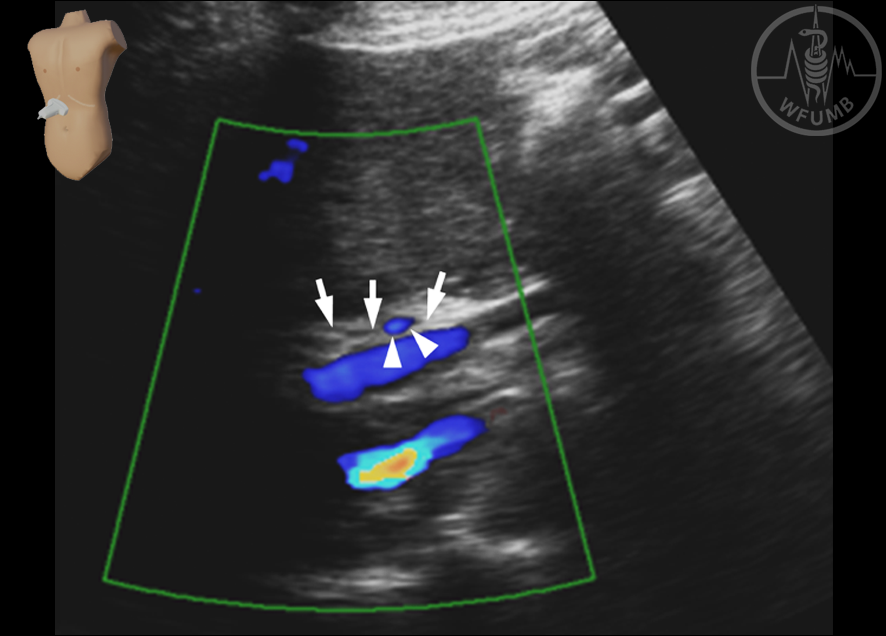
Fig 7.7
Longitudinal scan of the common duct. Note that the right hepatic artery (arrowheads) with color flow crosses over the common duct (arrows) anteriorly
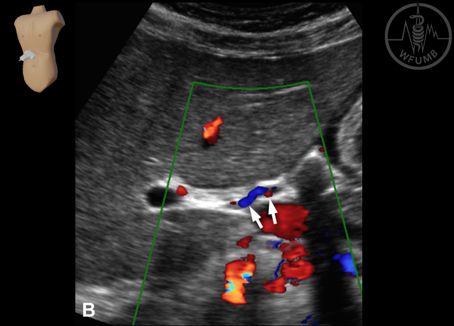
Fig 7.8
Longitudinal scan of the common duct. Note the right hepatic artery (arrowheads) crosses behind to the portal vein (arrows)
Fig 7.9a
Longitudinal scan of
the hepatic artery in
B-mode ultrasound.
The hepatic artery
appears slightly tortuous
Fig 7.9b
Longitudinal scans of the hepatic artery in color Doppler ultrasound. The hepatic artery appears slightly tortuous, and can be discriminated by demonstrating color flow at color Doppler ultrasound
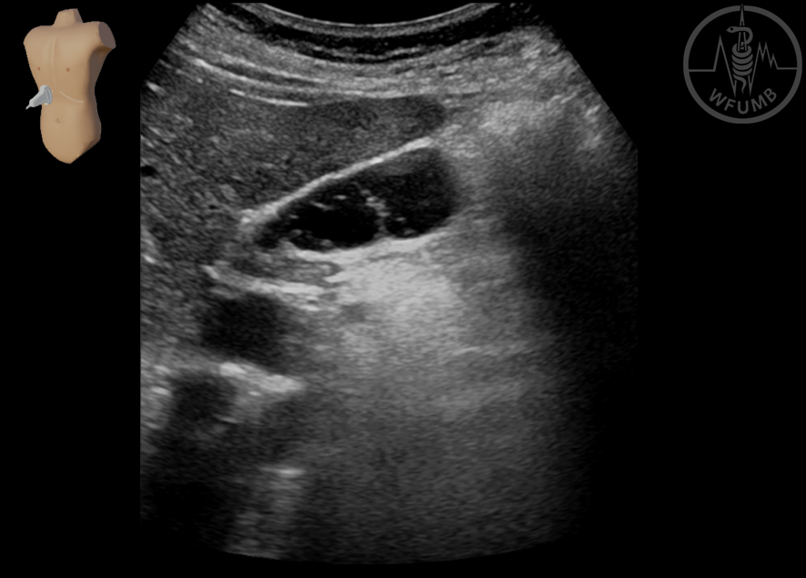
Fig 7.10
A case of phrygian cap deformity. Longitudinal scan of the gallbladder shows that bottom of the gallbladder folds over the rest of it
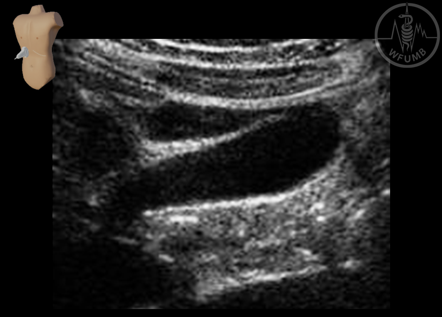
Fig 7.11
A case of sepate gallbladder. Longitudinal scan of the gallbladder shows a single septum dividing the gallbladder
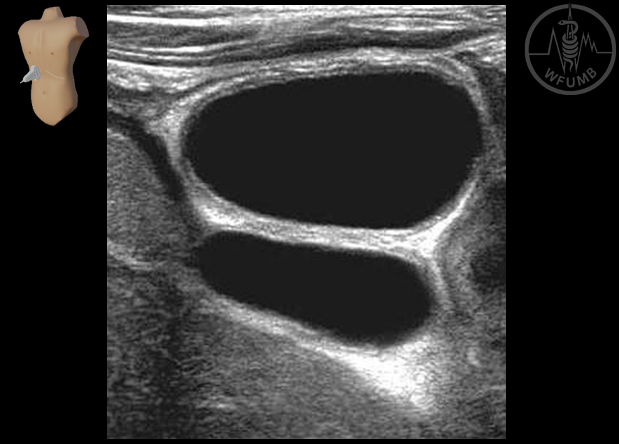
Fig 7.12a
A case of multiseptate gallbladder. Longitudinal scan of the gallbladder shows several septa in the gallbladder lumen
Fig 7.12b
A case of multiseptate gallbladder. Longitudinal scan of the gallbladder shows several septa in the gallbladder lumen
Chapter Videos
This website uses cookies to improve your experience. By using this website you agree to our Data Protection Policy.