WFUMB Course Book
Gallbladder and Bile Duct System - Sonopathology - Chaper 14 Media Library
Close window and return to Chapter 14
Chapter Images
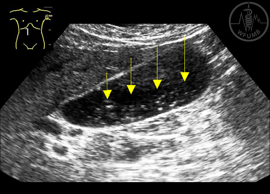
Fig 14.1 Biliary sludge – echoic material (arrow) inside the gallbladder with no posterior shadowing
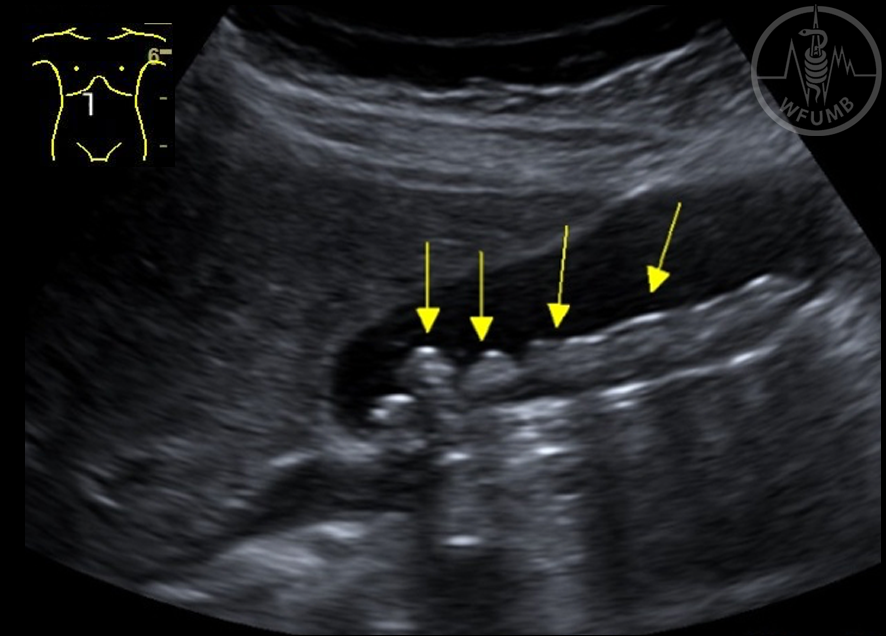
Fig 14.2 Biliary sludge – (arrows) - right anterior oblique section
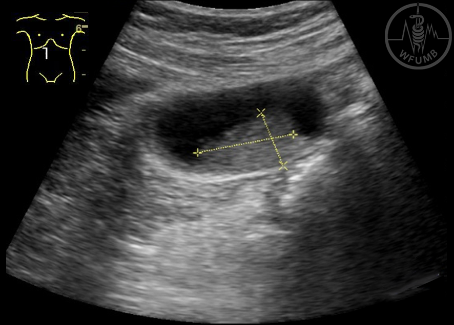
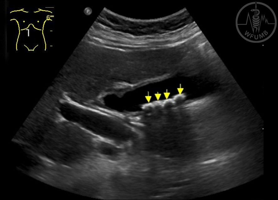
Fig 14.3 Biliary sludge (arrows) - Orthostasis
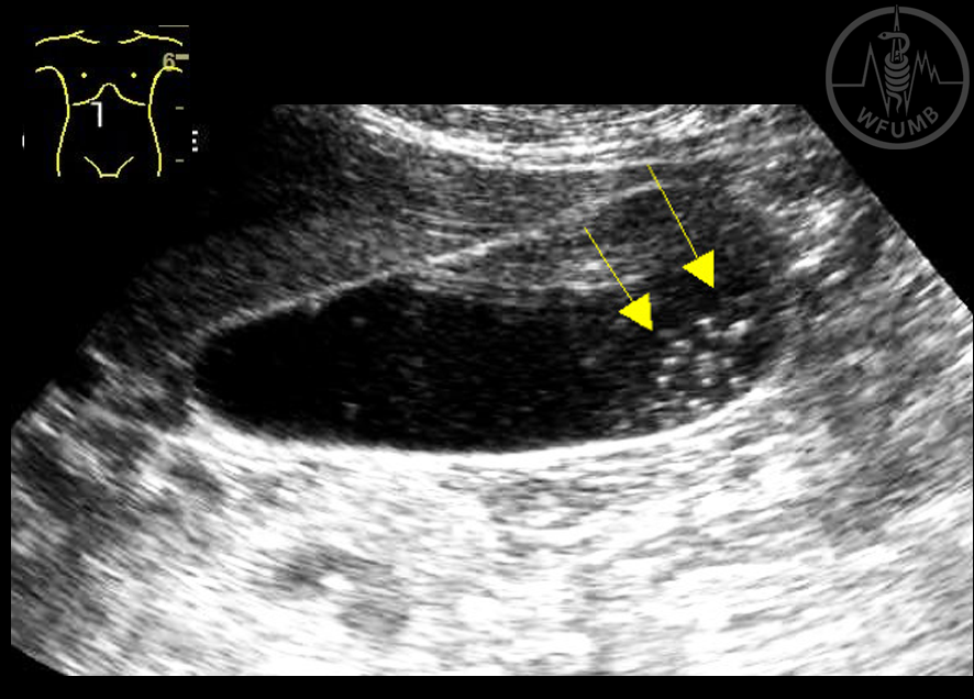
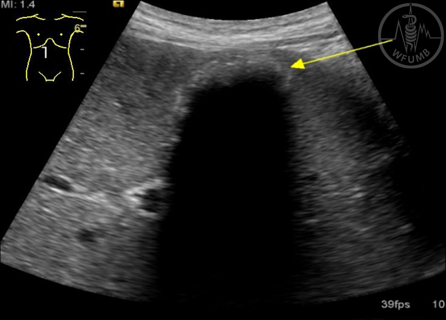
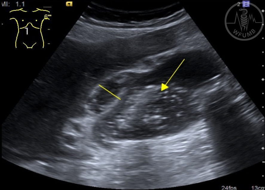
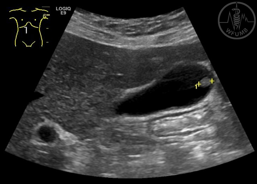
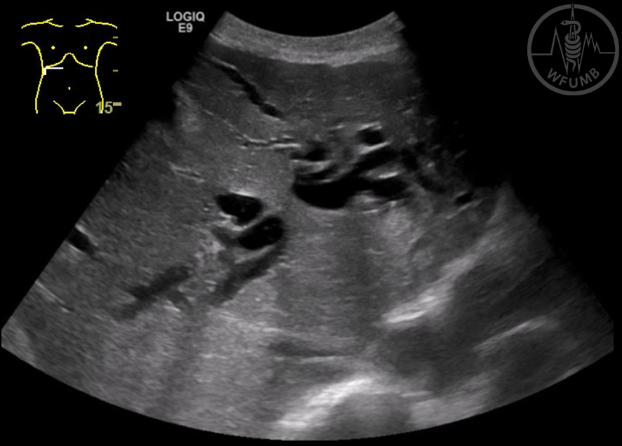
Fig 14.4 “Tumor like” biliary sludge
Fig 14.5 Gallbladder stones – hyperechoic calculi (arrows) with posterior shadowing
Fig 14.6 Gallbladder stones (arrows)
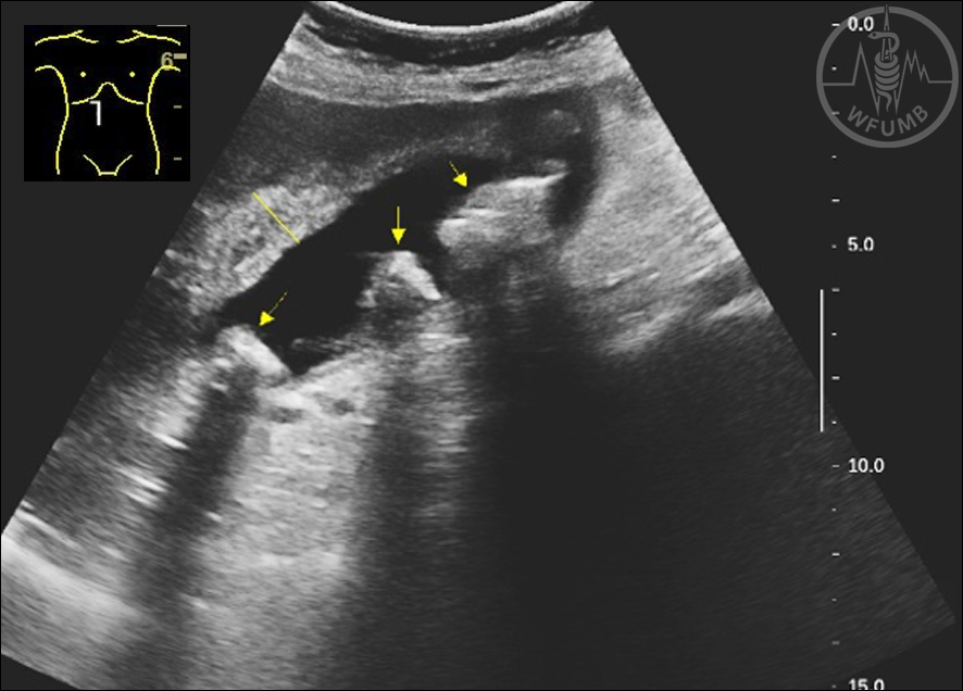
Fig 14.7 Small stones (arrows), not posterior shadowing behind all stones
Fig 14.8 Gallbladder stones - shell sign (arrow)
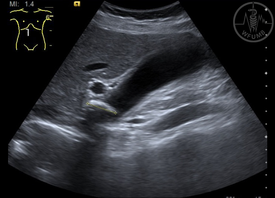
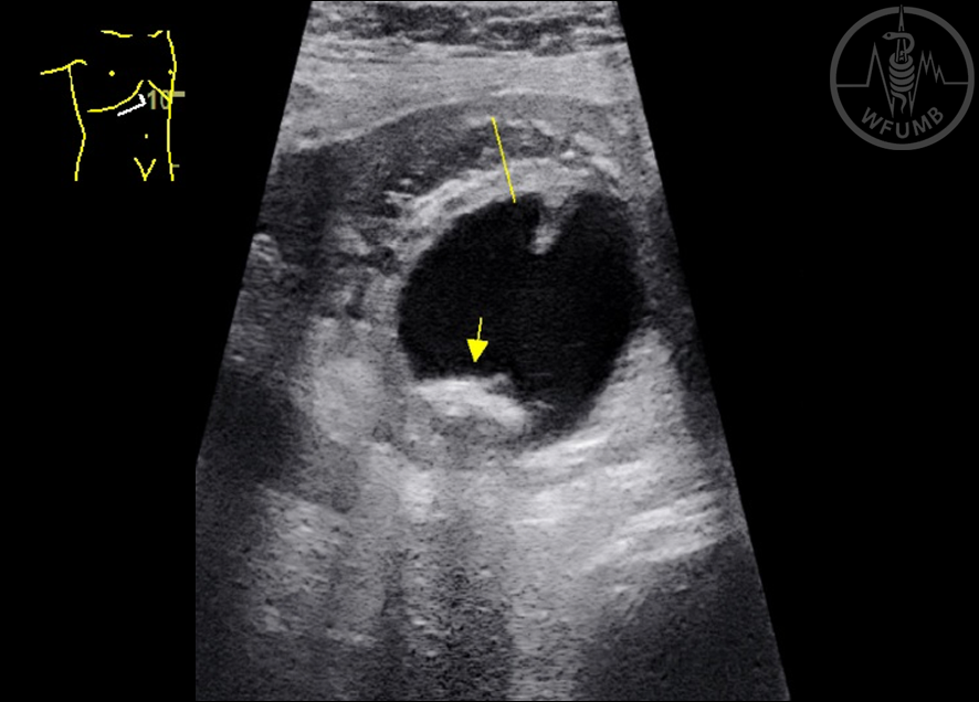
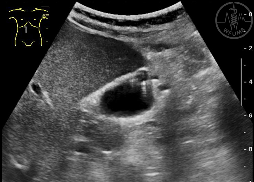
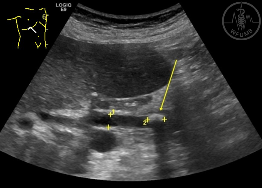
Fig 14.9 A stone in the infundibulum of the gallbladder
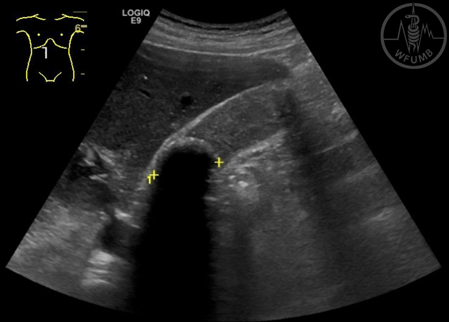
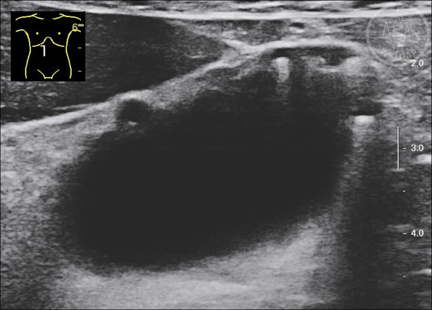
Fig 14.10 Stone in the gallbladder infundibulum, the gallbladder is filled with sludge
Fig 14.11 Acute cholecystitis – thick wall, hyperechoic material in the gallbladder (arrow)
Fig 14.12 Acute cholecystitis – a thick wall with edema, gallbladder stones (arrows)
Fig 14.13 Acute cholecystitis – a thick wall with edema in transverse section with gallbladder stones (arrow)
Fig 14.14 Chronic cholecystitis – a shrunken gallbladder with thickening of the wall and calculus
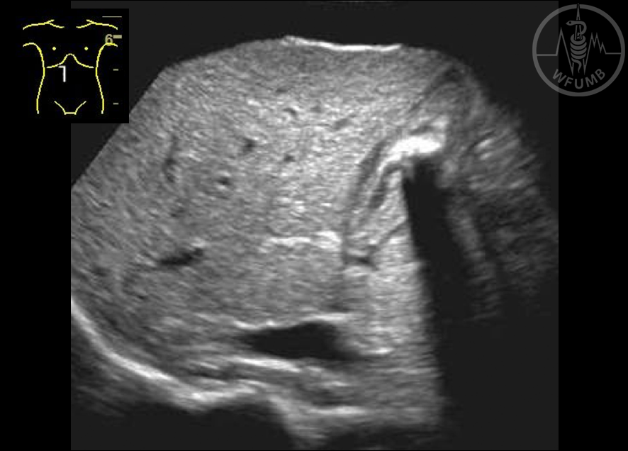
Fig 14.15 Chronic cholecystitis
Fig 14.16 Gallbladder adenoma – round structure with the same echogenicity as the gallbladder wall, no posterior shadowing
Fig 14.17 Gallbladder carcinoma – A CEUS study, the tumor enhances after contrast, the arrow shows a stone
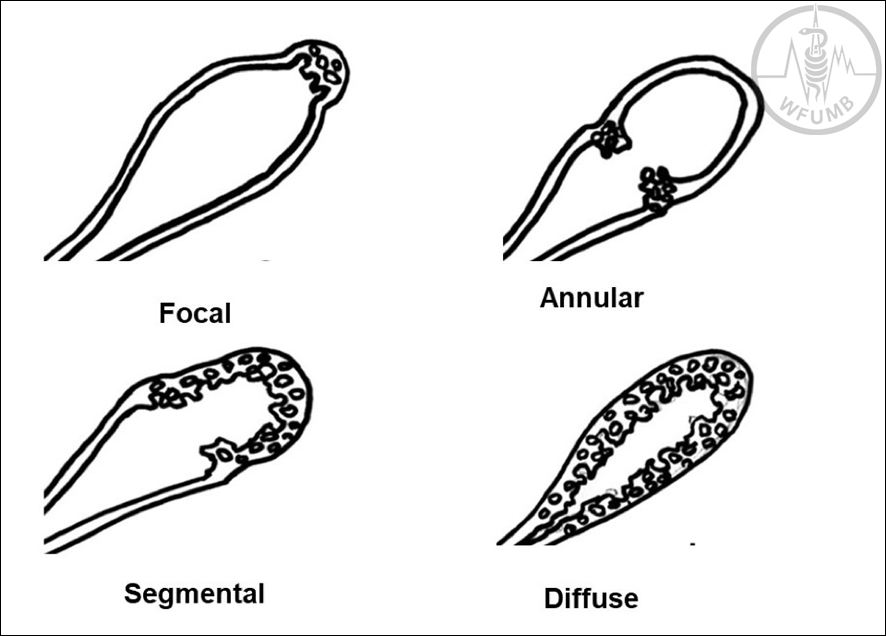
Fig 14.18 The various locations of gallbladder adenomyomatosis
Fig 14.19 Focal adenomyomatosis
Fig 14.20 Segmental adenomyomatosis
Fig 14.21 Dilated intrahepatic biliary ducts – the ”spider” appearance in the hilum
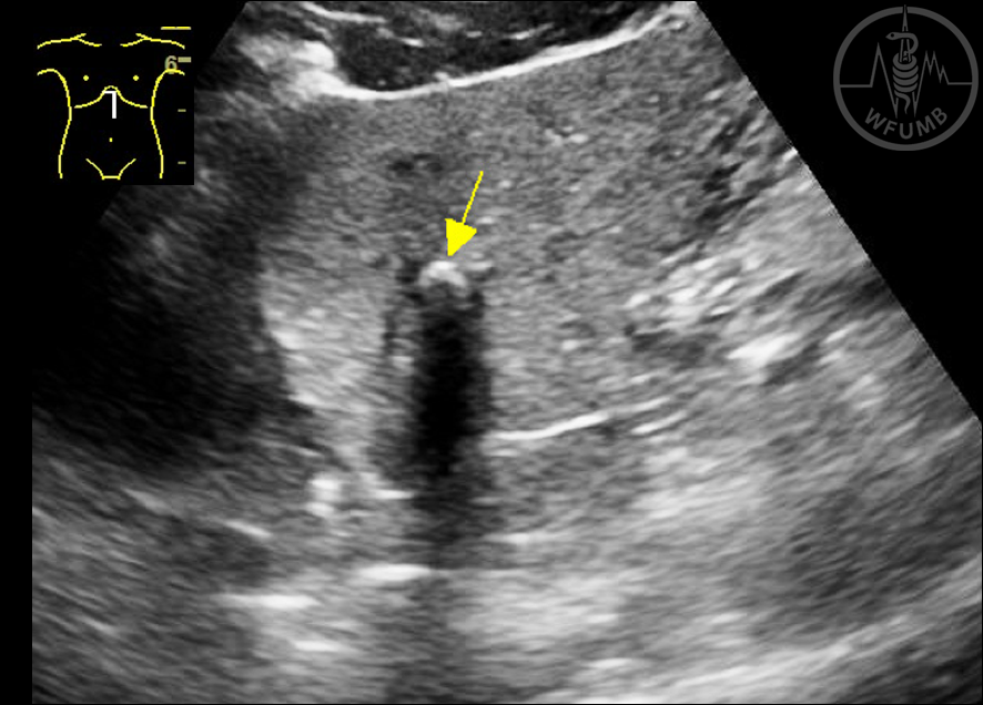
Fig 14.22 Choledocholithiasis (arrow) with dilated common bile duct
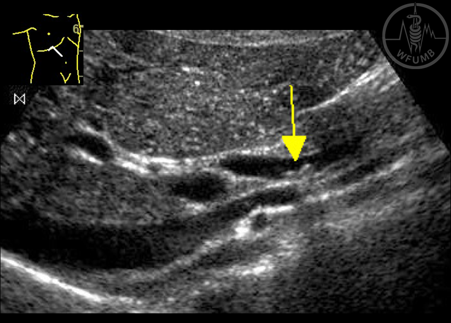
Fig 14.23 Intrahepatic lithiasis (arrow)
Fig 14.24 Choledocholithiasis (arrow) without dilatation of the biliary tract
Chapter Videos
Translate »