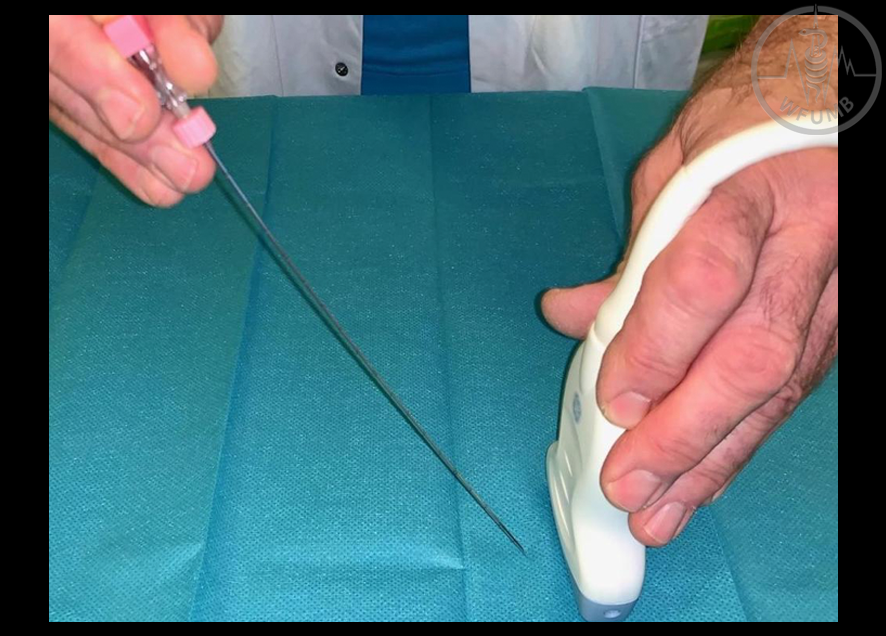
Fig 31.1b
Needle guide attached to a transducer - on US image the theoretical needle path is displayed as double line
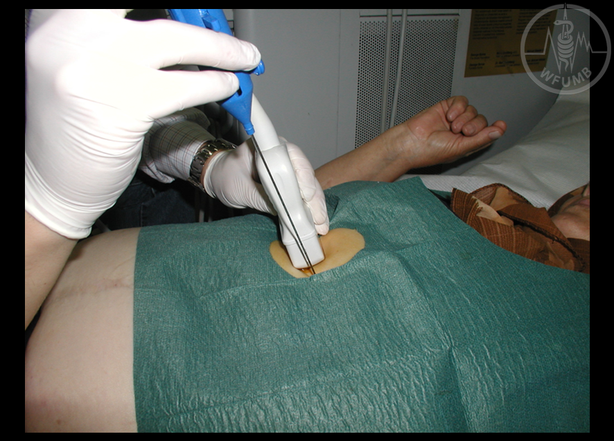
Fig 31.2a
Free hand technique - in plane
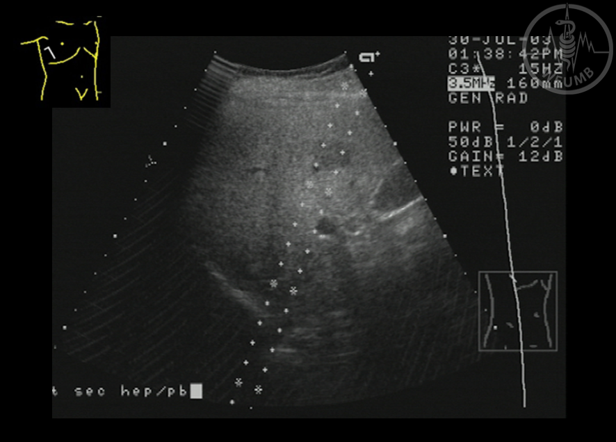
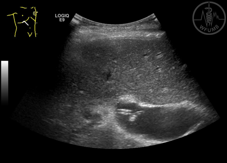
Fig 31.2b
Free hand technique. US guided parenchymal liver biopsy
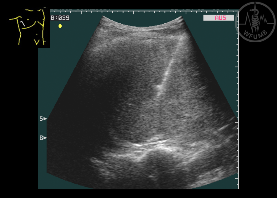
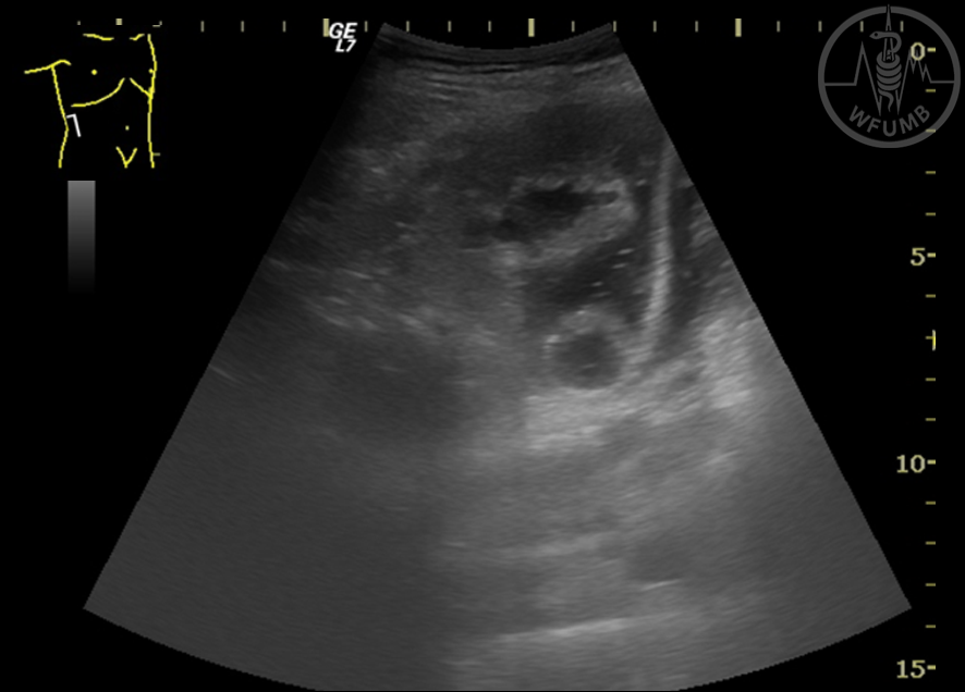
Fig 31.3
Free hand technique - out of plane
Fig 31.4a
Bleeding after a liver biopsy - intrahepatic haematoma (note that in early phases after bleeding the blood is hyper or isoechoic in respect to the liver)
Fig 31.4b
Bleeding after a liver biopsy - perihepatic blood (note that in early phases after bleeding the blood is hyper- or isoechoic in respect to the liver)
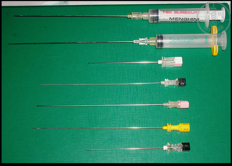
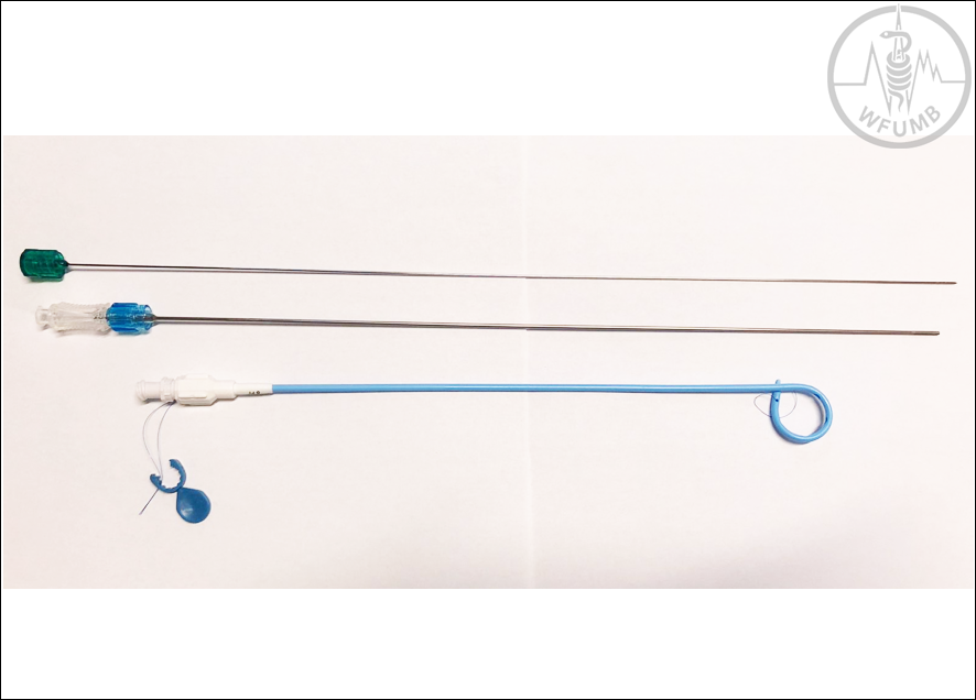
Fig 31.5
Types of fine aspiration needles for puncture and FNA
Fig 31.6a
Automatic gun with TruCut needle
Fig 31.6b
TruCut principle
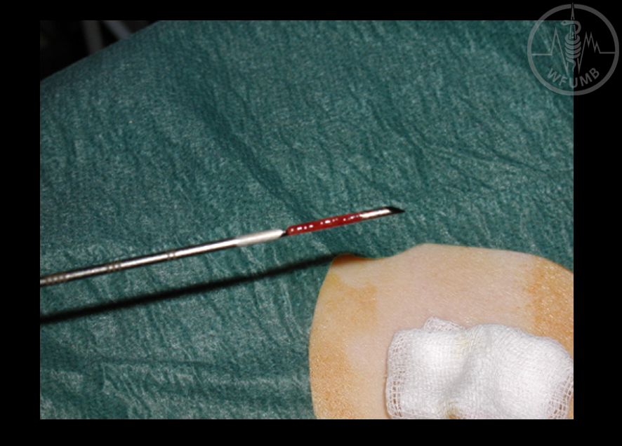
Fig 31.6c
Redish specimen in the distal notch
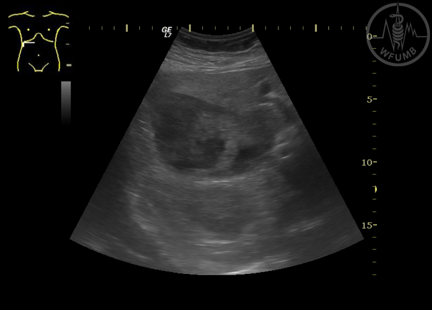
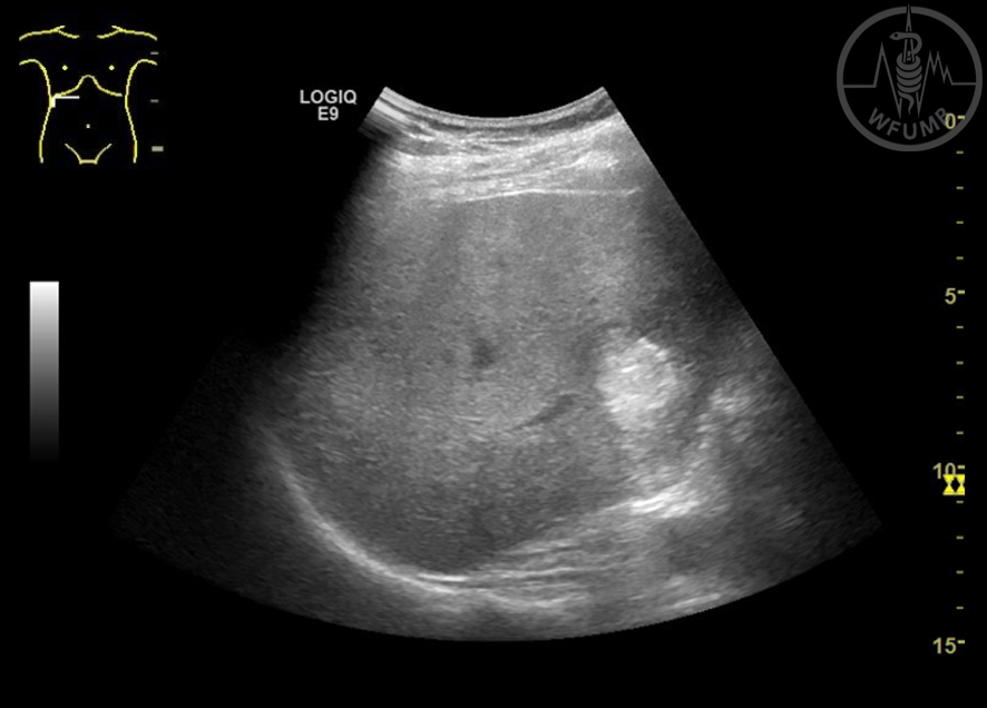
Fig 31.7
Anechoic structure inside the liver. Diagnostic puncture with aspiration. The tip of the needle is seen inside the lesion and almost the whole shaft
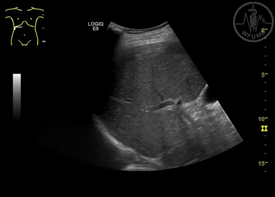
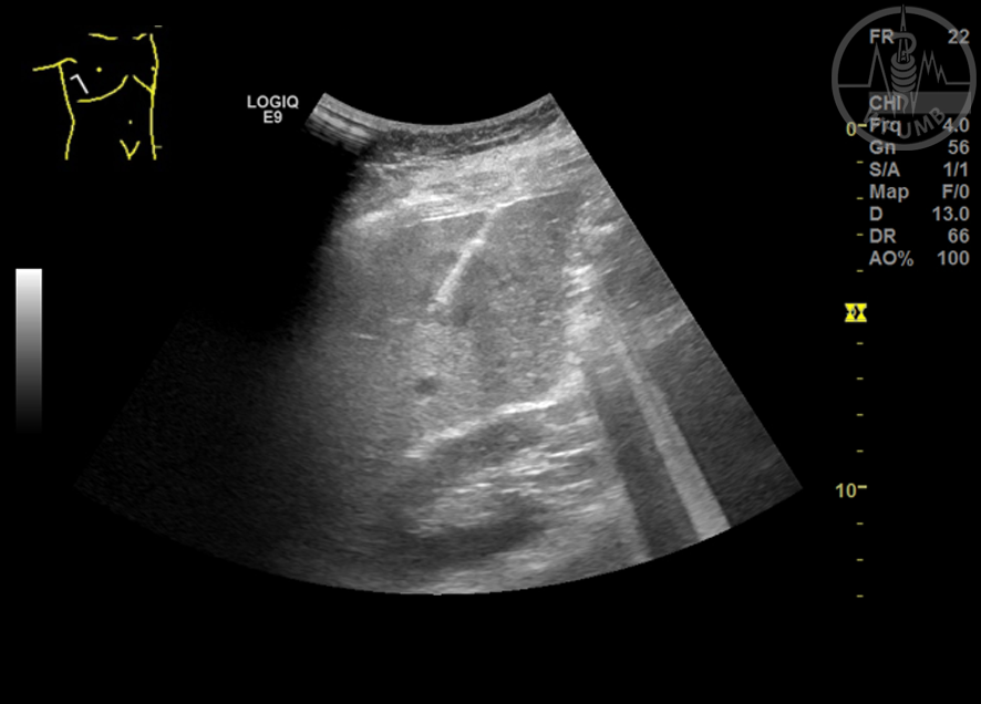
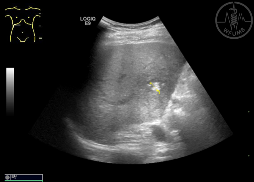
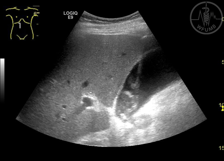
Fig 31.8
US guided biopsy of a hepatic tumor
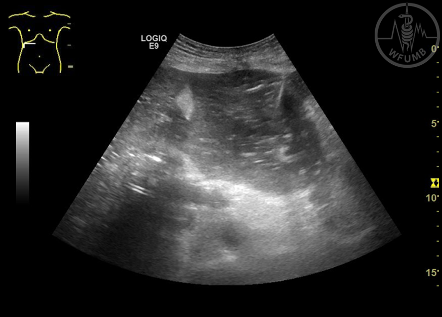
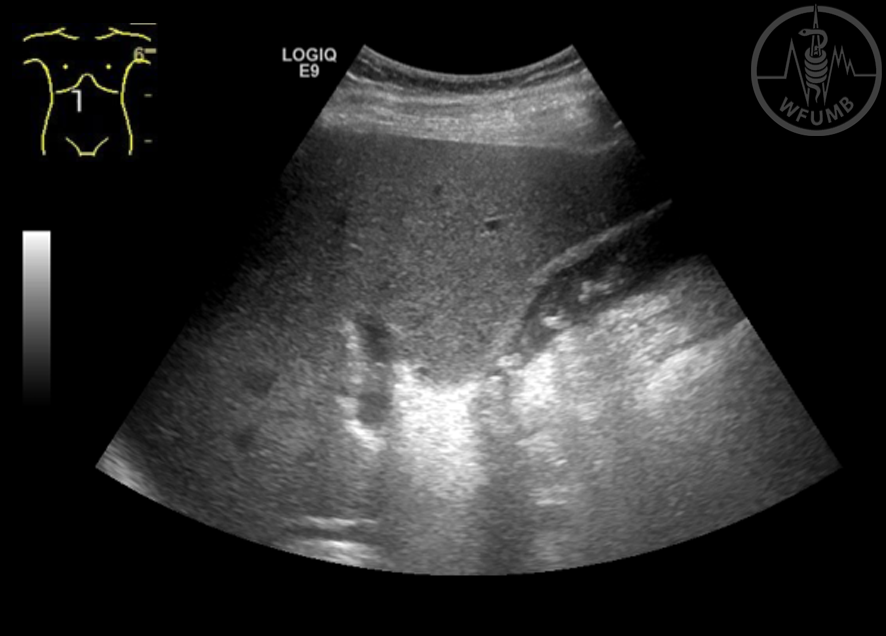
Fig 31.9
US guided biopsy of a retroperitoneal tumor
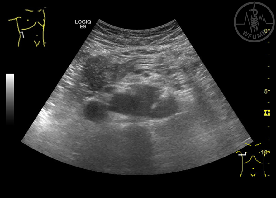
Fig 31.10
Liver biopsy with CEUS guidance in a large necrotic lesion
Fig 31.11a
Treatment of a small liver abscess with needle puncturing
Fig 31.11b
Treatment of a small liver abscess with needle puncturing - aspiration and lavage
Fig 31.11c
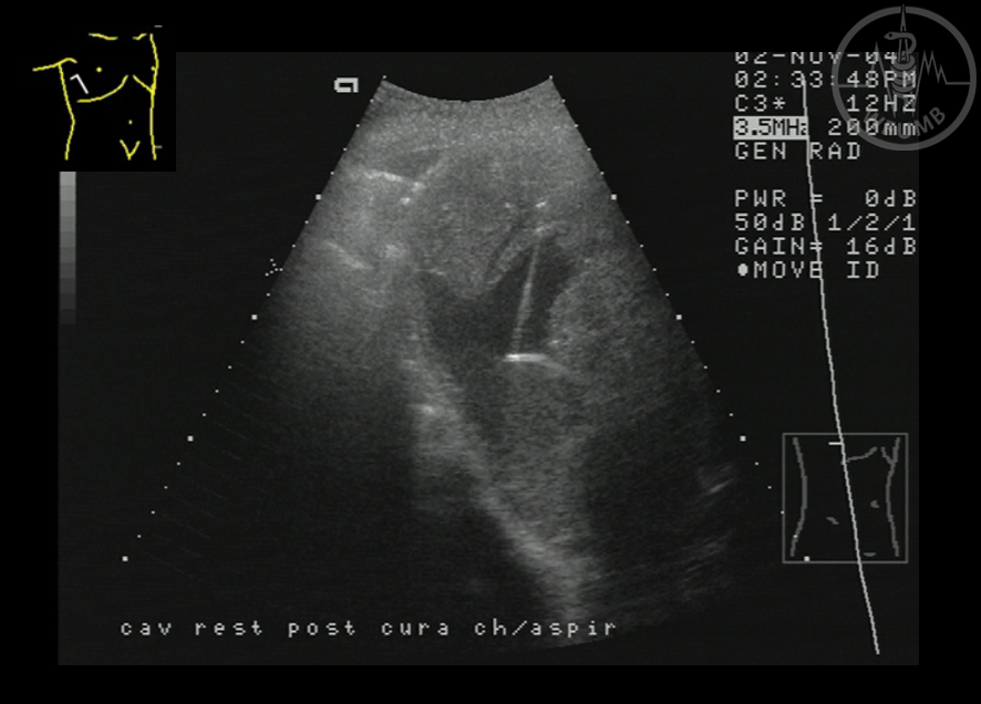
Treatment of a small liver abscess with needle puncturing. The result of the treatment is a small echogenic residual lesion
Fig 31.12
8 Fr pigtail catheter with internal fixation (string)
Fig 31.13a
Large liver abscess treated by catheter drainage
Fig 31.13b
Large liver abscess treated by catheter drainage - residual image after 10 days drainage
Fig 31.14
US guided drainage for an infected ascites. The pigtail of the catheter is well seen in the infected ascites
Fig 31.15a
US guided external biliary drainage. Puncture of a dilated peripheral bile duct
Fig 31.15b
US guided external biliary drainage. The guide wire is seen inside the left hepatic duct
Fig 31.15c
US guided external biliary drainage. The catheter inside the dilated common bile duct
Fig 31.16
Percutaneous nephrostomy. The catheter (>) is well seen in the dilated calyx
Fig 31.17a
Acute calculous cholecystitis. One step percutaneous cholecystomy using a 10 F pigtail catheter
Fig 31.17b
Acute calculous cholecystitis. US aspect after drainage
Fig 31.18a
RFA ablation of a small HCC in segment 6
Fig 31.18b
RFA ablation of a small HCC - an echogenic cloud is seen covering the lesion
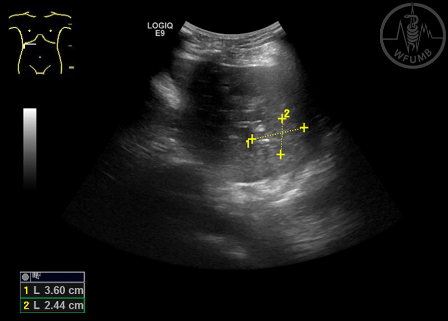
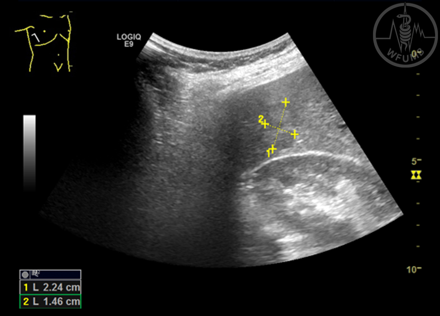
Fig 31.19a
MWA ablation of a 2.2 cm liver met located in segment 6 - US image
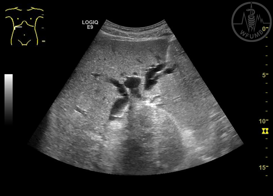
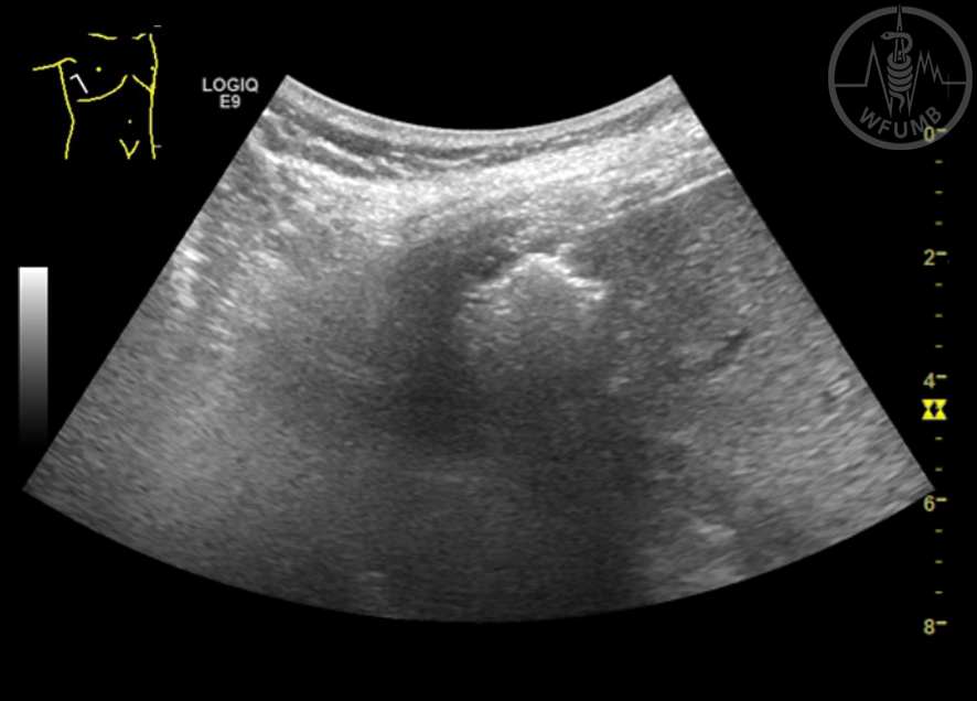
Fig 31.19b
MWA ablation of a 2.2 cm liver met located in segment 6 - US image during ablation with the echogenic cloud
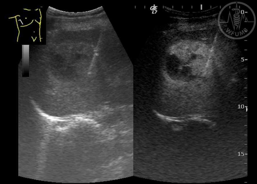
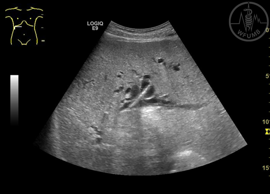
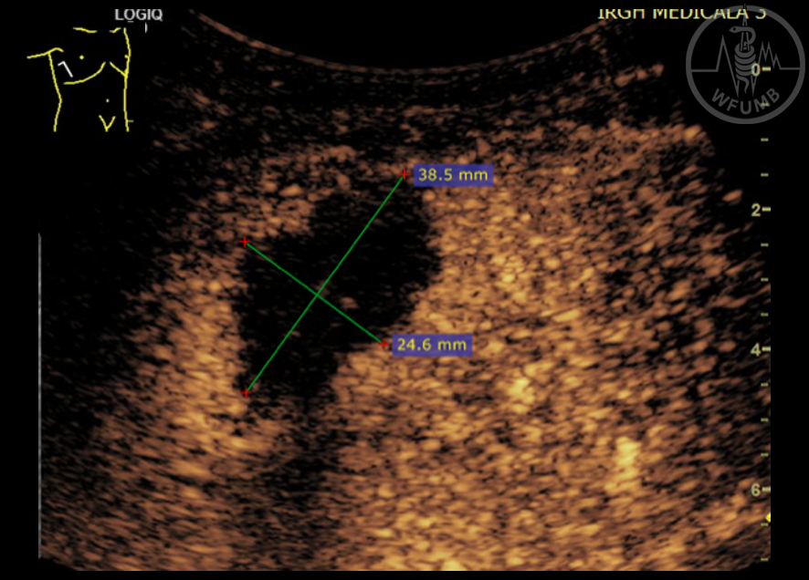
Fig 31.19c
MWA ablation of a 2.2 cm liver met located in segment 6 - CEUS performed 5 min after the procedure showing a 3.9/2.5 cm unenhanced necrotic area
Chapter Video
This website uses cookies to improve your experience. By using this website you agree to our Data Protection Policy.