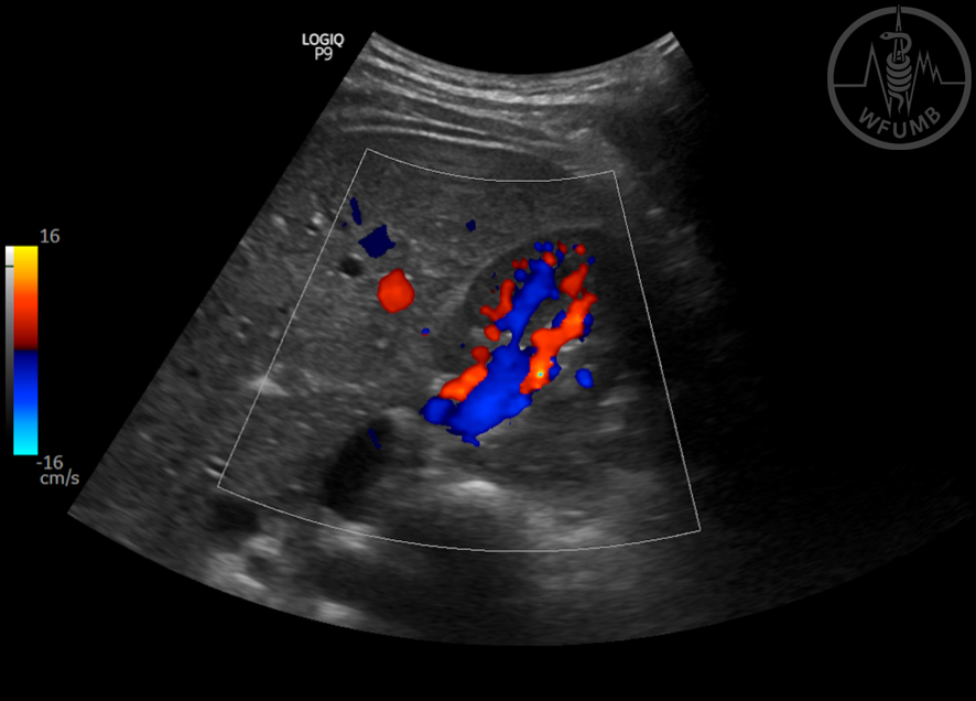
Fig 10.4
Normal vascularization of kidney in CD long view
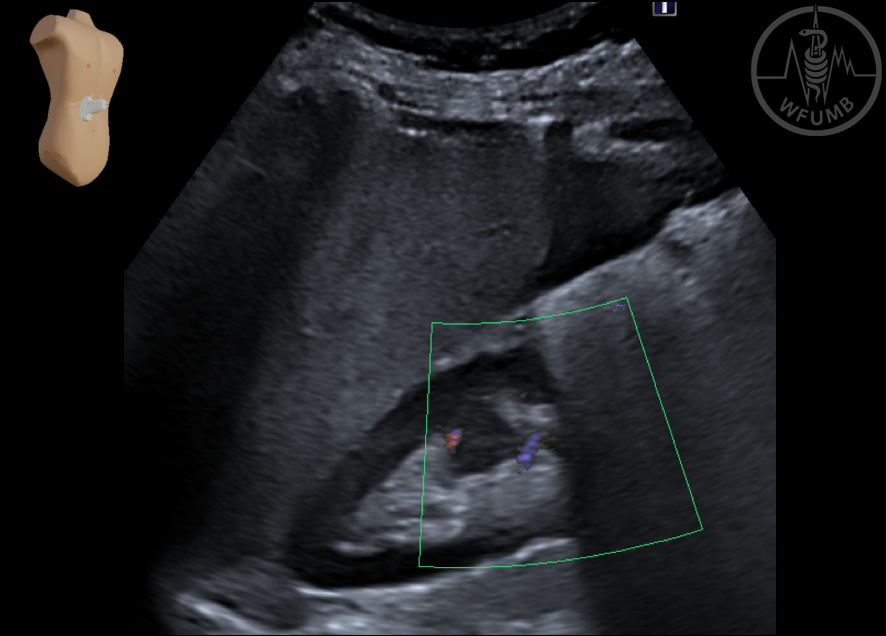
Fig 10.5
Normal kidney vascularization in CD transverse view of left kidney
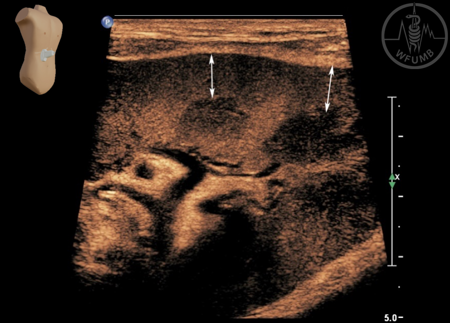
Fig 10.6
Hypertrophy of Bertin renal column
Fig 10.7
Renal junctional defect
Fig 10.8
Renal hypoplasia due to vesicoureteral reflux
Fig 10.9
Compensatory hypertrophy in unilateral surgical kidney longit. 15 cm (post-nephrectomy)
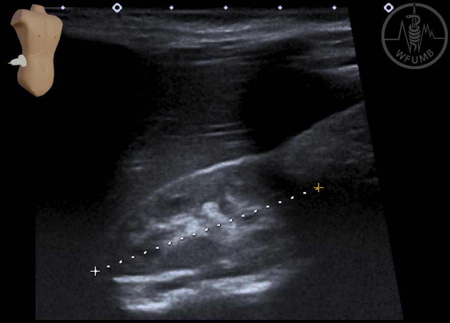
Fig 10.10
Ectopic Kidney located in right iliac fossa
Fig 10.11
Horseshoe kidney with midline isthmus demonstrated
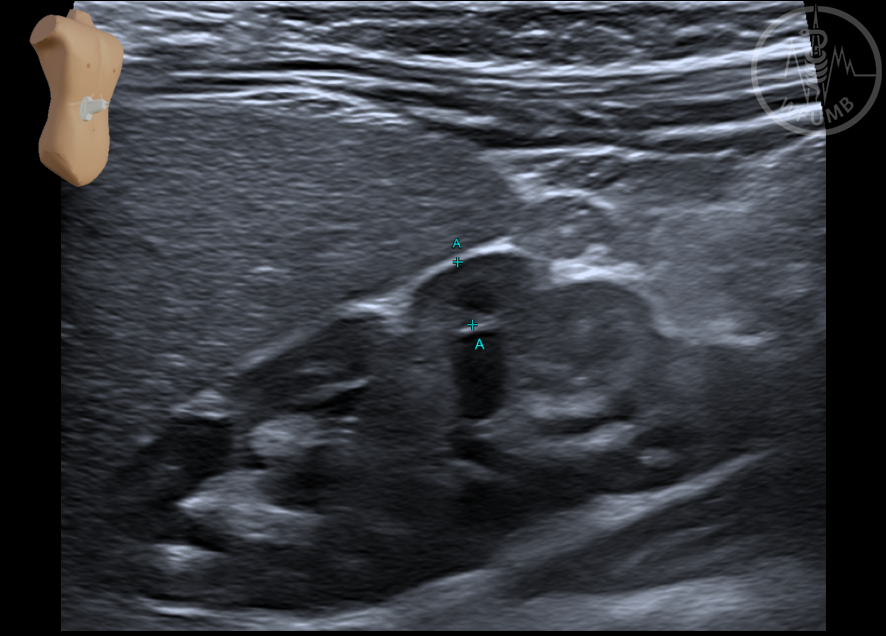
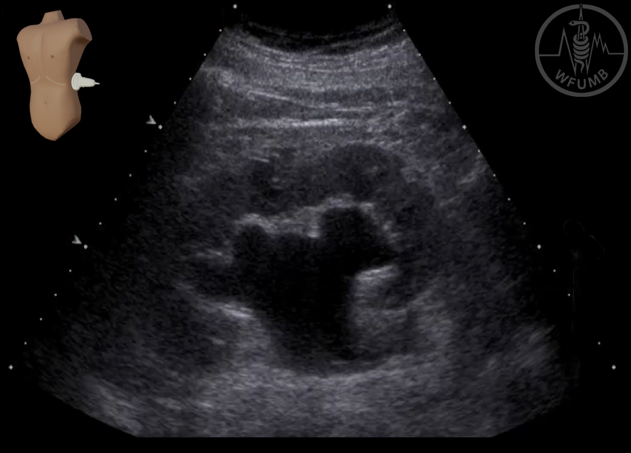
Fig 10.12
Duplication of collecting system with minimal lower pole dilatation (Dilatation /hydronephrosis stage 1)
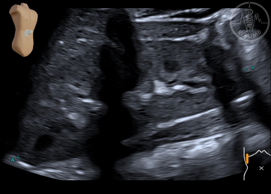
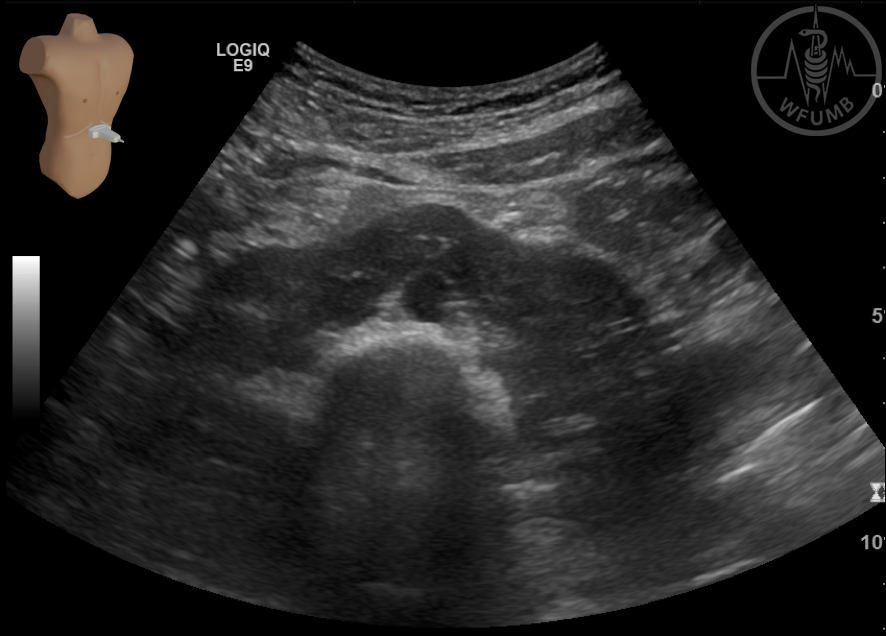
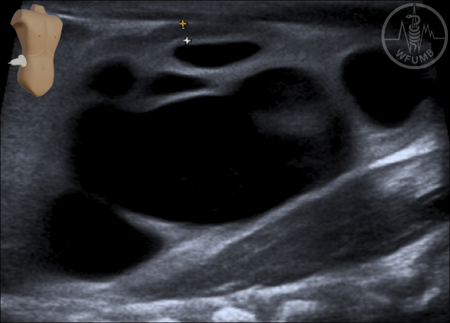
Fig 10.13
Ureteropelvic junction obstruction with gross dilatation of the renal pelvis and calyces and cortical thinning (objectified on the right side) (Dilatation/hydronephrosis stage 4)
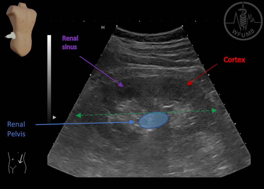
Fig 10.14
Normal ri. Kidney and renal pelvis, no dilatation of echorich pelvis
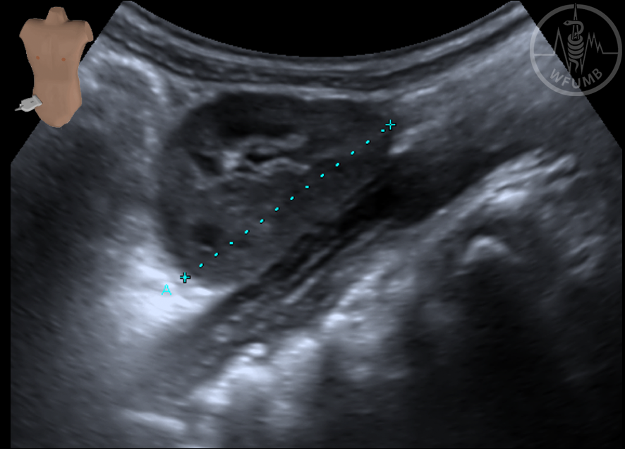
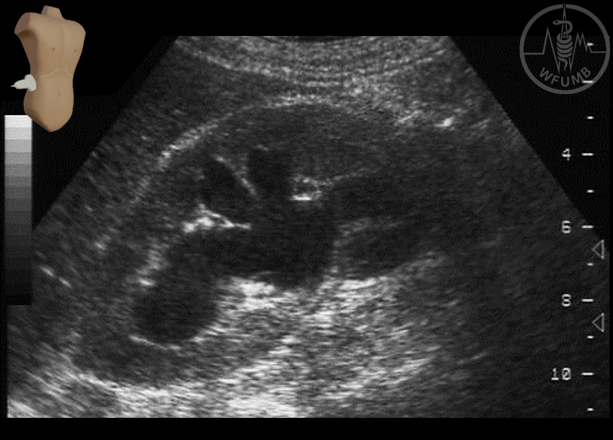
Fig 10.15
Dilatation 2nd degree of the left kidney because of distal ureter stone
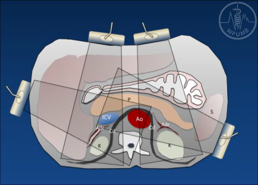
Fig 10.16
Cross sectional of the abdomen at the level of the adrenal glands – relationship with surrounding organs
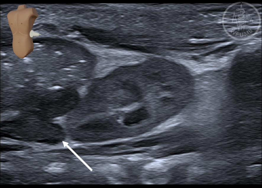
Fig 10.17
Normal right adrenal gland in longit. Scan using the liver as an acoustic window
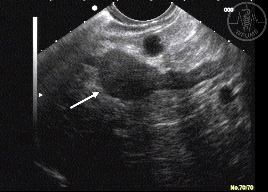
Fig 10.18
Normal left adrenal gland in EUS
Fig 10.19
Hypoechoic left adrenal hyperplasia in newborn
Fig 10.20
Left adrenal focal hyperplasia in EUS
Chapter Videos
This website uses cookies to improve your experience. By using this website you agree to our Data Protection Policy.