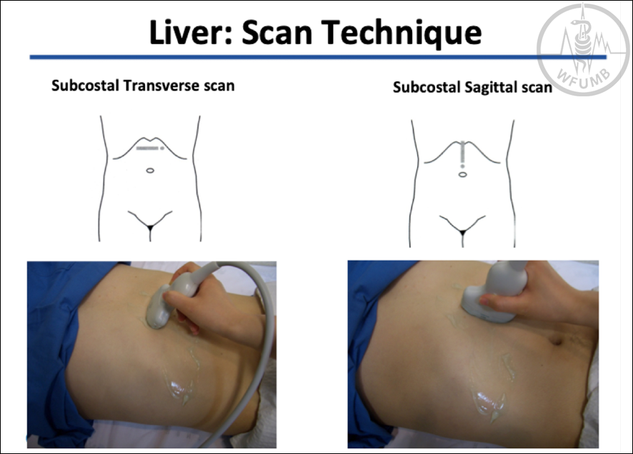
Fig 6.1
Liver scan technique, left liver lobe. Subcostal longitudinal (sagittal) and transverse scanning
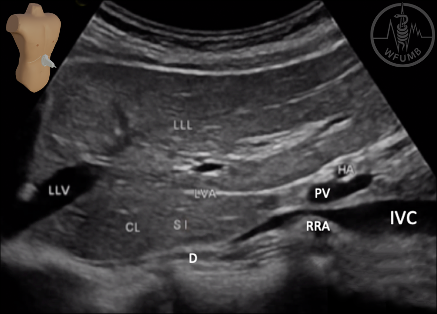
Fig 6.2.
Longitudinal (sagittal) ultrasound aspect of the Left Liver Lobe (LLL): LLV - Left liver vein; LVA - Ligamentum venosum Arantii; CL - Caudate lobe (SI, Segment 1); IVC - Inferior vena cava; D – Diaphragma; RRA - Right renal artery; PV - Portal vein; HA – hepatic artery
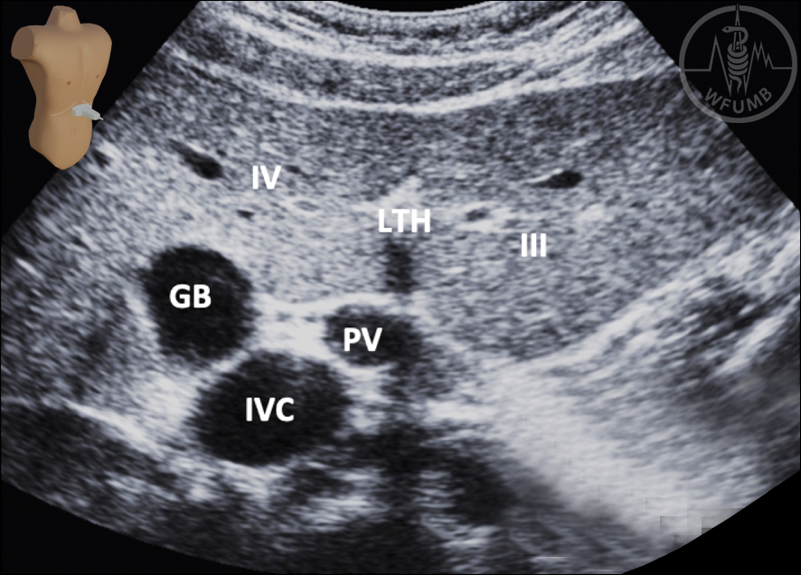
Fig 6.3.
Transverse ultrasound aspect of the Left Liver Lobe (LLL): IVC - Inferior vena cava; PV - Portal vein; LTH - Ligamentum teres hepatis; GB – Gallbladder; III - Liver segment III; IV - Liver segment IV.
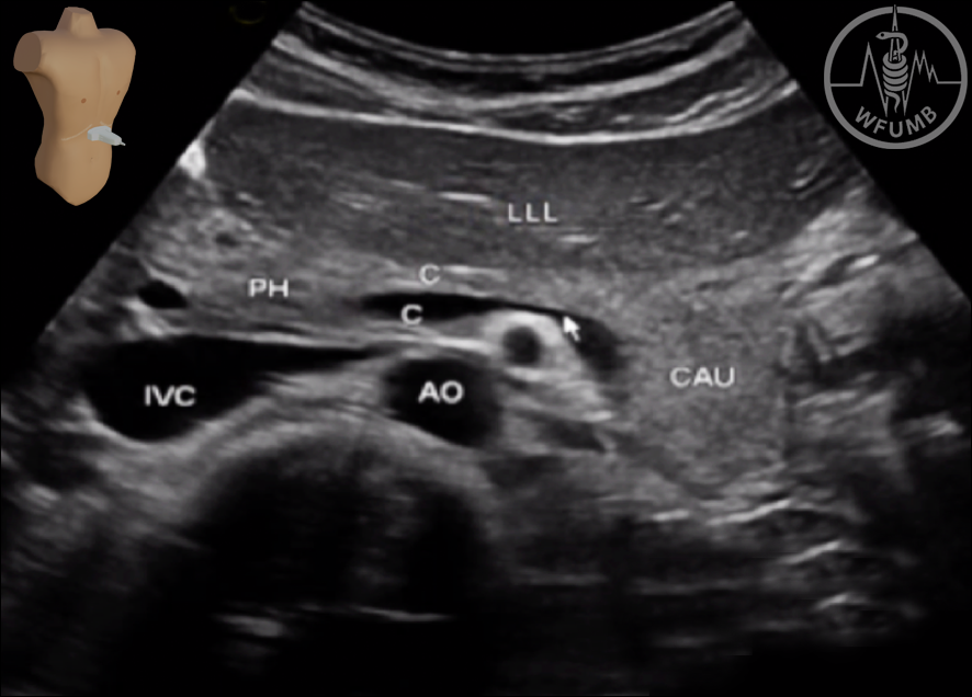
Fig 6.4.
Transverse ultrasound aspect of the Left Liver Lobe (LLL): IVC - Inferior vena cava; AO – Aorta;
PH – Pancreatic head;
C – Pancreatic body;
CAU – pancreatic tail.
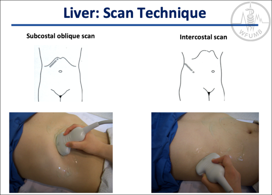
Fig 6.5
Liver scan technique, right liver lobe. Subcostal oblique and subcostal and intercostal umbilicus - shoulder scanning
Fig 6.6
Ultrasound aspect of the right liver lobe (RLL) in a subcostal oblique scan: RLV - Right liver vein; MLV - Intermediate liver vein; LLV - Left liver vein; VCI - Vena cava inferior; C - Confluens of MLV and LLV.
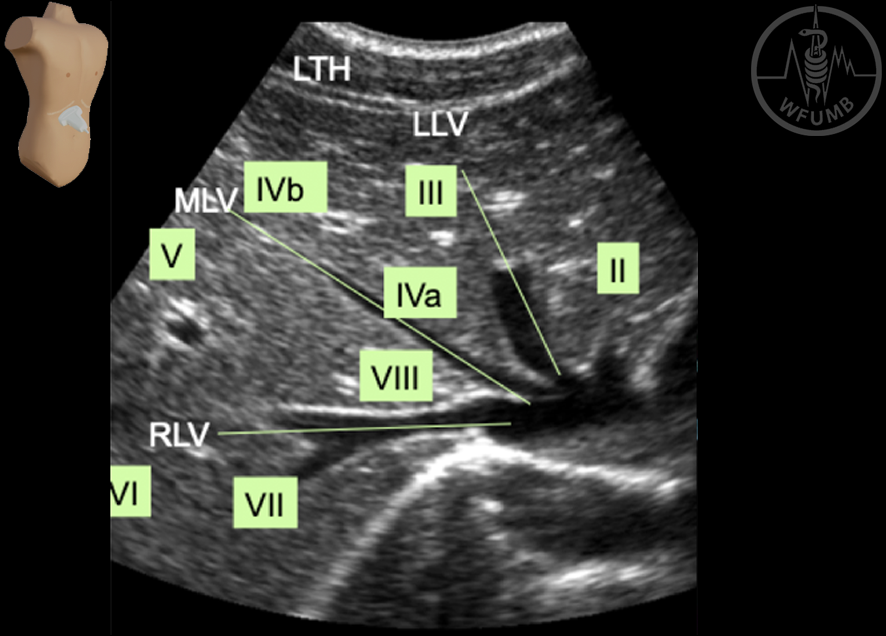
Fig. 6.7a
Ultrasound aspect of the right liver lobe in a subcostal oblique scan: RLV - Right liver vein; MLV - Intermediate liver vein; LLV - Left liver vein; LTH - Ligamentum teres hepatis. The liver segments II, III, IVa, IVb, V, VI, VII and VIII are indicated as well.
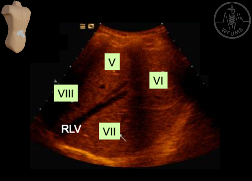
Fig. 6.7b
Ultrasound aspect of the right liver lobe in a subcostal oblique scan: RLV - Right liver vein; The liver segments V, VI, VII and VIII are indicated as well
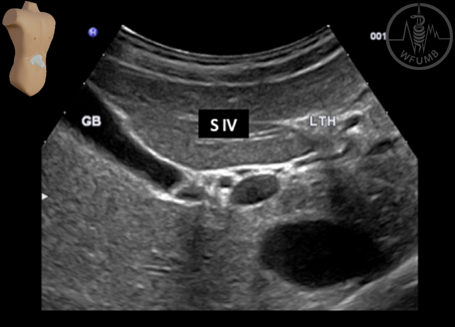
Fig. 6.8a
Ultrasound aspect of the right liver lobe in a
subcostal oblique scan:
GB – gallbladder,
LTH (ligamentum teres hepatis); The liver segment IV is indicated as well
Fig. 6.8b
Ultrasound aspect of the right liver lobe in a subcostal oblique scan: A - Anterior portal vein branch;
P - Posterior portal vein branch; U - Pars umbilicalis of portal vein
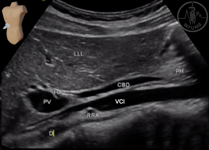
Fig. 6.9
Ultrasound aspect of the right liver lobe (RLL) in a subcostal and intercostal umbilicus - shoulder scanning. LLL - Left Liver Lobe; VCI - Inferior vena cava; D – Diaphragma; RRA - Right renal artery; PV - Portal vein; HA – hepatic artery; CBD – common bile duct; PH – pancreatic head
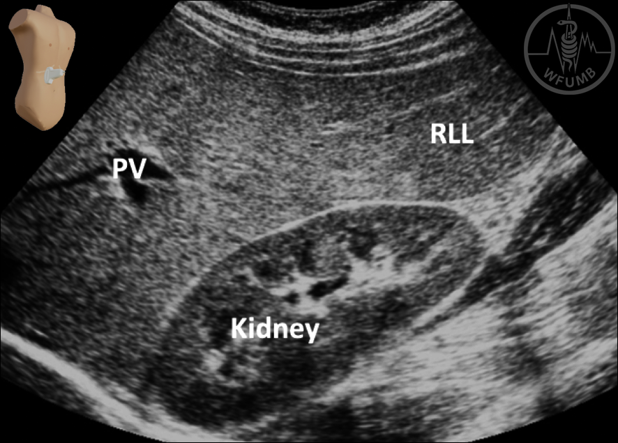
Fig. 6.10
Ultrasound aspect of the right liver lobe (RLL) in a subcostal and intercostal umbilicus - shoulder scanning. PV - Portal vein