WFUMB Course Book
Liver – Diffuse Liver Diseases - Chaper 13.1 Media Library
Close window and return to Chapter 13.1
Chapter Images
Fig 13.1.1 Gallbladder wall thickening in acute hepatitis
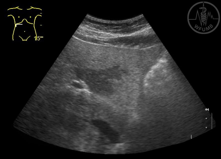
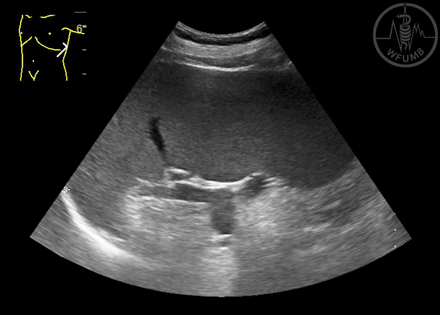
Fig 13.1.2 Mild splenomegaly
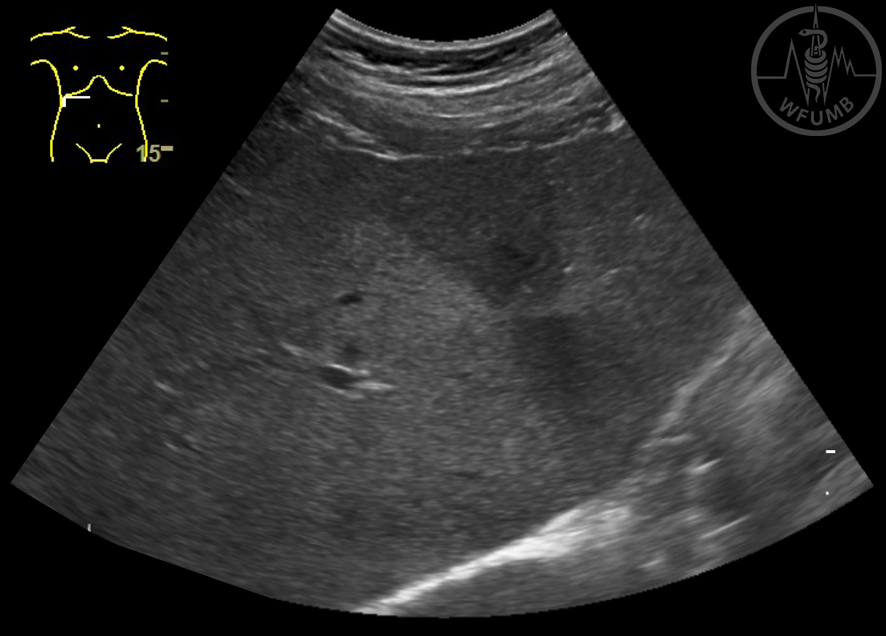
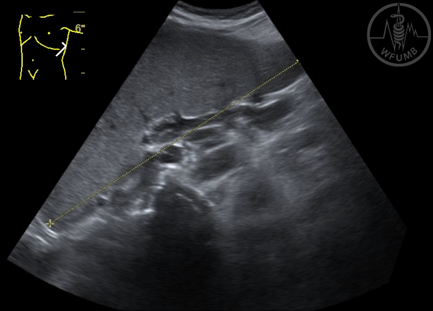
Fig 13.1.4 Significant splenomegaly (spleen long axis 16,5 cm)
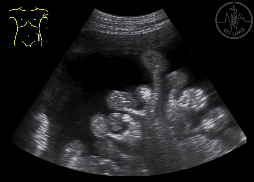
Fig 13.1.5 Hilar oval adenopathy in chronic hepatitis C
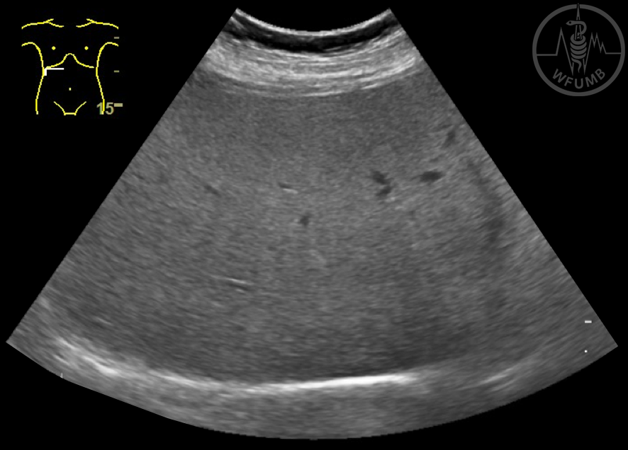
Fig 13.1.6 Mild liver steatosis (“bright liver”)
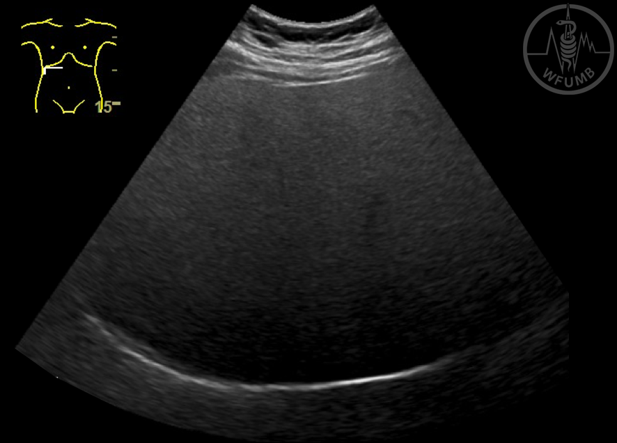
Fig 13.1.7 Liver steatosis (“Bright liver” with posterior attenuation)
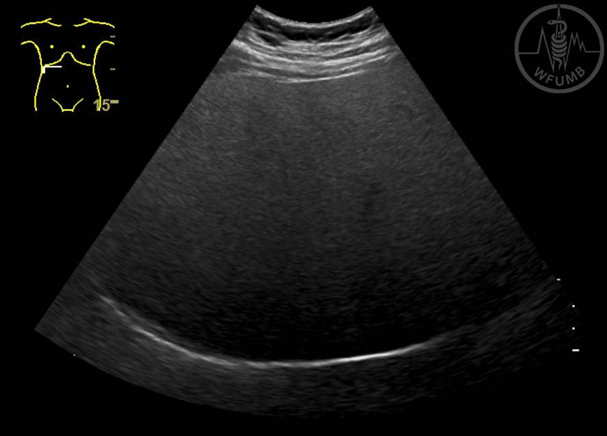
Fig 13.1.8 Severe liver steatosis (“Bright liver” with important posterior attenuation)
Fig 13.1.9 Liver steatosis (increased hepato-renal index)
Fig 13.1.10 Liver steatosis (“Bright liver” with increased hepato-renal index)
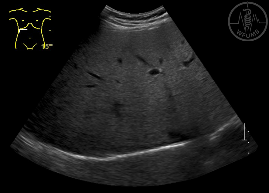
Fig 13.1.11 Liver steatosis
Fig 13.1.12 Hypoechoic area near the gallbladder – fatty sparing lesion
Fig 13.1.13 Fatty sparing lesion (“fatty free area”) - hypoechoic area with map-like delineation in the left liver lobe
Fig 13.1.14 Fatty sparing lesion (“fatty free area”) - hypoechoic area with map-like delineation in the right liver lobe
Fig 13.1.15 Fatty sparing lesion (“fatty free area”) - hypoechoic area left liver lobe
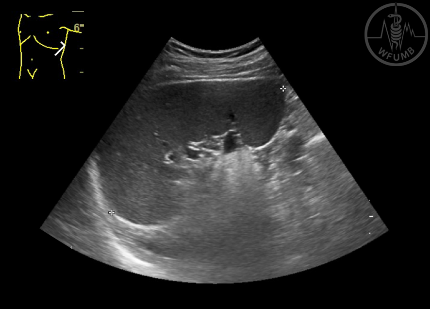
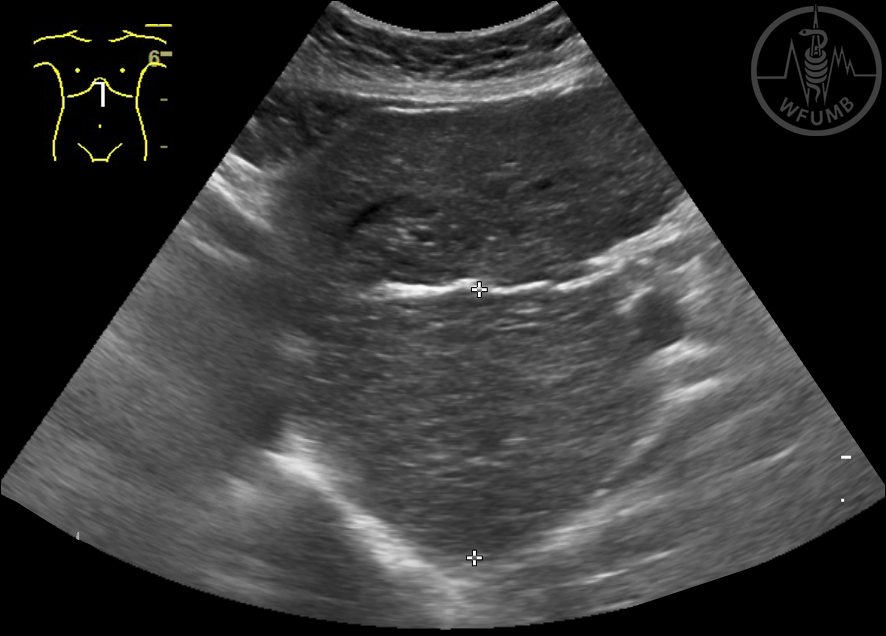
Fig 13.1.16 Caudate lobe larger than 35 mm in a patient with liver cirrhosis
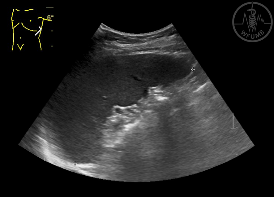
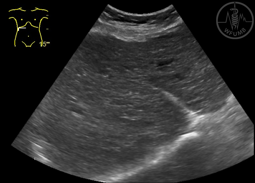
Fig 13.1.17 Increased size of caudate lobe. Perihepatic ascites
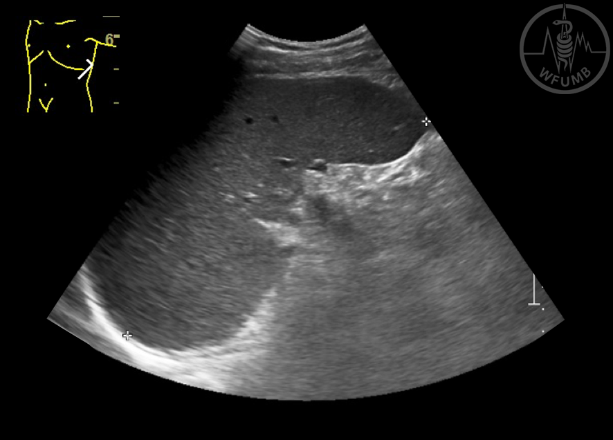
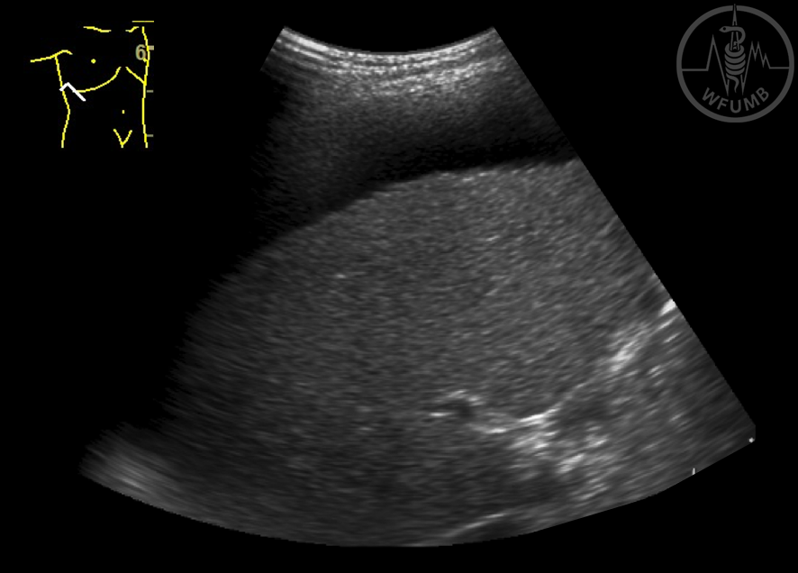
Fig 13.1.18 Very large caudate lobe in a patient with liver cirrhosis
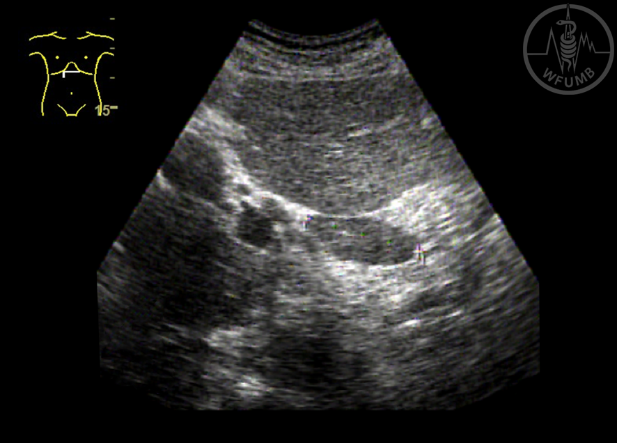
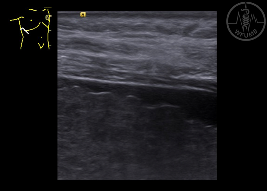
Fig 13.1.19 Hepatic heterogeneity in a patient with liver LC
Fig 13.1.20 Irregular liver surface and ascites in a patient with LC
Fig 13.1.21 Irregular liver surface and ascites in a patient with LC using high frequency transducer
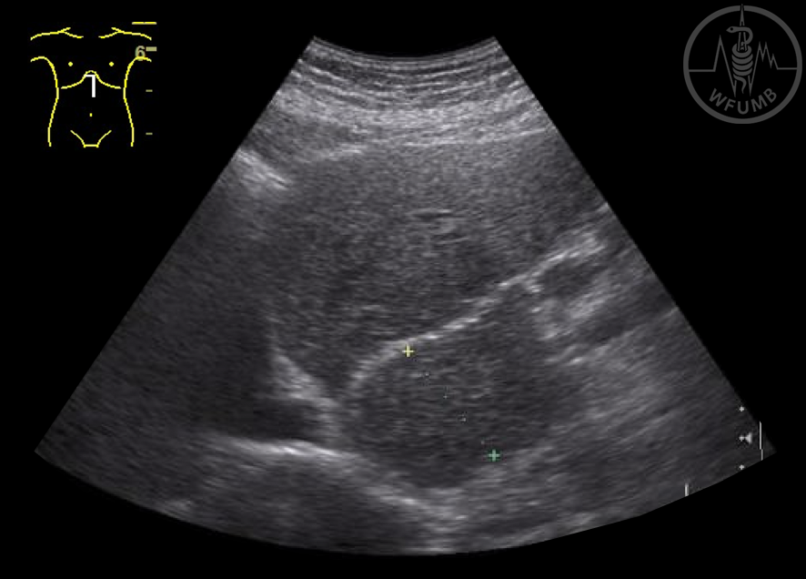
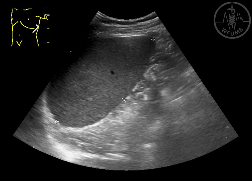
Fig 13.1.22 Splenomegaly
Fig 13.1.23 Splenomegaly with portal hypertension
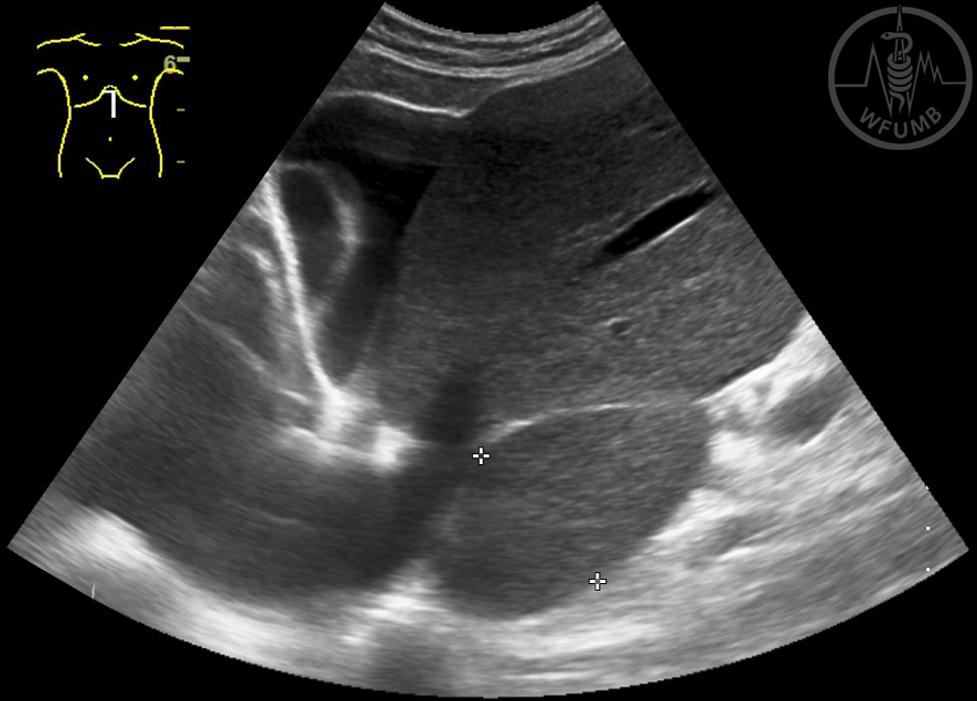
Fig 13.1.24 Globulous spleen
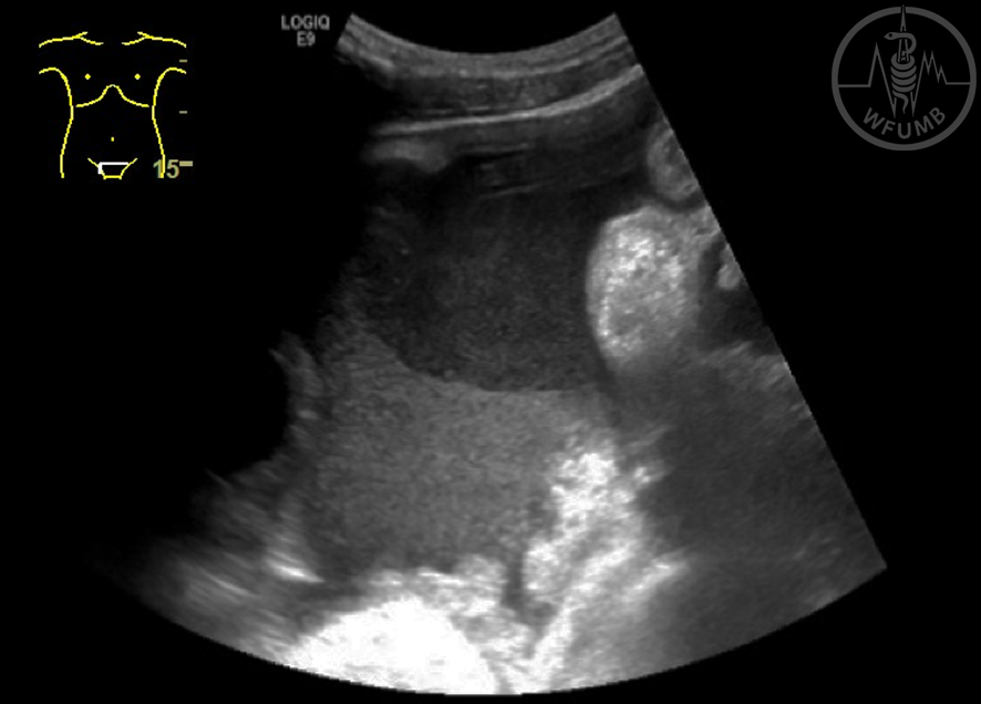
Fig 13.1.25 Bowel loops in ascites
Fig 13.1.26 Perihepatic ascites and heterogeneous liver echotexture in a patient with LC
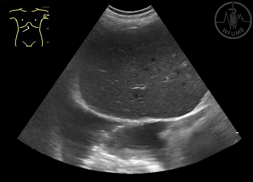
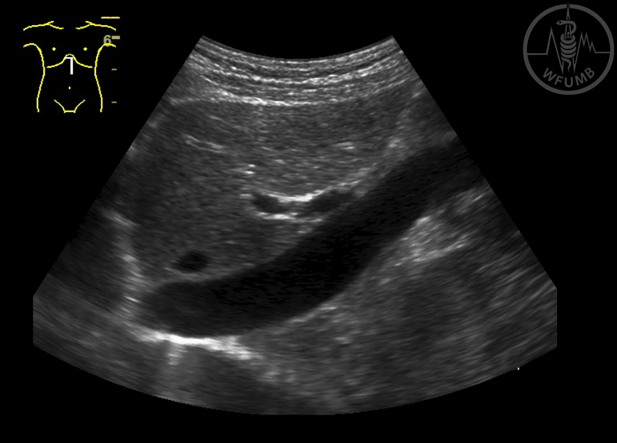
Fig 13.1.27 ”Dense” ascites
Fig 13.1.28 Pleural effusion (fluid outside diafragma)
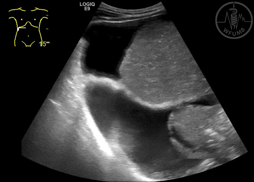
Fig 13.1.29 Ascites and pleural effusion (diafragma between them)
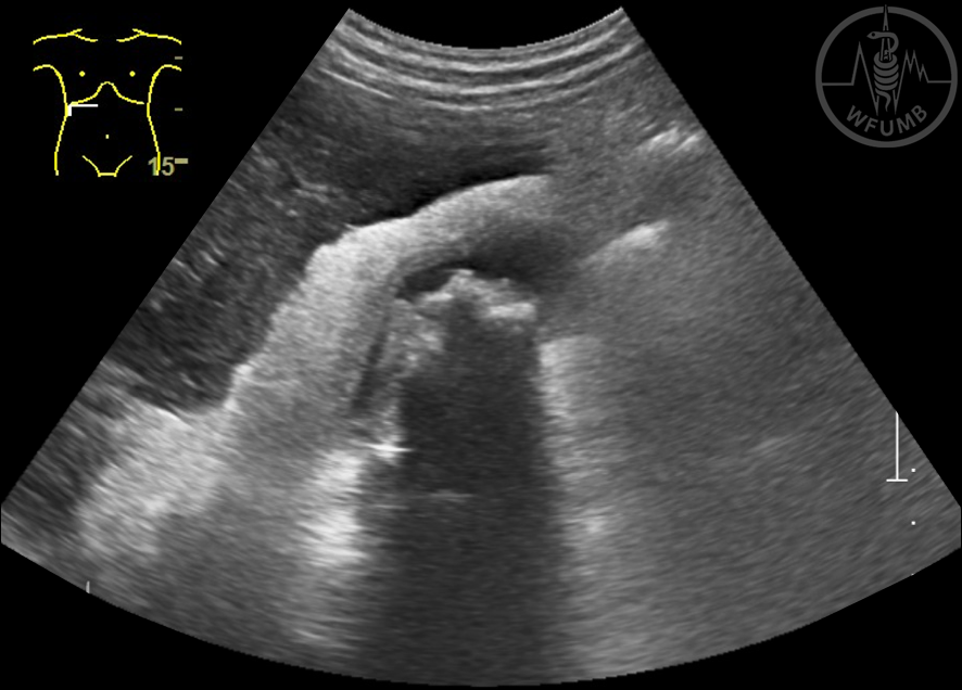
Fig 13.1.30 Portal hypertension.Tortuous splenic varices
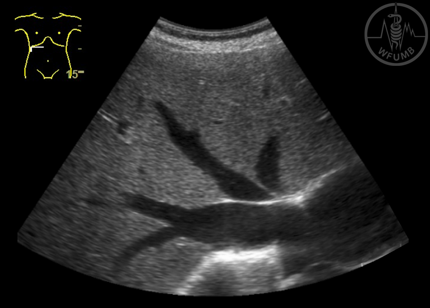
Fig 13.1.31 Splenic varices
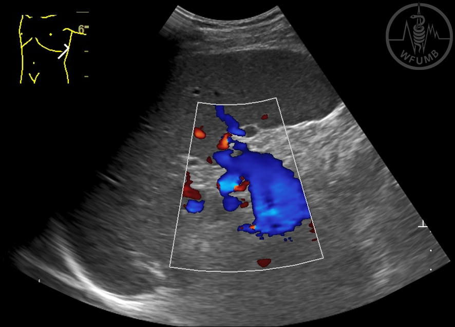
Fig 13.1.32 Splenic varices. Doppler examination
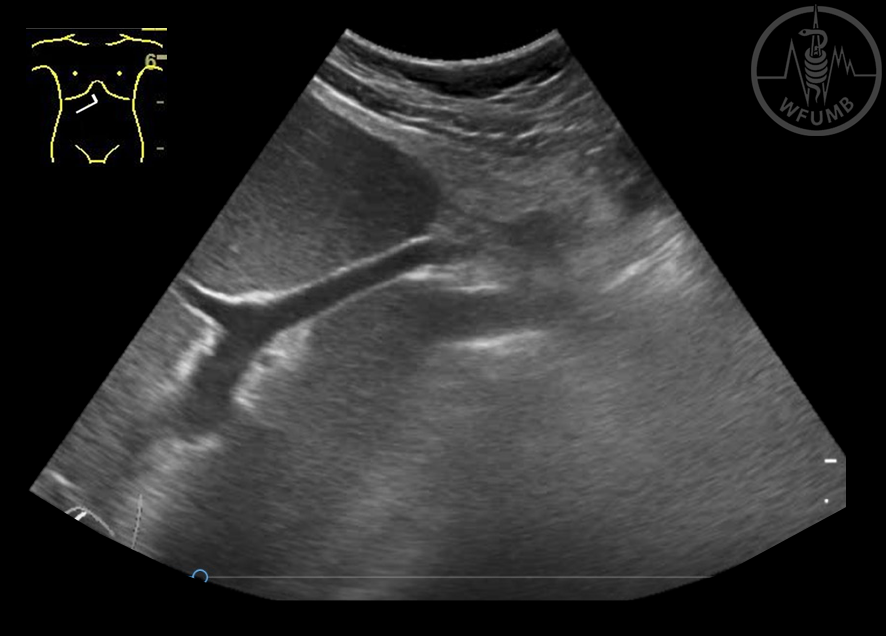
Fig 13.1.33 Repermeabilization of paraumbilical vein
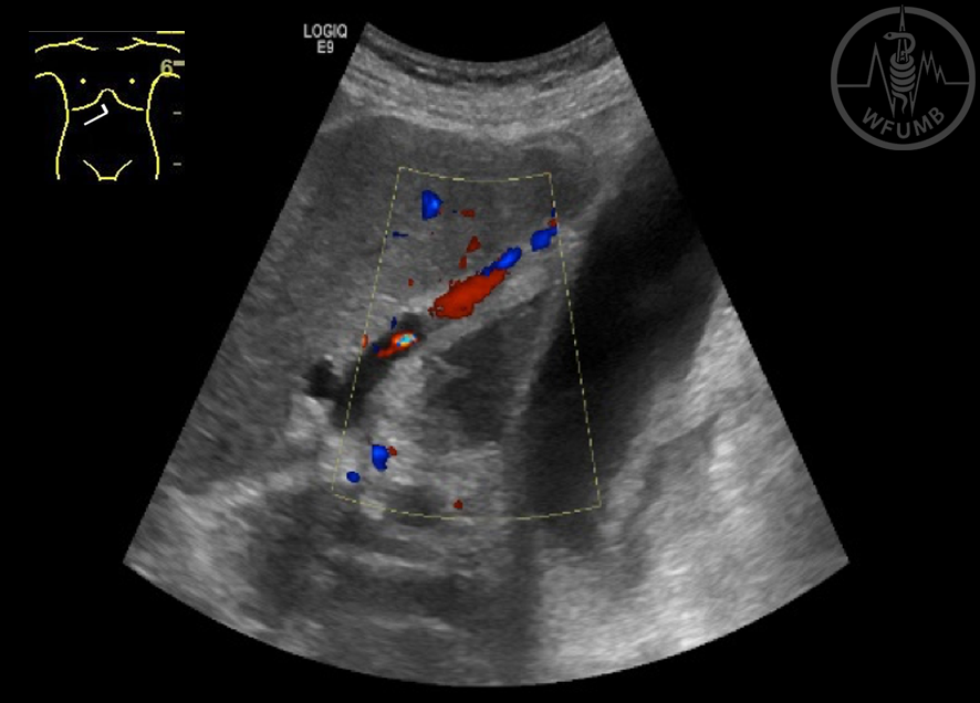
Fig 13.1.34 Dilatation of the paraumbilical vein. Color Doppler
Fig 13.1.35 Thickened gall bladder wall in a case of liver cirrhosis with stones
Fig 13.1.36 Dilation of hepatic veins
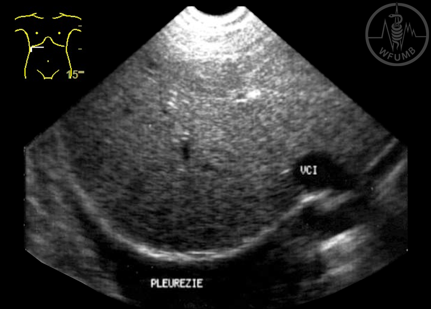
Fig 13.1.37 Dilation of the inferior vena cava
Fig 13.1.38 Pleural effusion
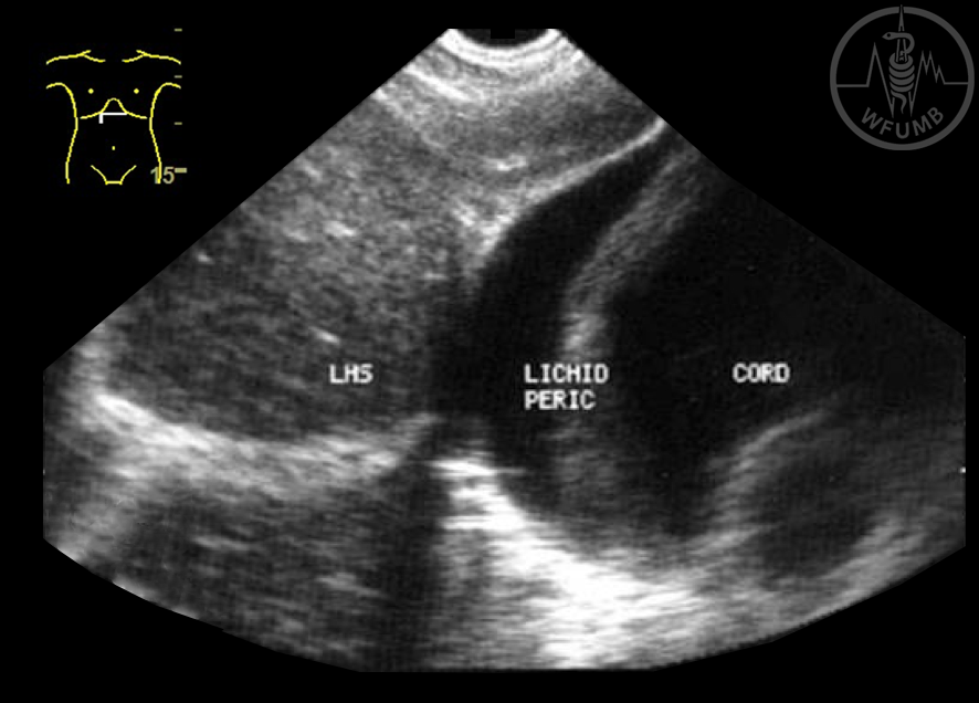
Fig 13.1.39 Pericardial effusion
Chapter Videos
This website uses cookies to improve your experience. By using this website you agree to our
Data Protection Policy
.
Read more
Accept all
Translate »