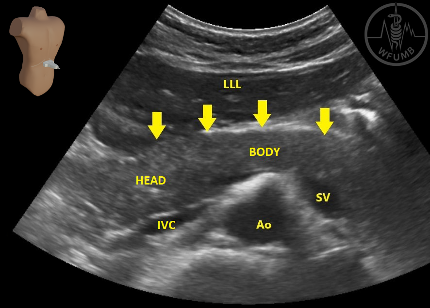
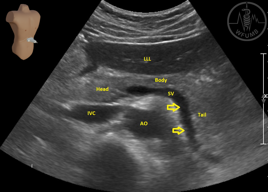
Fig 8.4
Transverse section with all vascular structures
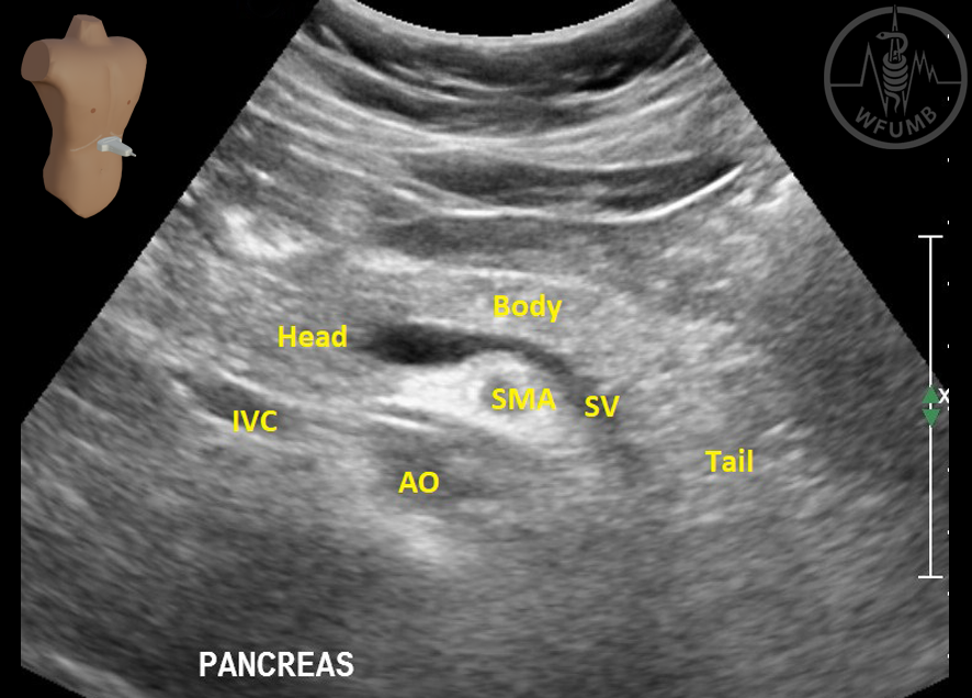
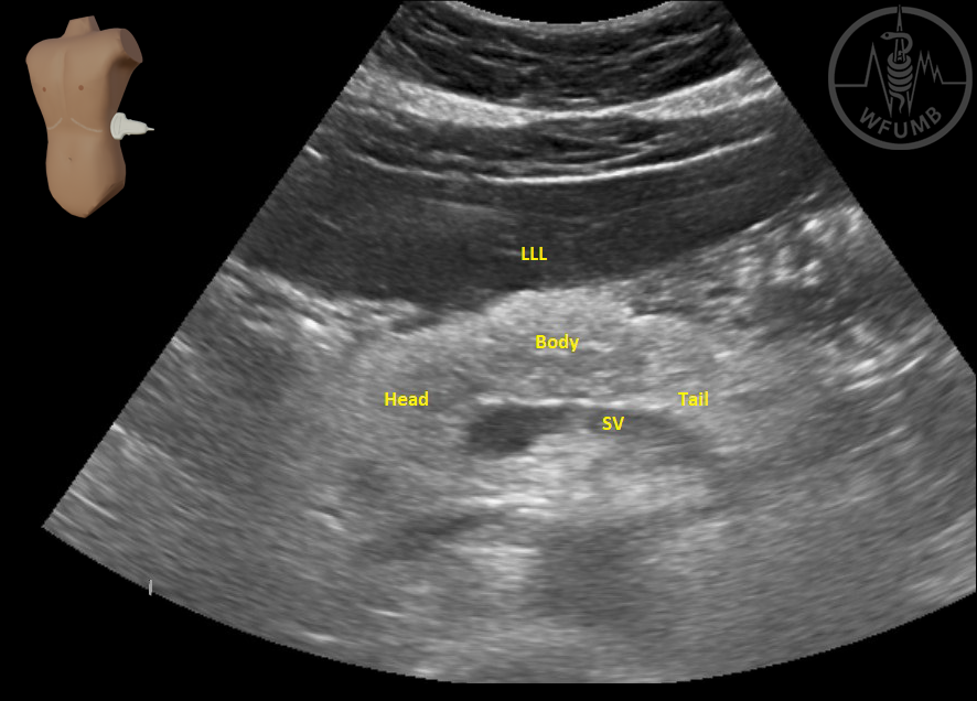
Fig 8.5
Pancreatic tail with splenic vein
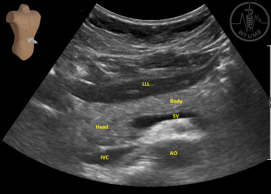
Fig 8.6a
Pancreatic head and uncinate processus
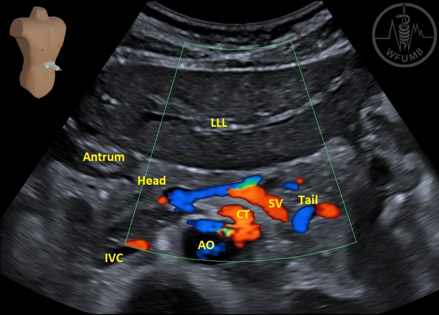
Fig 8.6b
Doppler examination of epigastric region
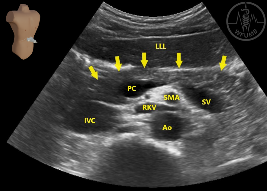
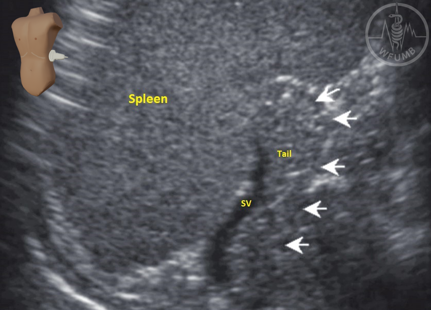
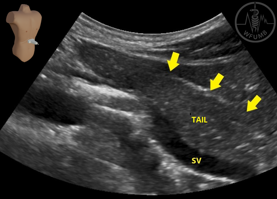
Fig 8.7
Use of the spleen as an acoustic window to visualize the tail of the
pancreas (arrows).
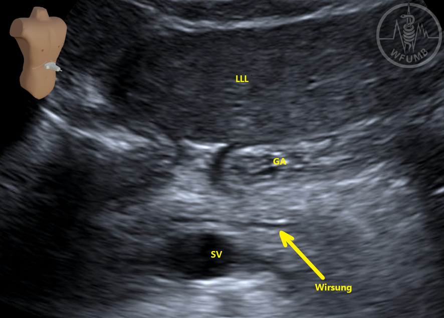
Fig 8.8
The systematic use of
color Doppler (Fig 8.6b) and of oblique section
(Fig 8.8) may also aid in the precise identification of the pancreatic landmark.
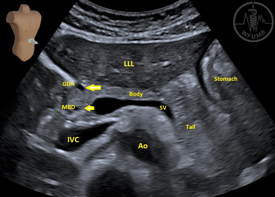
Fig 8.9
Oblique section with main biliary duct (MBD) and the head of the pancreas.