Quiz-summary
0 of 2 questions completed
Questions:
- 1
- 2
Information
You have already completed the quiz before. Hence you can not start it again.
Quiz is loading...
You must sign in or sign up to start the quiz.
You have to finish following quiz, to start this quiz:
Results
0 of 2 questions answered correctly
Time has elapsed
Categories
- Not categorized 0%
-
That is Case 01 completed – to move on to Case 02 ~ click here
- 1
- 2
- Answered
- Review
-
Question 1 of 2
1. Question
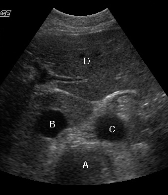
Clinical Description:
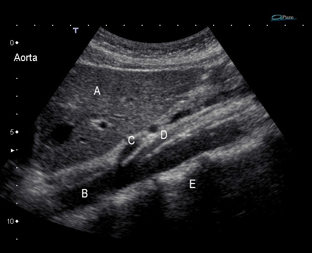
Review the anatomy of the longitudinal and transverse images of the proximal abdominal aorta
Match the following structures with their labels
Please sort the answers in right order A first through to D last with the “Move” Button which will show when you hover over the answers.
-
Vertebra
-
IVC
-
Aorta
-
Liver
Correct
Incorrect
-
-
Question 2 of 2
2. Question
Clinical Description:
Review the anatomy of the longitudinal and transverse images of the proximal abdominal aorta

Match the following structures with their labels
Please sort the answers in the right order, A first through to E last, with the “Move” Button which will show when you hover over the answers.
-
Liver
-
Aorta
-
Coeliac axis
-
SMA
-
Vertebra
Correct
The aorta is located to the left of the IVC. It has 2 anterior branches, the coeliac axis and the SMA, which arise from the anterior aspect of the aorta inferior to the liver at the level of T12 (so the coeliac axis may not be seen in the unfasted patient). The IVC is thinner walled. It has 1 structure draining anteriorly, the middle hepatic vein, which enters in the liver parenchyma just inferior to the junction with the right atrium.
The IVC is found to the right of the aorta (except in very rare congenital anomalies), but note that both pulsate.
Incorrect
The aorta is located to the left of the IVC. It has 2 anterior branches, the coeliac axis and the SMA, which arise from the anterior aspect of the aorta inferior to the liver at the level of T12 (so the coeliac axis may not be seen in the unfasted patient). The IVC is thinner walled. It has 1 structure draining anteriorly, the middle hepatic vein, which enters in the liver parenchyma just inferior to the junction with the right atrium.
The IVC is found to the right of the aorta (except in very rare congenital anomalies), but note that both pulsate.
-

