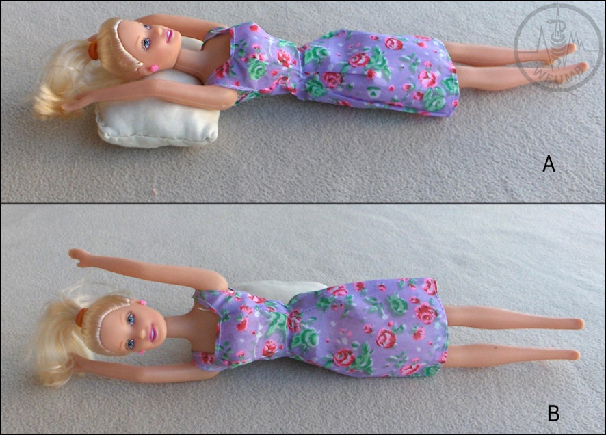
Fig 9.2
Spleen can be examined in the supine position (A) or from a coronal plane with the patient’s left side up (B)
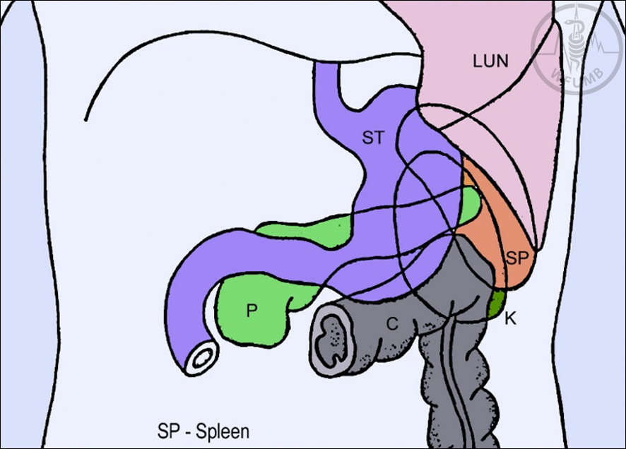
Fig 9.3
The front of the spleen is completely covered by the stomach, ribs, lung, and left colic flexure
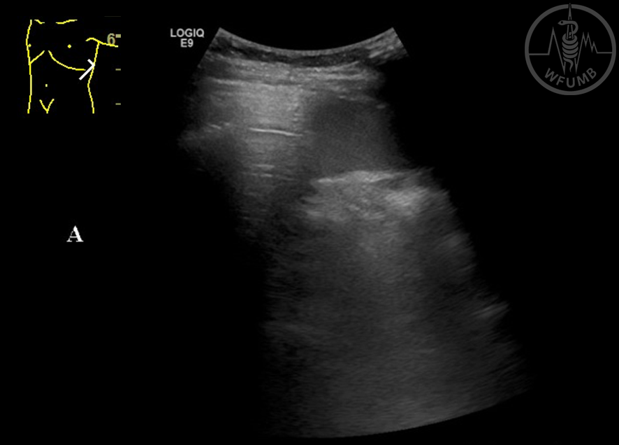
Fig 9.4a
Examination during inspiration difficult to see spleen (A).
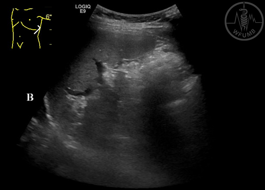
Fig 9.4b
Examination during expiration - spleen can be seen easily (B)
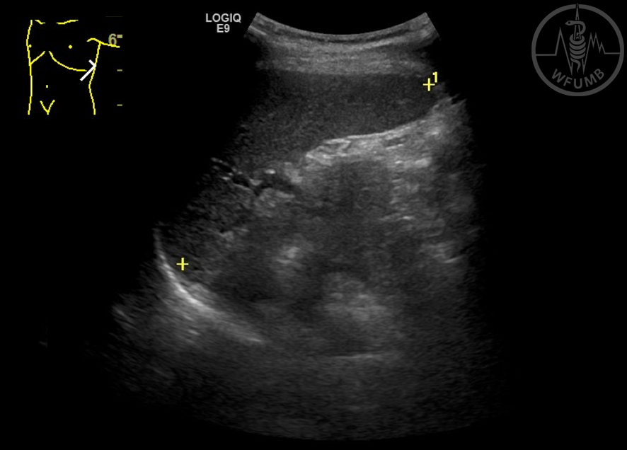
Fig 9.5
Ultrasound picture of normal spleen
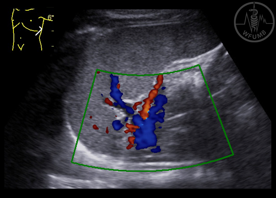
Fig 9.6
Color Doppler of spleen vessels
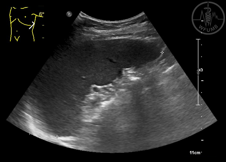
Fig 9.7
Maximum length of the spleen
Fig 9.8
Embryological components of the spleen may present different shapes of spleen
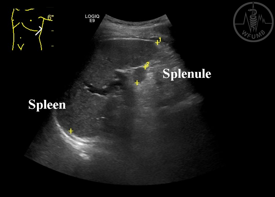
Fig 9.9
Accessory spleens are not uncommon and are seen at the hilum region
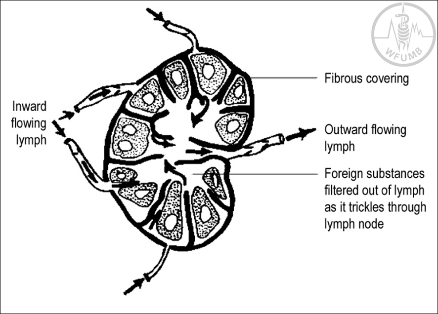
Fig 9.10
The lymph enters a lymph node by several afferent vessels and is filtered and analyzed on its way through the lymph node and goes out by efferent vessel
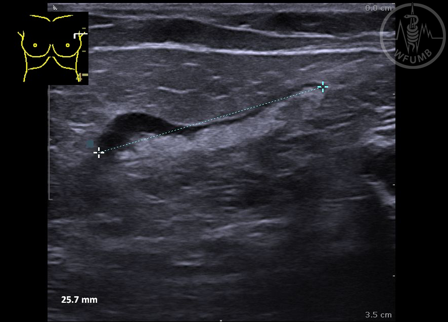
Fig 9.11
Inflammatory enlargement of axillary lymph nodes is common
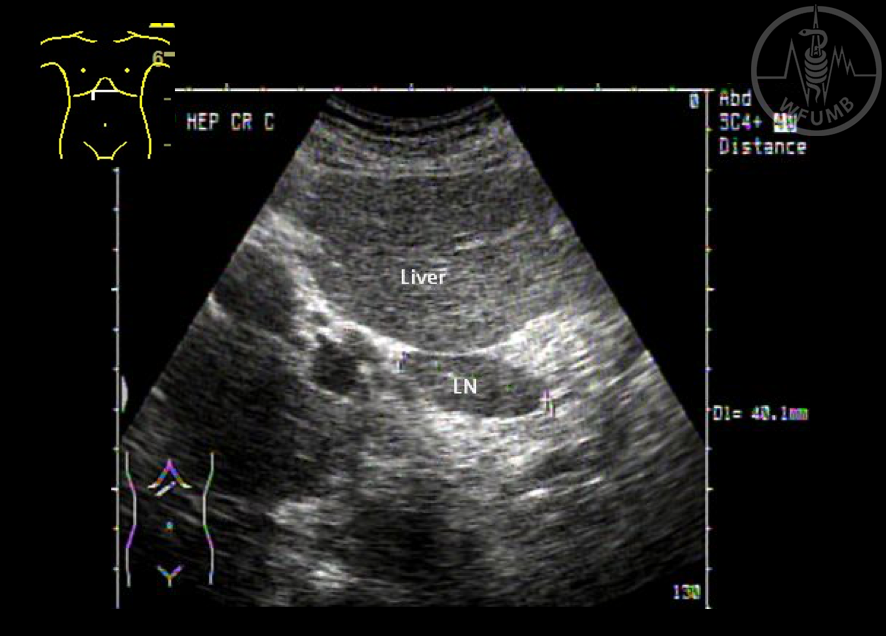
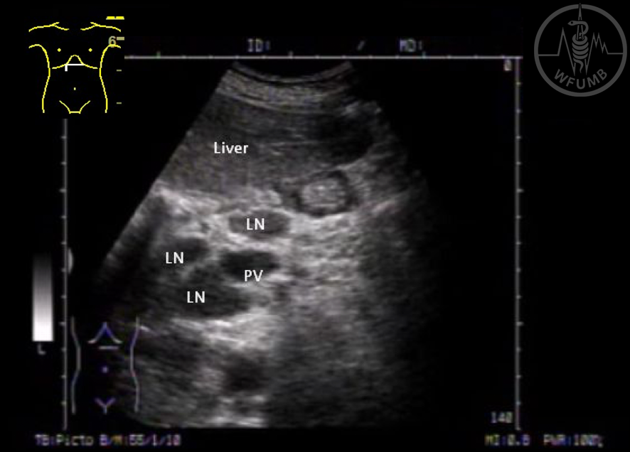
Fig 9.12
Inflammatory liver diseases commonly cause increase size of lymph nodes
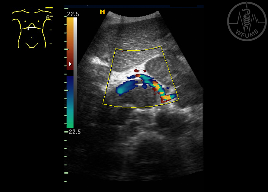
Fig 9.13
Massive central mesenteric lymph node enlargement is a typical feature of lymphoma
Fig 9.14
Enlarged para-aortic lymph nodes are typically low and uniformly echogenic, sometimes almost anechoic
Chapter Videos
This website uses cookies to improve your experience. By using this website you agree to our Data Protection Policy.