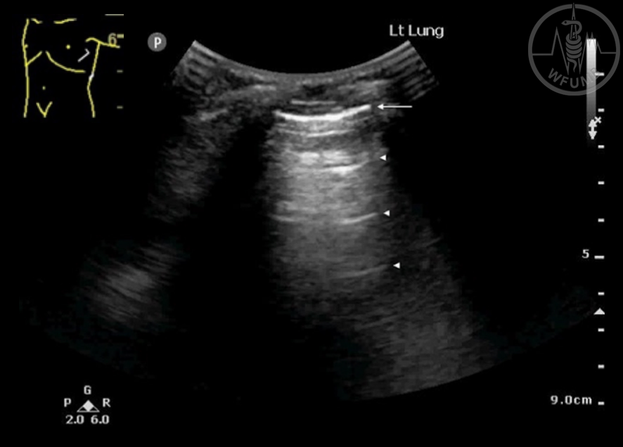
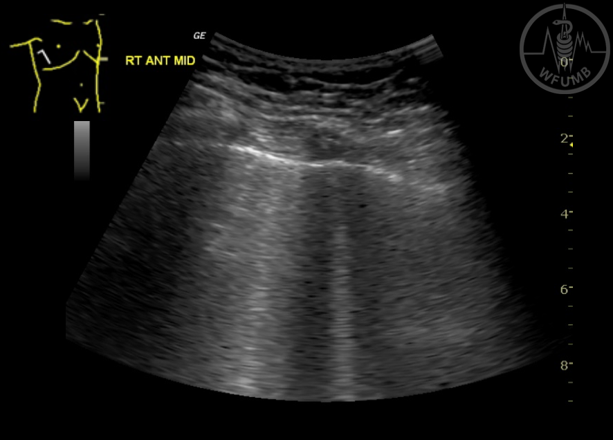
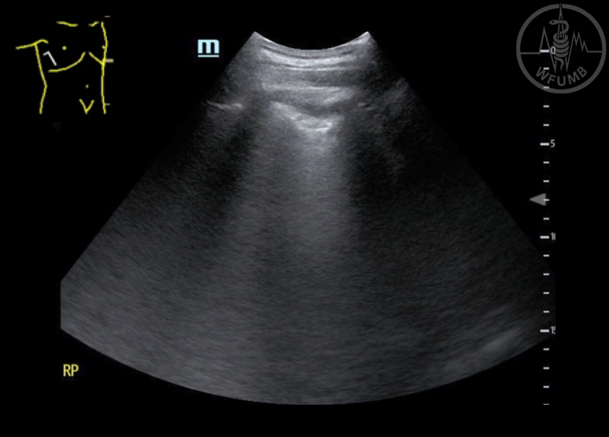
Fig 27.2
Normal bright, equally spaced “A lines” (arrowheads) evenly spaced below the air interface line (arrow)
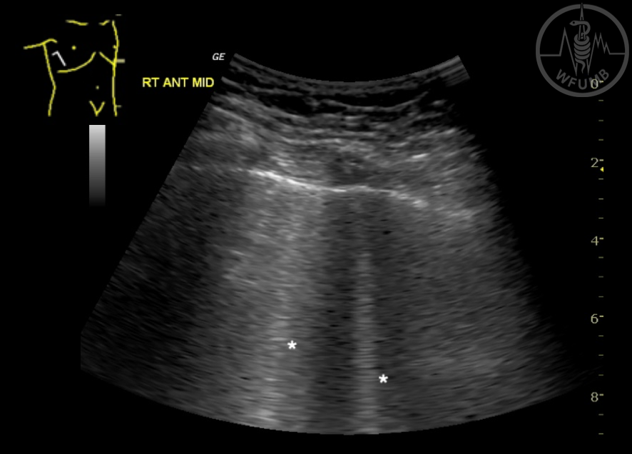
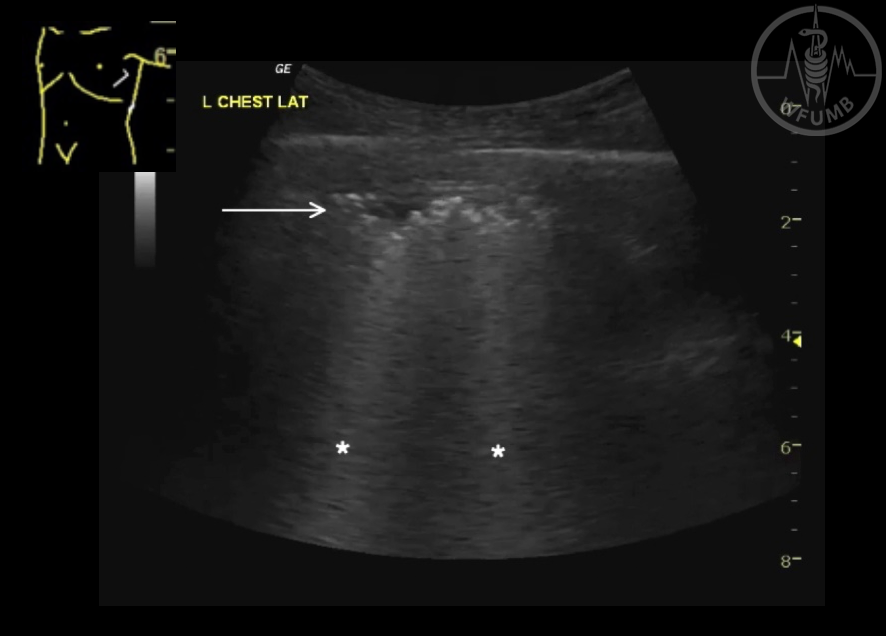
Fig 27.3
B-lines arising from the air interface line (*)
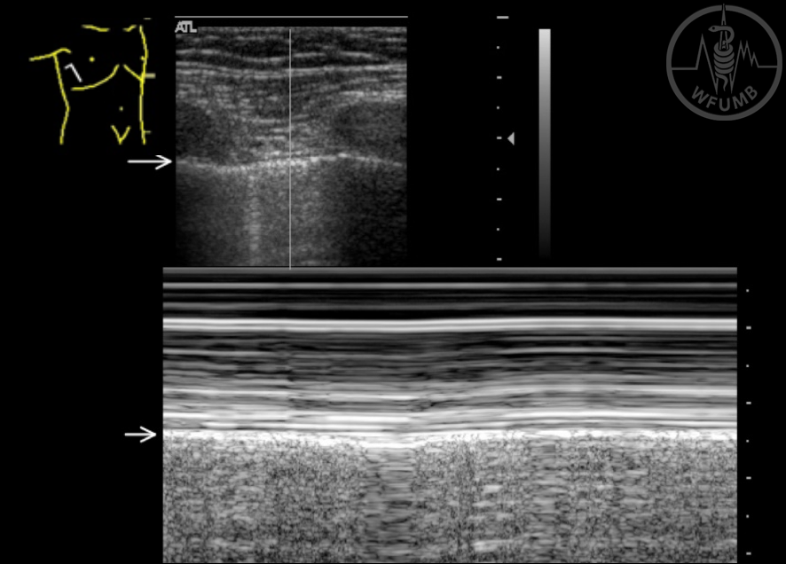
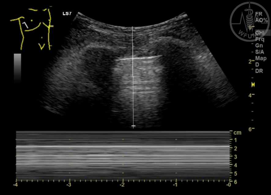
Fig 27.4
M-mode of normal lung creating a ‘seashore’ appearance
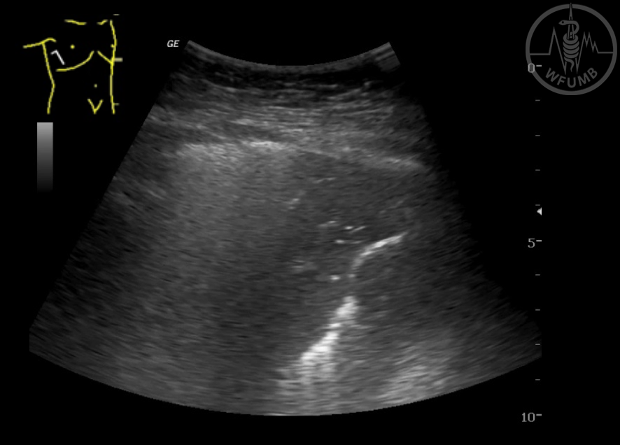
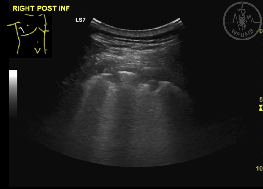
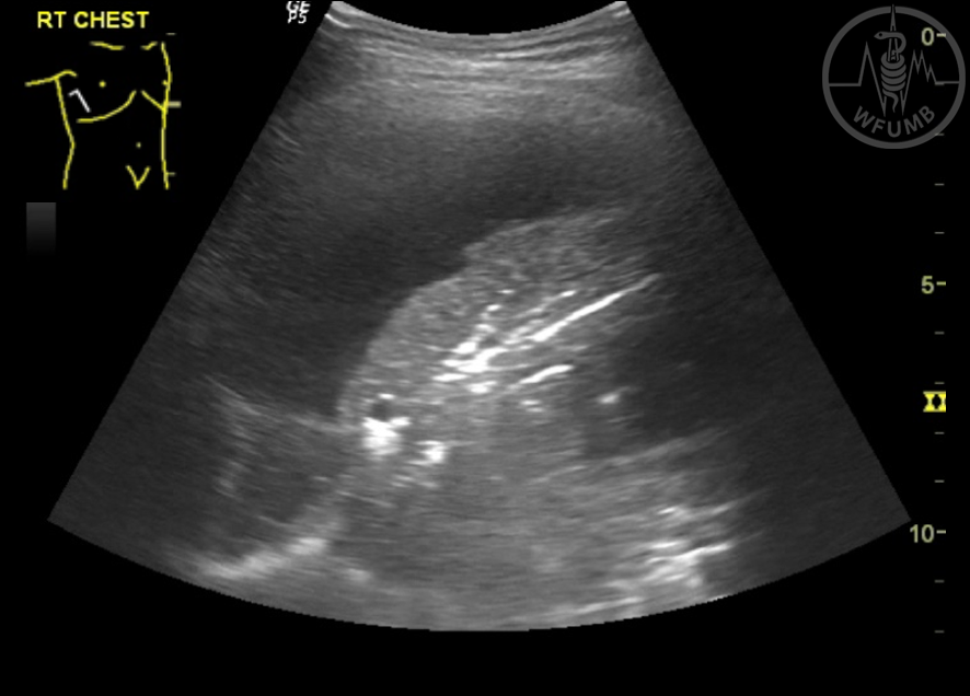
Fig 27.5
Consolidated lung with air bronchogram
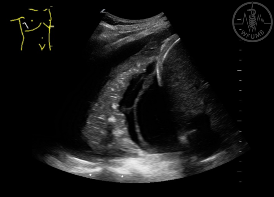
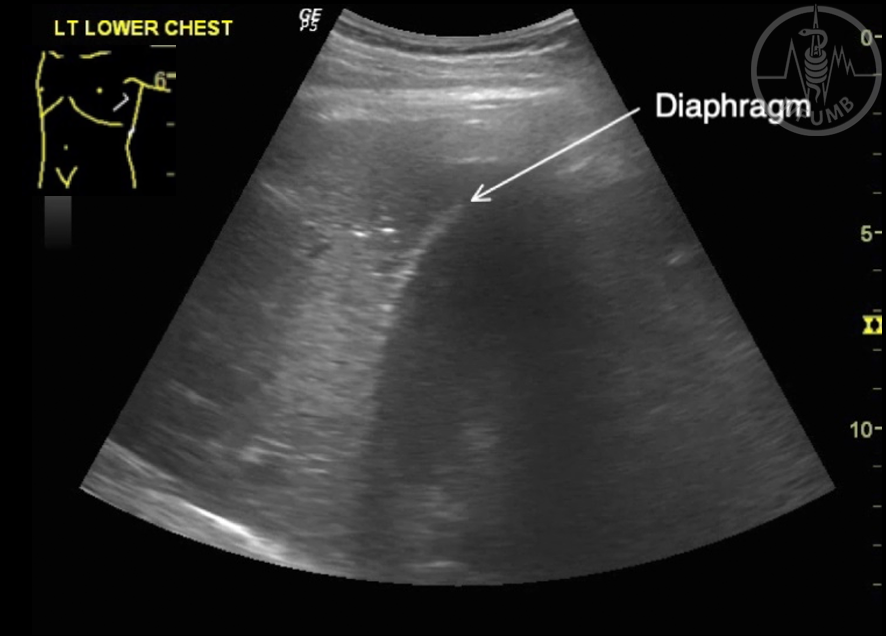
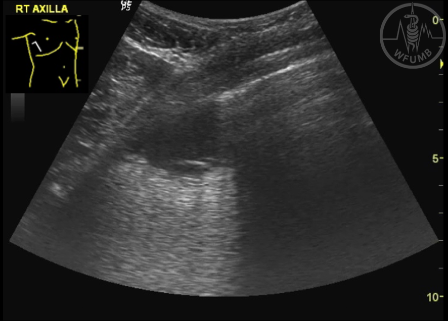
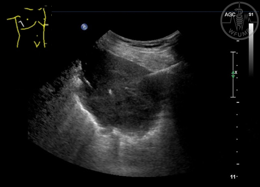
Fig 27.6
Pleural effusion allowing the thoracic vertebrae (*) to be visualised
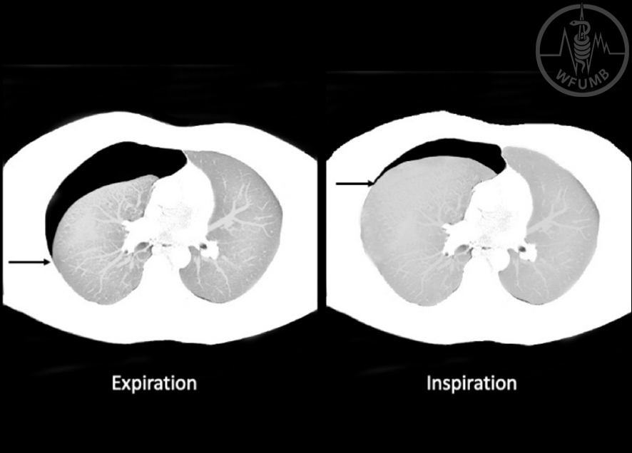
Fig 27.7
Illustration of how the ‘lung point’ moves with respiration
Fig 27.8
M-mode of a pneumothorax shows a ‘barcode’ or ‘stratosphere’ pattern
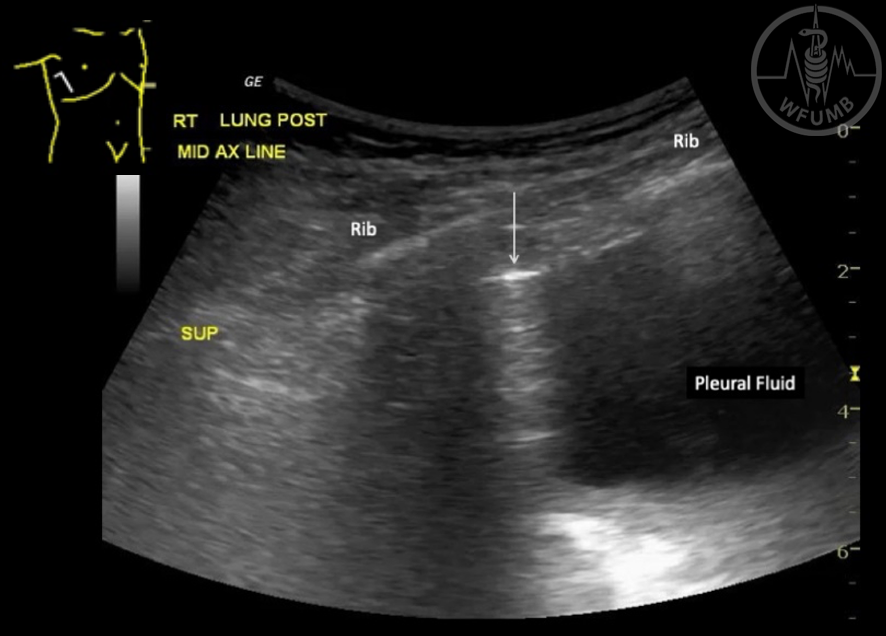
Fig 27.9
Hydropneumothorax. Arrow shows air interface of pneumothorax above pleural fluid
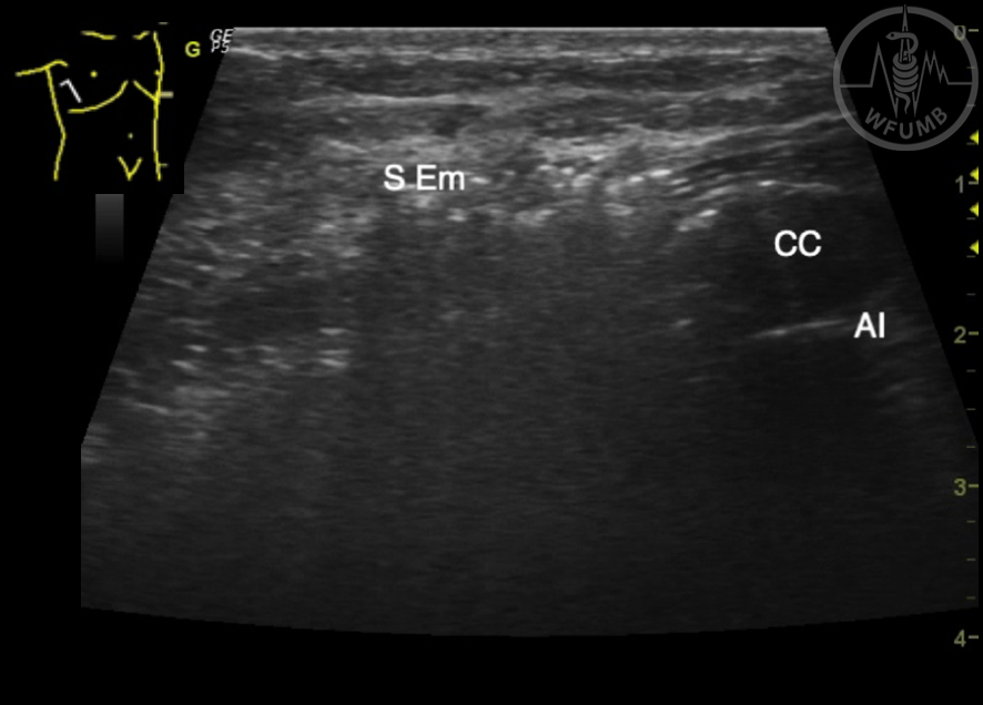
Fig 27.10
Subcutaneous emphysema (S Em). Gas in the tissue prevents any deeper structures being seen. CC = costal cartilage, AI = Air interface line
Fig 27.11
Two B lines and a smooth air interface line in congestive cardiac failure
Fig 27.12
B lines (*) and a thickened irregular air interface in interstitial lung disease
Fig 27.13
Lobar consolidation with 2 small brights ‘dots’ of gas
Fig 27.14
Bronchopneumonia with irregular, ‘shredded’ air interface
Fig 27.15
Coalesced B lines causing ‘waterfall’ or ‘lightbeam’ in SARS-CoV2
Fig 27.16
Subpleural consolidation and irregular air interface line in SARS-CoV2
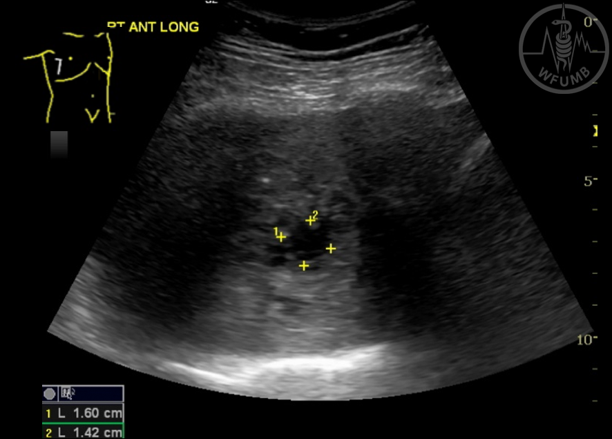
Fig 27.17
Lung abscess within consolidation
Fig 27.18
Small subpleural consolidation in TB
Fig 27.19
Larger area of consolidation in TB
Fig 27.20
Pleural effusion with secondary lung atelectasis
Fig 27.21
Rounded area of lung infarction due to pulmonary embolus
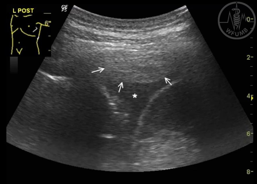
Fig 27.22
Pleural thickening (arrows) with a small pleural effusion (*)
Fig 27.23
Rounded, hypoechoic lung malignancy with a feeding vessel
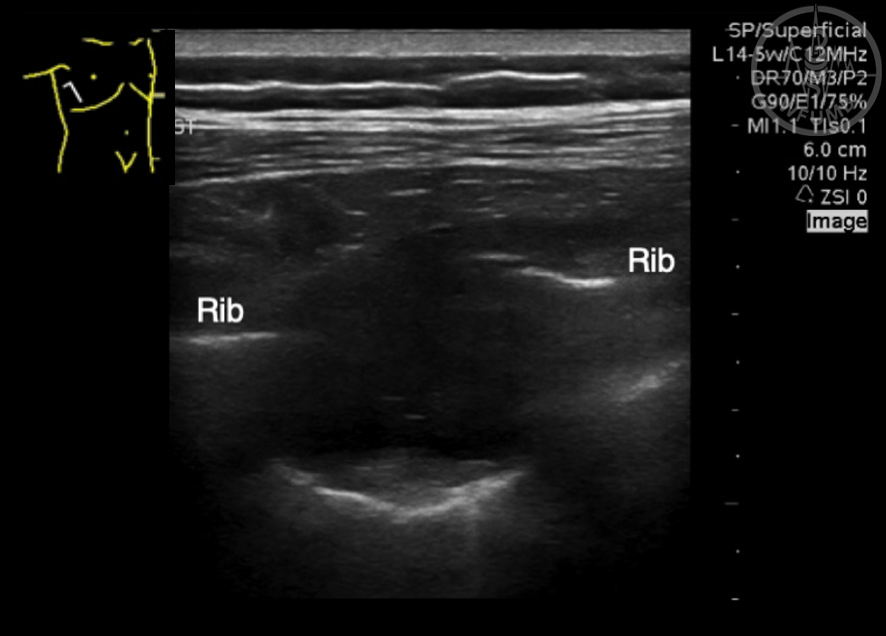
Fig 27.24
Rib metastasis resulting in disruption of the cortical surface (probe aligned with rib)
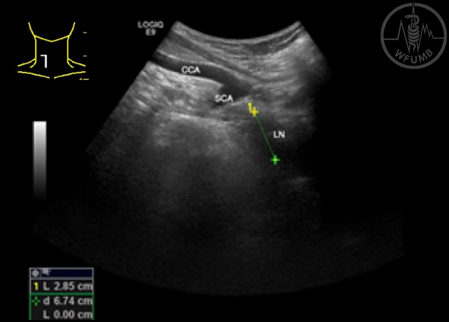
Fig 27.25
Right paratracheal and subcarinal lymphadenopathy (LN) in sarcoidoisis. Suprasternal approach shows the common carotid (CCA) and subclavian (SCA) arteries
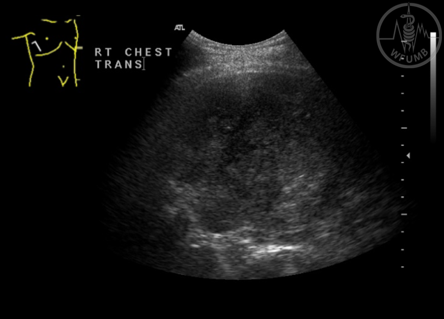
Fig 27.26
Solid tumour filling the pleural space
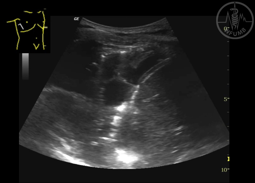
Fig 27.27
Multiloculated pleural effusion
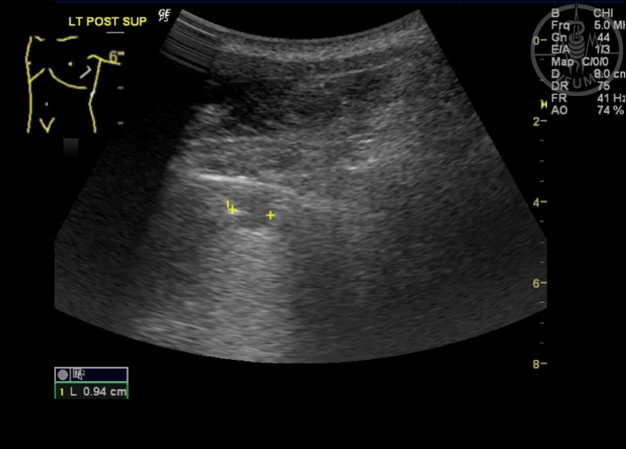
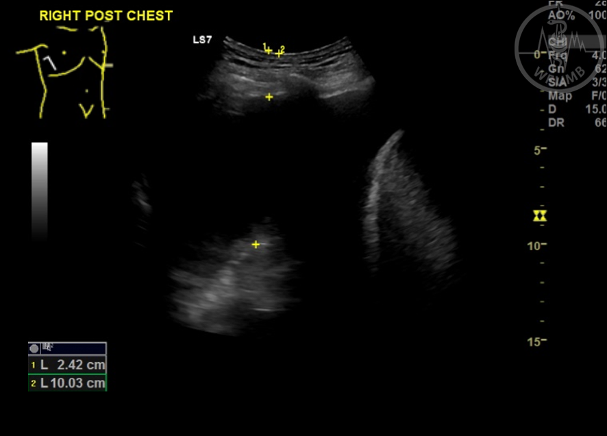
Fig 27.28
Pleural effusion measurements prior to pleural aspiration
Fig 27.29
Direct visualisation of biopsy of lung lesion
Chapter Videos
This website uses cookies to improve your experience. By using this website you agree to our Data Protection Policy.