WFUMB Course Book
Thyroid and neck - Chapter 28 Media Library
Close window and return to Chapter 28
Chapter Images
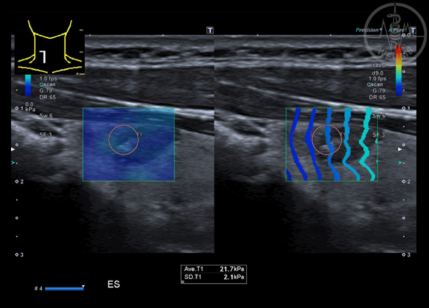
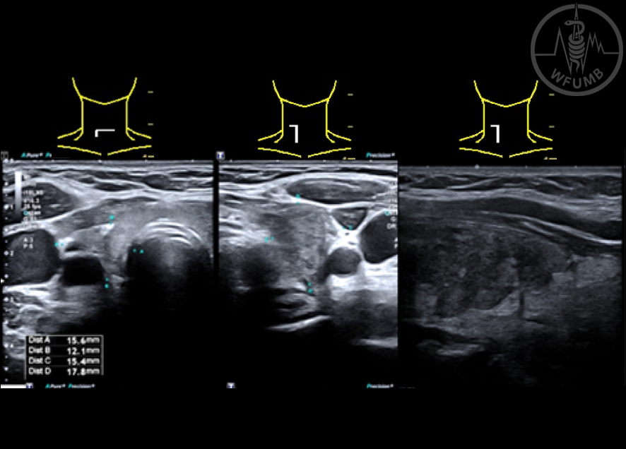
Fig 28.1a A solid, wider than tall, hypoechoic, well marginated nodule of the right thyroid lobe is depicted
Fig 28.1b CD shows peripheral and internal vascularity
Fig 28.1c At strain elastography (SE), the nodule appears soft
Fig 28.1d At shear waves elastography (SWE), the nodule appears soft
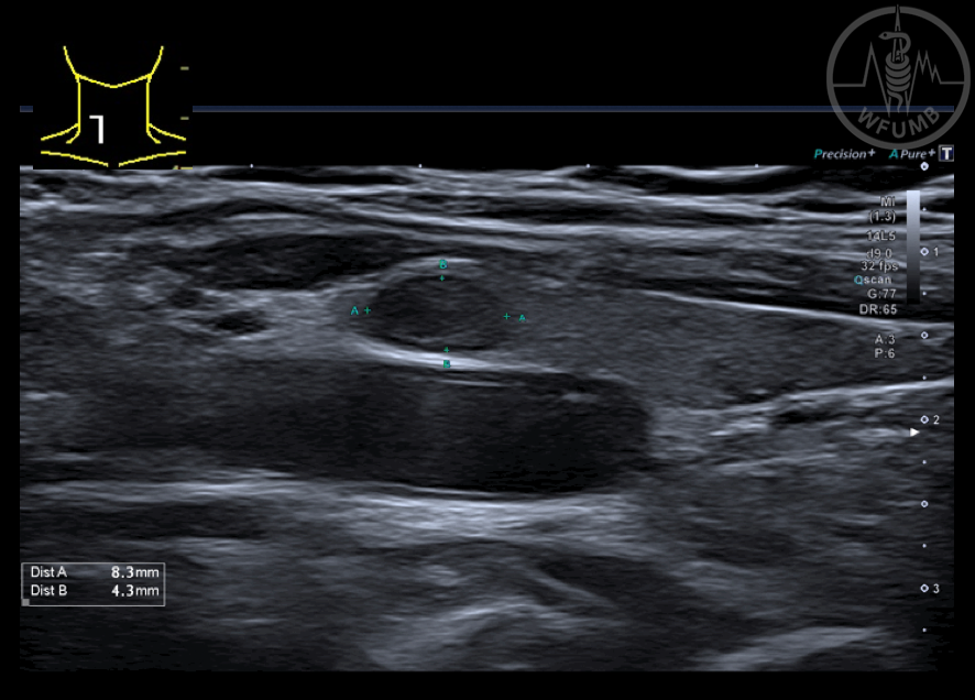
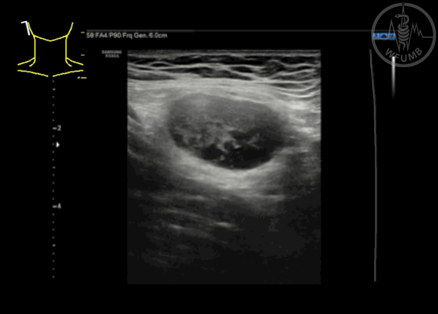
Fig 28.2a Solid, wider than tall hypoechoic nodule is visualized in the right thyroid lobe
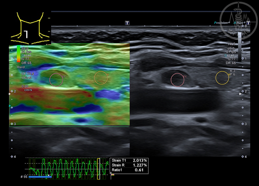
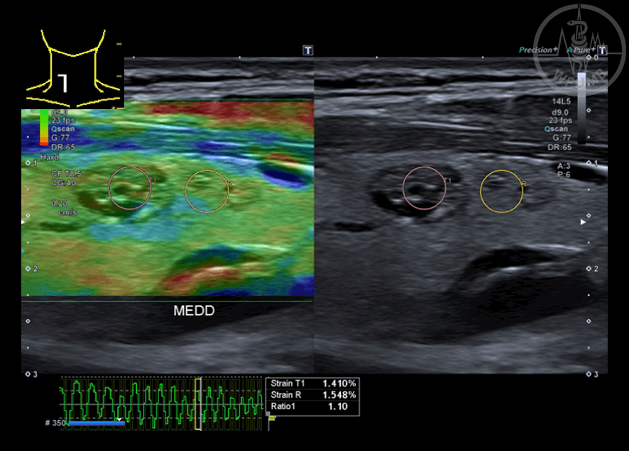
Fig 28.2b Thyroid nodule which appears soft at SE (SR: 1.4)
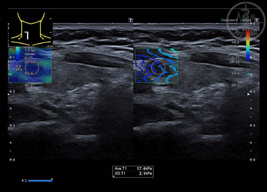
Fig 28.2c Thyroid nodule which appears soft at SWE (20-30 kPa)
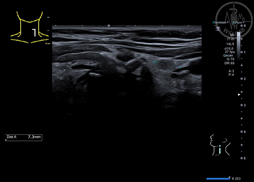
Fig 28.3a A left paraisthmic, mainly solid, hypoechoic, well marginated nodule is depicted
Fig 28.3b Left paraisthmic nodule - at SE it has a soft appearance
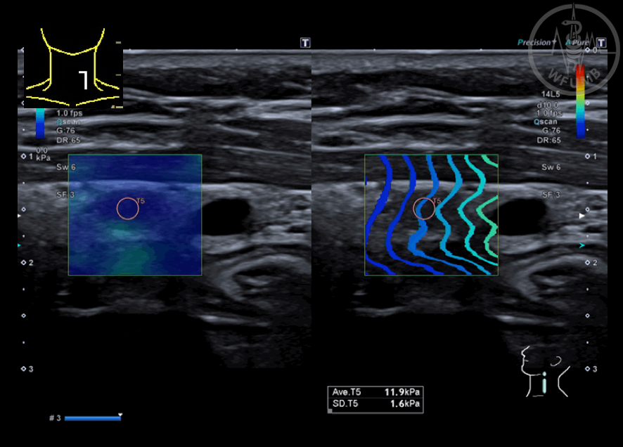
Fig 28.3c Left paraisthmic nodule - at SWE, 11.9 kPA is obtained
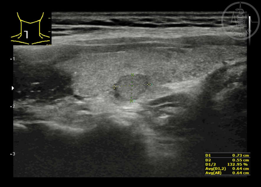
Fig 28.4a Wider than tall, solid, hypoechoic, well marginated nodule of the right thyroid lobe
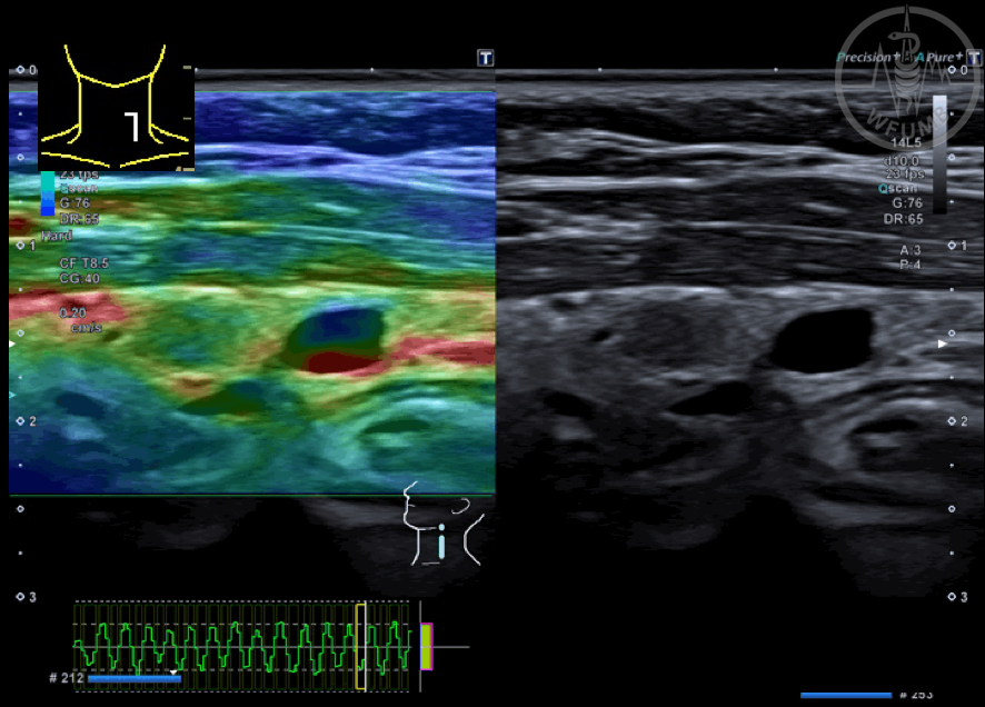
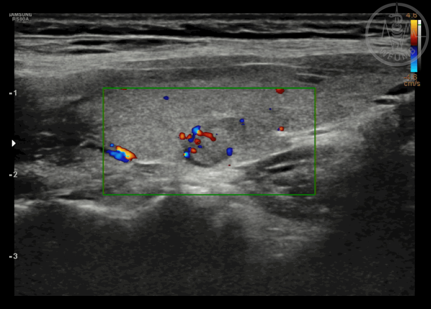
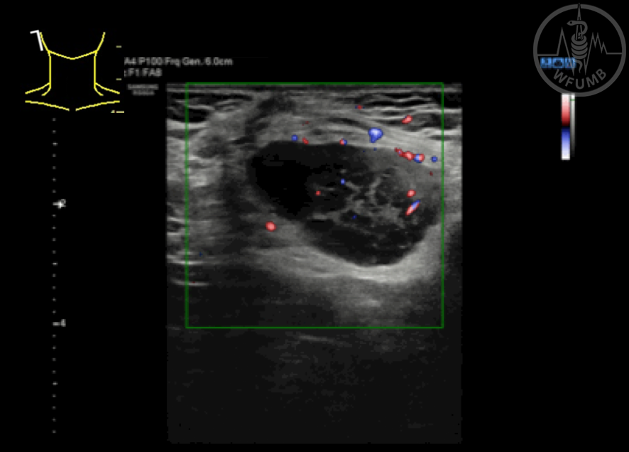
Fig 28.4b Nodule of the right thyroid lobe shows peripheral and internal vascularity
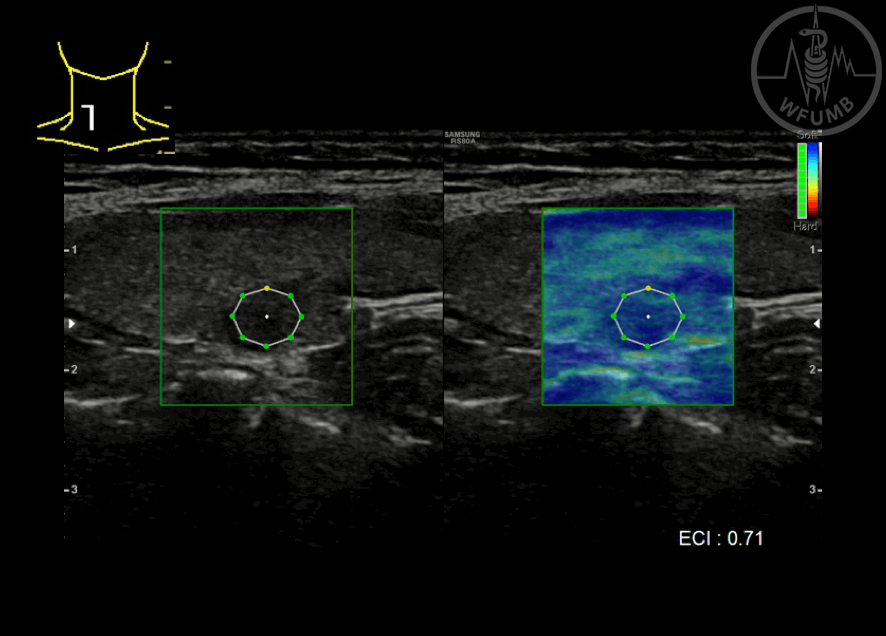
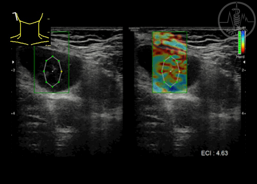
Fig 28.4c Nodule of the right thyroid lobe - at elastography, the nodule appears soft (ECI: 0.71)
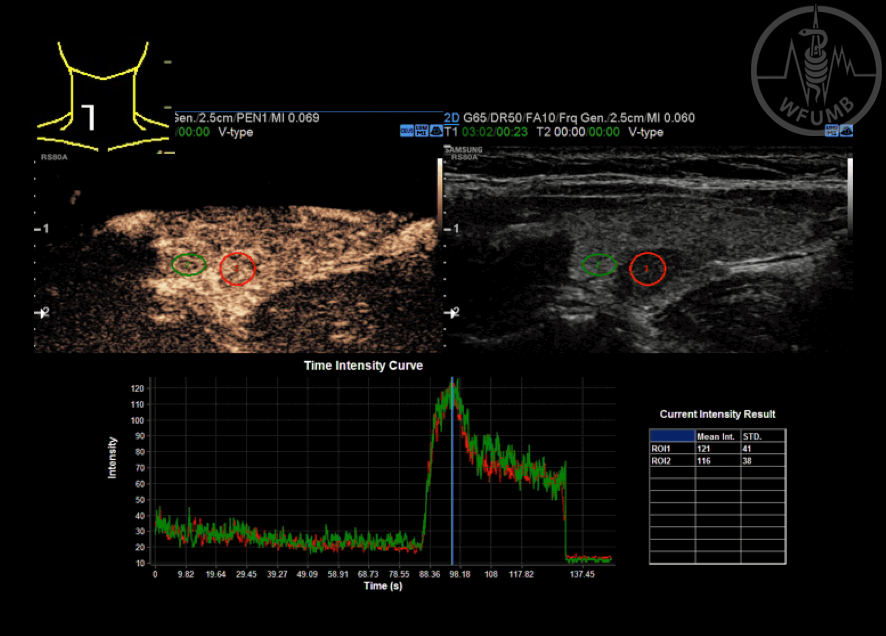
Fig 28.4d Nodule of the right thyroid lobe - at CEUS, the nodule quickly enhaces and quickly washes out, in a similar way to the parenchyma
Fig 28.5a A diffusely enlarged thyroid gland containing a large, taller than wide, solid, very hypoechoic, polylobulated nodule
Fig 28.5b Thyroid nodule appears of intermediate stiffness at elastography, but with a high SR
Fig 28.5c Thyroid nodule appears stiff at SWE
Fig 28.6a Enlarged retroauricolar oval shaped, well marginated LN, with scarcely visible hilum
Fig 28.6b The LNs shows multiple peripheral and internal vascular spots
Fig 28.6c The LN shows hard appearance and high ECI value
Chapter Videos
This website uses cookies to improve your experience. By using this website you agree to our
Data Protection Policy
.
Read more
Accept all
Translate »