April 2023 from Sudan
September 12, 2019ISUOG Virtual World Congress on Ultrasound in Obstetrics and Gynecology 2020
September 19, 2019Affiliation
Christoph F Dietrich. Medizinische Klinik 2, Caritas-Krankenhaus Bad Mergentheim, Germany.
Video 1 and 2
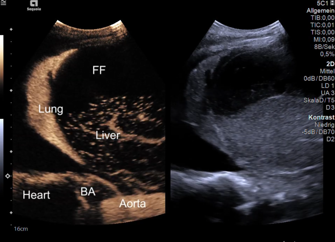
The contrast enhancement of the heart and lung after intravenous injection follows certain anatomical rules. First the right atrium (video 1, RA), right ventricle (video 1, RV), pulmonary artery, lung parenchyma (video 2, LUNG) and pulmonary veins are enhancing followed by the left atrium (video 1, LA), left ventricle (video 1, LV), coronary arteries & myocardium (video 1, MYO), aorta (video 2, AORTA), bronchial arteries (BA) and the systemic vessels (video 2 including hepatic arteries, portal venous system and liver parenchyma). In other words the venous blood from the heart to the lung parenchyma is featured, which is mandatory for the gaseous exchange. Thereafter, the systemic arterial vascular system is enhancing. In the lung the analysis of the dual blood supply allows the differentiation of lung emboli (pulmonary artery vascular supply followed by pulmonary vein washout) and neoplasia (bronchial artery vascular supply followed by bronchial vein washout). In the liver the dual blood supply allows the differentiation of malignant and benign focal liver lesion. Herewith we demonstrate the contrast enhanced ultrasound enhancement step by step in real-time [1, see also EFSUMB Case of the Month “Amyloidosis”, www.efsumb.org].
Take Home Message
The videos demonstrate the contrast phases in the lung, heart, aorta, bronchial arteries and liver in real-time.
Featured reference
Dietrich CF, Averkiou M, Nielsen MB, Barr RG, Burns PN, Calliada F, et al. How to perform Contrast-Enhanced Ultrasound (CEUS). Ultrasound Int Open. 2018;4(1):E2-E15.

