February 2023 from Sudan
November 11, 2019Why We Need Infection Control Guidelines for Ultrasound Practice
December 12, 2019Intermittent pain in the lower right quadrant of the abdomen.
Quiz-summary
0 of 1 questions completed
Questions:
- 1
Information
View the December Case below, answer the question and then click check >
You have already completed the quiz before. Hence you can not start it again.
Quiz is loading...
You must sign in or sign up to start the quiz.
You have to finish following quiz, to start this quiz:
Results
0 of 1 questions answered correctly
Your time:
Time has elapsed
You have reached 0 of 0 points, (0)
Categories
- Not categorized 0%
- 1
- Answered
- Review
-
Question 1 of 1
1. Question
Intermittent pain in the lower right quadrant of the abdomen
By Meidahl S, Ewertsen C,
Department of Radiology, Rigshospitalet – Copenhagen University Hospital, DenmarkClinical History:
A 55-year-old female presented to our clinic with a history of intermittent pain in the lower right quadrant of the abdomen and also an intermittent impression of a lump. The pain was described as a feeling of heaviness and fullness including a dull pain radiating to the flank with varying duration – especially after long periods of standing or walking and also after meals. Clinical examination revealed no palpable lump in the right lower quadrant. An appendicectomy scar was noted medially.Ultrasound:
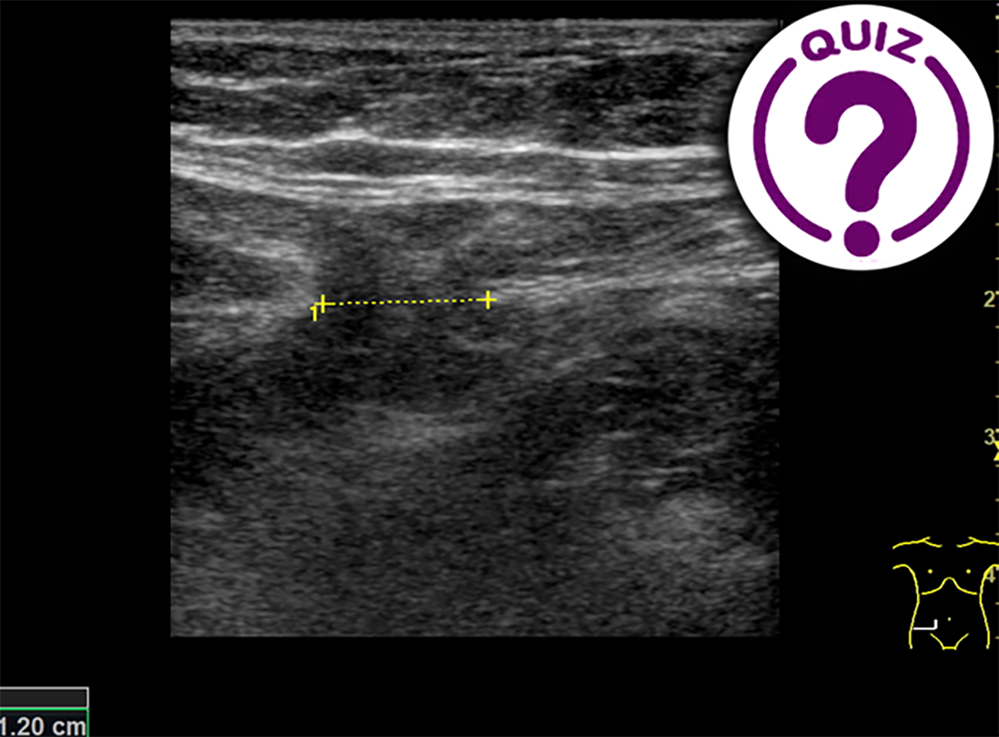
The patient was examined in the supine and standing position including with and without the Valsalva maneuver. Images from the examination: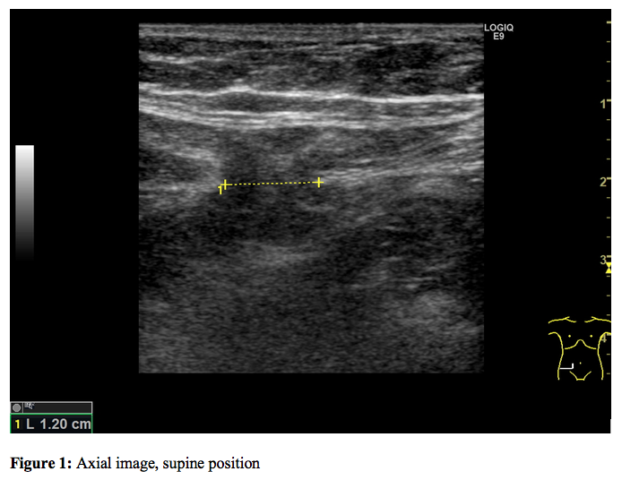

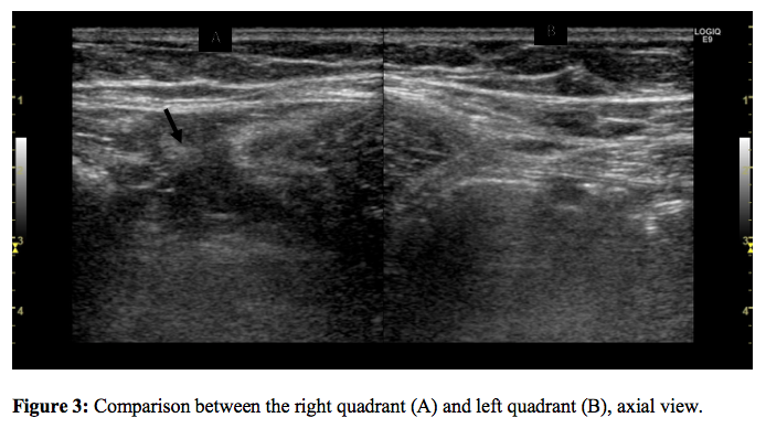
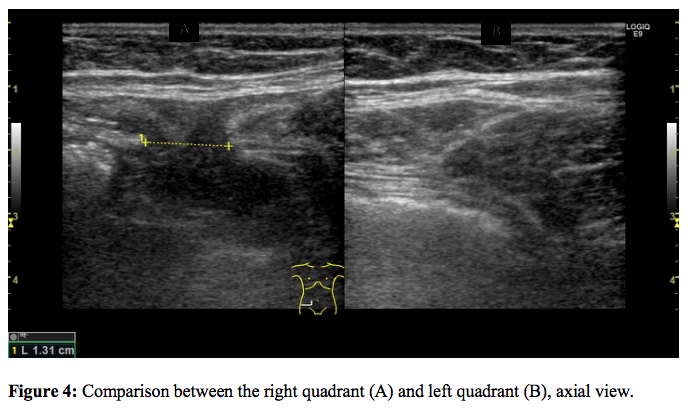
Question: What is the diagnosis of the patient?
Correct
CORRECT ANSWER EXPLAINED BELOW 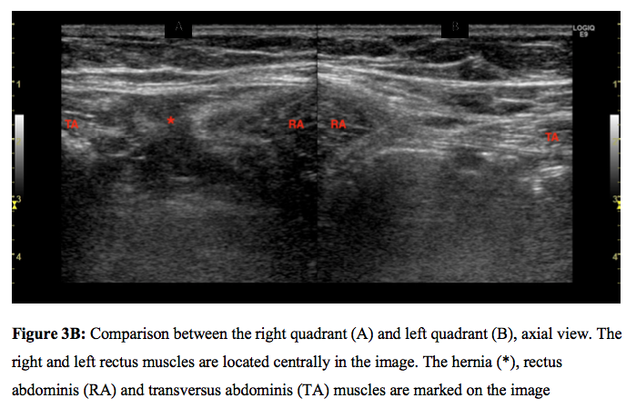

Imaging Findings:
The examination revealed a hernia between the rectus abdominis and transversus abdominis muscles at the level of the anterior superior iliac spine – a lateral, ventral hernia or Spigelian hernia. The dimension of the hernial orifice was 1.3 x 0.7 cm and the hernia contained preperitoneal fat without any bowel loops. There was no change in size or content of the hernia on standing or Valsalva.
Discussion:
The Spigelian fascia is located lateral to the rectus abdominis muscle along the semilunar line.1–3 Spigelian hernias penetrate through this fascia and are very rare (approximately 1% of all abdominal hernias1). The location of the hernia gives the diagnosis. Cicatricial hernia are located in scars from previous surgery, inguinal hernia are located in the groin and umbilical hernia in the umbilicus.In some cases, the Spigelian hernia only penetrates the transverse abdominal aponeurosis, but since this is tightly bound to the internal oblique muscle, the hernia usually penetrates both3. In addition to the hernias often being obscured by at least one aponeurotic layer, they tend to be small, and they can therefore be very difficult to see and feel3. If doubt persists after radiological examinations with ultrasound or CT then a diagnostic laparoscopy is usually performed due to the high risk of strangulation2.
Teaching points:
Although Spigelian hernias are very rare, it is important to remember them when dealing with a patient with vague symptoms and recurring lower abdominal pain. Diagnosis from clinical presentation is often problematic, but can be made by ultrasound or CT with high accuracy1,2.REFERENCES:
- https://radiopaedia.org/articles/spigelian-hernia-1
- Mittal T, Kumar V, Khullar R et al. Diagnosis and management of Spigelian hernia: A review of literature and our experience. J Minim Access Surg. 2008;4:95-8.
- Spangen L. Spigelian hernia. World J Surg. 1989;13:573-80.
Incorrect
CORRECT ANSWER EXPLAINED BELOW 

Imaging Findings:
The examination revealed a hernia between the rectus abdominis and transversus abdominis muscles at the level of the anterior superior iliac spine – a lateral, ventral hernia or Spigelian hernia. The dimension of the hernial orifice was 1.3 x 0.7 cm and the hernia contained preperitoneal fat without any bowel loops. There was no change in size or content of the hernia on standing or Valsalva.
Discussion:
The Spigelian fascia is located lateral to the rectus abdominis muscle along the semilunar line.1–3 Spigelian hernias penetrate through this fascia and are very rare (approximately 1% of all abdominal hernias1). The location of the hernia gives the diagnosis. Cicatricial hernia are located in scars from previous surgery, inguinal hernia are located in the groin and umbilical hernia in the umbilicus.In some cases, the Spigelian hernia only penetrates the transverse abdominal aponeurosis, but since this is tightly bound to the internal oblique muscle, the hernia usually penetrates both3. In addition to the hernias often being obscured by at least one aponeurotic layer, they tend to be small, and they can therefore be very difficult to see and feel3. If doubt persists after radiological examinations with ultrasound or CT then a diagnostic laparoscopy is usually performed due to the high risk of strangulation2.
Teaching points:
Although Spigelian hernias are very rare, it is important to remember them when dealing with a patient with vague symptoms and recurring lower abdominal pain. Diagnosis from clinical presentation is often problematic, but can be made by ultrasound or CT with high accuracy1,2.REFERENCES:
- https://radiopaedia.org/articles/spigelian-hernia-1
- Mittal T, Kumar V, Khullar R et al. Diagnosis and management of Spigelian hernia: A review of literature and our experience. J Minim Access Surg. 2008;4:95-8.
- Spangen L. Spigelian hernia. World J Surg. 1989;13:573-80.

