
Quiz-summary
0 of 2 questions completed
Questions:
- 1
- 2
Information
You have already completed the quiz before. Hence you can not start it again.
Quiz is loading...
You must sign in or sign up to start the quiz.
You have to finish following quiz, to start this quiz:
Results
0 of 2 questions answered correctly
Time has elapsed
Categories
- Not categorized 0%
-
That is Case 10 completed – to move on to Case 11 ~ click here
- 1
- 2
- Answered
- Review
-
Question 1 of 2
1. Question
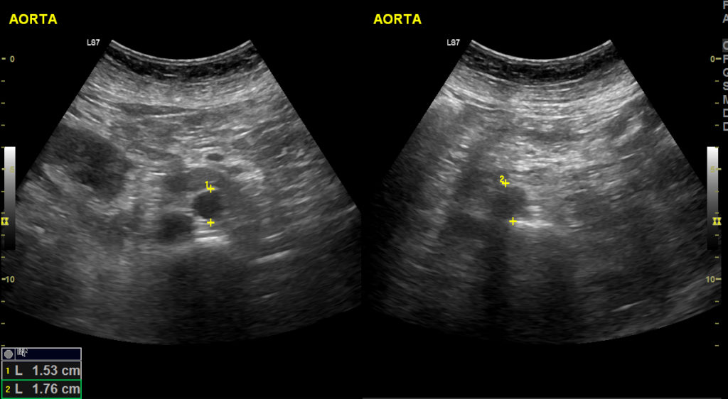
Clinical Description:
A 65 yr old lady presented with right flank pain for 2 weeks. Her aorta was scanned as part of a renal scan.
A short segment of bowel gas initially blocked the view of the mid aorta (loop AAA10_01). How was this overcome?
Correct
The gas causing the shadowing was only a short segment and it appeared a window without gas was present to the (patient’s) left of this. The probe was therefore moved to the left and angled back towards the aorta, allowing visualisation of the full length of the aorta in transverse. Constant pressure had already been applied but the gas had not moved.
Incorrect
-
Question 2 of 2
2. Question
Clinical Description:
A 65 yr old lady presented with right flank pain for 2 weeks. Her aorta was scanned as part of a renal scan.
What structure is seen when the probe is fanned to the patient’s right in the longitudinal view (middle of loop AAA_02)?
Correct
The clip shows the ultrasound image fan across from the aorta to the IVC (which also pulsates) then back to the aorta. If there is any doubt that the image plane has ‘slipped’ off the aorta when rotating to longitudinal, then the IVC should also be viewed to ensure that the correct vascular structure is being measured. Returning to the transverse image and re-rotating can also help.
Incorrect