Abdominal Aortic Aneurysm Quiz- Case 20
April 9, 2020Webinar: Ultrasound in COVID-19 – global experience
May 11, 2020Jaundice in an infant
Rogerio Augusto Pinto da Silva, MD
Belo Horizonte, Minas Gerais State – Brazil
Email: ecosala1@gmail.com
Clinical History:
A 94-days-old boy referred to ultrasound imaging due to the development of jaundice.
Quiz-summary
0 of 1 questions completed
Questions:
- 1
Information
View the May Case below, answer the question and then click check >
You have already completed the quiz before. Hence you can not start it again.
Quiz is loading...
You must sign in or sign up to start the quiz.
You have to finish following quiz, to start this quiz:
Results
0 of 1 questions answered correctly
Your time:
Time has elapsed
You have reached 0 of 0 points, (0)
Categories
- Not categorized 0%
- 1
- Answered
- Review
-
Question 1 of 1
1. Question
Question: What is the most probable diagnosis based on the ultrasound findings?
Correct
CORRECT ANSWER EXPLAINED BELOW Explanation
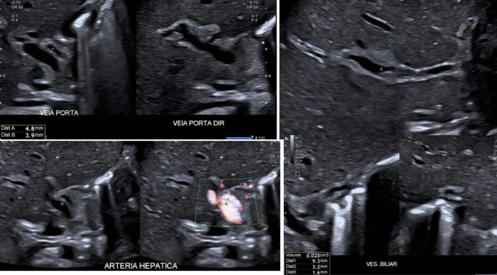
Biliary atresia can be diagnosed based on biliary hilar thickening (the so called triangular cord sign), hypoplastic gallbladder, and enlarged hepatic artery – the relation between it and the right hepatic portal branch is = 0.66 (Normal < 0.50).
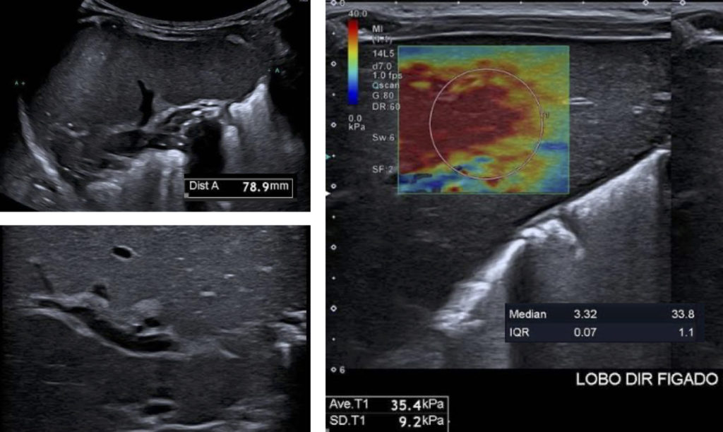
Except for the periportal thickening, the liver keeps its usual sonographic features. Indirect signs of liver disease as ascites and splenomegaly are the only suspicious findings on B-mode ultrasound.
Elastography, specially 2D-SWE, has an important role as it can show increased liver stiffness, usually above 20 kPa. This is due to biliary atresia as well as to liver cirrhosis that is already present at this age. The child was referred to liver transplantation.
Expected findings related to the alternative answers
Answer 1: Neonatal hepatitis – Liver has normal sonographic features. Liver stiffness can be slightly increased, around 10 to 15 kPa.
Answer 2: Obstruction due to biliary sludge – in this setting there is biliary dilation. The sludge can be seen inside the common bile duct.
Answer 3: Congenital cirrhosis – due to multiple causes, as congenital infections, metabolic disorders, gestational alloimmune liver disease, haemophagocytic lymphohistiocytosis, and ischaemic injury. If cirrhosis develops during pregnancy, the newborn shows signs of liver failure. Portal hypertension signals can be depicted by Doppler. In this setting the ductus venosus does not close, and can be seen with flow at color Doppler. Usually, the liver has no other suspicious findings
References
- Ciocca M, Álvarez F. Neonatal acute liver failure: a diagnosis challenge. Arch Argent Pediatr. 2017;115:175-180.
- Duan X, Peng Y, Liu W, Yang L, Zhang J. Does Supersonic Shear Wave Elastography Help Differentiate Biliary Atresia from Other Causes of Cholestatic Hepatitis in Infants Less than 90 Days Old? Compared with Grey-Scale US. Biomed Res Int. 2019;9036362.
- Leschied JR, Dillman JR, Bilhartz J, Heider A, Smith EA, Lopez MJ. Shear wave elastography helps differentiate biliary atresia from other neonatal/infantile liver diseases. Pediatr Radiol. 2015;45:366-75.
- Neonatal Liver Failure and Congenital Cirrhosis due to Gestational Alloimmune Liver Disease: A Case Report and Literature Review. Roos Mariano da Rocha C, Rostirola Guedes R, Kieling CO, Rossato Adami M, Cerski CT, Gonçalves Vieira SM. Case Reports in Pediatrics. 2017;7432859
- Wang X, Qian L, Jia L, Bellah R, Wang N, Xin Y, Liu Q. Utility of Shear Wave Elastography for Differentiating Biliary Atresia From Infantile Hepatitis Syndrome. J Ultrasound Med. 2016;35:1475-9.
- Zhou L, Shan Q, Tian W, Wang Z, Liang J, Xie X. Ultrasound for the Diagnosis of Biliary Atresia: A Meta-Analysis. AJR Am J Roentgenol. 2016;206:W73-82.
- Zhou LY, Wang W, Shan QY, Liu BX, Zheng YL, Xu ZF, Xu M, Pan FS, Lu MD, Xie XY. Optimizing the US Diagnosis of Biliary Atresia with a Modified Triangular Cord Thickness and Gallbladder Classification. Radiology. 2015;277:181-91.
Incorrect
CORRECT ANSWER EXPLAINED BELOW Explanation
Biliary atresia can be diagnosed based on biliary hilar thickening (the so called triangular cord sign), hypoplastic gallbladder, and enlarged hepatic artery – the relation between it and the right hepatic portal branch is = 0.66 (Normal < 0.50).
Except for the periportal thickening, the liver keeps its usual sonographic features. Indirect signs of liver disease as ascites and splenomegaly are the only suspicious findings on B-mode ultrasound.
Elastography, specially 2D-SWE, has an important role as it can show increased liver stiffness, usually above 20 kPa. This is due to biliary atresia as well as to liver cirrhosis that is already present at this age. The child was referred to liver transplantation.
Expected findings related to the alternative answers
Answer 1: Neonatal hepatitis – Liver has normal sonographic features. Liver stiffness can be slightly increased, around 10 to 15 kPa.
Answer 2: Obstruction due to biliary sludge – in this setting there is biliary dilation. The sludge can be seen inside the common bile duct.
Answer 3: Congenital cirrhosis – due to multiple causes, as congenital infections, metabolic disorders, gestational alloimmune liver disease, haemophagocytic lymphohistiocytosis, and ischaemic injury. If cirrhosis develops during pregnancy, the newborn shows signs of liver failure. Portal hypertension signals can be depicted by Doppler. In this setting the ductus venosus does not close, and can be seen with flow at color Doppler. Usually, the liver has no other suspicious findings
References
- Ciocca M, Álvarez F. Neonatal acute liver failure: a diagnosis challenge. Arch Argent Pediatr. 2017;115:175-180.
- Duan X, Peng Y, Liu W, Yang L, Zhang J. Does Supersonic Shear Wave Elastography Help Differentiate Biliary Atresia from Other Causes of Cholestatic Hepatitis in Infants Less than 90 Days Old? Compared with Grey-Scale US. Biomed Res Int. 2019;9036362.
- Leschied JR, Dillman JR, Bilhartz J, Heider A, Smith EA, Lopez MJ. Shear wave elastography helps differentiate biliary atresia from other neonatal/infantile liver diseases. Pediatr Radiol. 2015;45:366-75.
- Neonatal Liver Failure and Congenital Cirrhosis due to Gestational Alloimmune Liver Disease: A Case Report and Literature Review. Roos Mariano da Rocha C, Rostirola Guedes R, Kieling CO, Rossato Adami M, Cerski CT, Gonçalves Vieira SM. Case Reports in Pediatrics. 2017;7432859
- Wang X, Qian L, Jia L, Bellah R, Wang N, Xin Y, Liu Q. Utility of Shear Wave Elastography for Differentiating Biliary Atresia From Infantile Hepatitis Syndrome. J Ultrasound Med. 2016;35:1475-9.
- Zhou L, Shan Q, Tian W, Wang Z, Liang J, Xie X. Ultrasound for the Diagnosis of Biliary Atresia: A Meta-Analysis. AJR Am J Roentgenol. 2016;206:W73-82.
- Zhou LY, Wang W, Shan QY, Liu BX, Zheng YL, Xu ZF, Xu M, Pan FS, Lu MD, Xie XY. Optimizing the US Diagnosis of Biliary Atresia with a Modified Triangular Cord Thickness and Gallbladder Classification. Radiology. 2015;277:181-91.


