Case of the Month August 2020- The tip of the iceberg
August 5, 2020Ultrasound the Best #06: Colour Doppler imaging of the bowel wall in inflammatory bowel disease
September 21, 2020Lucas Japiassu Mendonça Rocha1 ; Eduardo Davino Chiovatto1 ; Alessandra Rodrigues Silva Chiovatto1 ; Renato Davino Chiovatto1, 4; Maria Cristina Chammas1, 2; Wagner Iared1, 3
- Ultrasonography Improvement and Research Center Dr. Giovanni Guido Cerri, DASA, São Paulo, Brazil.
- Institute of Radiology – Hospital das Clínicas School of Medicine – University of São Paulo, São Paulo, Brazil
- Departament of Medicine – Federal University of São Paulo, São Paulo, Brazil.
- Institute of Heart – Hospital das Clínicas School of Medicine – University of São Paulo, São Paulo, Brazil
Clinical History:
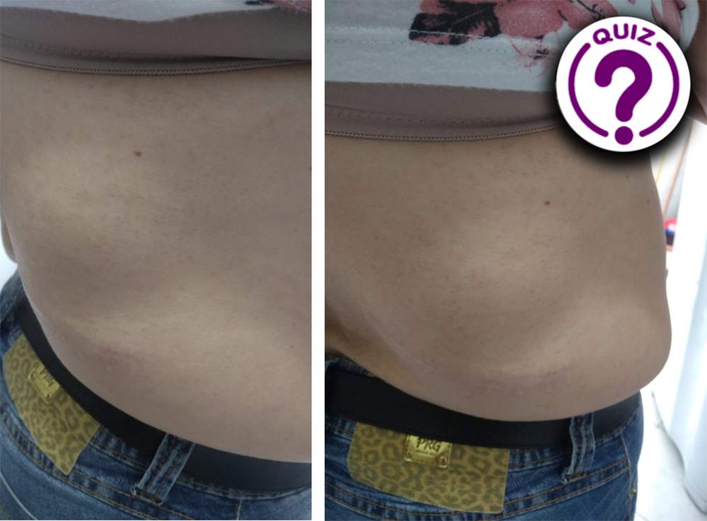
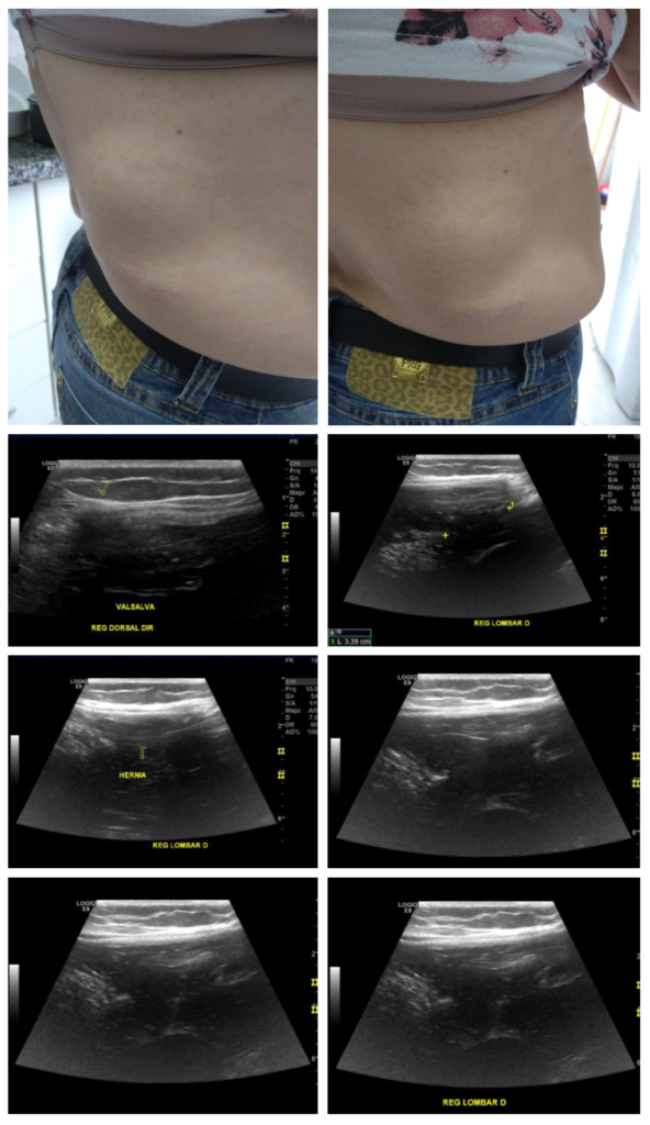
A 61-year-old woman with a previous history of right side lumbar herniorrhaphy was referred to our clinic for a diagnostic ultrasound exam. She had noticed a bulging in the right flank, associated with local pain 4 months previously. The clinical examination revealed a palpable lump more pronounced in standing position on the right flank upper quadrant. A surgical scar could be seen just below the examined site.
Quiz-summary
0 of 1 questions completed
Questions:
- 1
Information
View the September Case below, answer the question and then click check >
You have already completed the quiz before. Hence you can not start it again.
Quiz is loading...
You must sign in or sign up to start the quiz.
You have to finish following quiz, to start this quiz:
Results
0 of 1 questions answered correctly
Your time:
Time has elapsed
You have reached 0 of 0 points, (0)
Categories
- Not categorized 0%
- 1
- Answered
- Review
-
Question 1 of 1
1. Question
Question: Based on the images and clinical history provided, what is your diagnosis?
Correct
CORRECT ANSWER EXPLAINED BELOW Correct answer is:
Relapsed Grynfelt lumbar hernia.
Explanation:
Lumbar hernia is characterized by the presence of a failure in the transversal fascia or aponeurosis of the transverse muscle of the abdomen that results in the extrusion of intra-abdominal content through the defect in the posterolateral abdominal wall.
Lumbar hernias are quite rare, have a higher prevalence in males (about 2/3 of all reported cases) and the majority are unilateral, being twice as common on the left side than on the right side [1; 2]. Most diagnosed patients are in the sixth to seventh decade of life. [3, 4] There is a higher prevalence of lumbar hernias located in the upper lumbar triangle, probably because it is larger and located in a region with weaker musculature than the lower triangle [2].
The lumbar region can be anatomically defined as the area of the abdominal wall lateral to the midclavicular line, between the subcostal plane and the plane that connects the tubercles of the iliac crests.
Lumbar hernias are classified as superior or inferior according to their anatomical location. The upper lumbar or Grynfelt hernia is the one that appears through the anatomical triangle delineated superiorly by the 12th rib, laterally by the external oblique muscle, inferiorly by the internal oblique muscle and medially by the erector spinae muscle. [1, 5]
The lower lumbar or Petit’s hernia is located in the lower triangle, limited inferiorly by the iliac crest, medially by the latissimus dorsi muscle and laterally by the external oblique muscle [1, 5].
The diagnosis is suspected by clinical and physical examination, and may be confirmed by imaging methods such as ultrasound, computed tomography or magnetic resonance imaging. Like other types of hernias, lumbar hernias can evolve with complications such as incarceration and necrosis of intestinal loops [6]. In the present case, the patient presented recurrence after herniorrhaphy with mesh placement.
Take Home Message
Although lumbar hernias are very rare, it is an important differential diagnosis to remember. The suspected clinical diagnosis can be confirmed by ultrasound with high accuracy.
References
1.Heniford BT, Iannitti DA, Gagner M. Laparoscopic inferior and superior lumbar hernia repair. Arch Surg 1997;132: 1141-4.
2.Zhou X, Nve JO, Chen G (2004) Lumbar hernia: clinical analysis of 11 cases. Hernia 8:260–263.
3.Sharma, Lt Col P (2009) Lumbar hernia. MJAFI 65:178–179. 42.Baker ME, Weinerth JL, Andriani RT et al (1986) Lumbar hernia: diagnosis by CT. AJR Am J Roentgenol 148:565–567.
4.Baker ME, Weinerth JL, Andriani RT et al (1986) Lumbar hernia: diagnosis by CT. AJR Am J Roentgenol 148:565–567.
5. Stamatiou D, Skandalakis JE, Skandalakis LJ, Mirilas P. Lumbar hernia: surgical anatomy, embryology, and technique of repair. American Surgeon. 2009;75(3):202–207.
6. Christiano Marlo Paggi CLAUS1,2, Lucas Thá NASSIF1 , Yan Sacha AGUILERA. Reparo Laparoscópico de hérnia lombar (GRYNFELT): Descrição ténica. 2017;30(1):56-59.
Incorrect
CORRECT ANSWER EXPLAINED BELOW Correct answer is:
Relapsed Grynfelt lumbar hernia.
Explanation:
Lumbar hernia is characterized by the presence of a failure in the transversal fascia or aponeurosis of the transverse muscle of the abdomen that results in the extrusion of intra-abdominal content through the defect in the posterolateral abdominal wall.
Lumbar hernias are quite rare, have a higher prevalence in males (about 2/3 of all reported cases) and the majority are unilateral, being twice as common on the left side than on the right side [1; 2]. Most diagnosed patients are in the sixth to seventh decade of life. [3, 4] There is a higher prevalence of lumbar hernias located in the upper lumbar triangle, probably because it is larger and located in a region with weaker musculature than the lower triangle [2].
The lumbar region can be anatomically defined as the area of the abdominal wall lateral to the midclavicular line, between the subcostal plane and the plane that connects the tubercles of the iliac crests.
Lumbar hernias are classified as superior or inferior according to their anatomical location. The upper lumbar or Grynfelt hernia is the one that appears through the anatomical triangle delineated superiorly by the 12th rib, laterally by the external oblique muscle, inferiorly by the internal oblique muscle and medially by the erector spinae muscle. [1, 5]
The lower lumbar or Petit’s hernia is located in the lower triangle, limited inferiorly by the iliac crest, medially by the latissimus dorsi muscle and laterally by the external oblique muscle [1, 5].
The diagnosis is suspected by clinical and physical examination, and may be confirmed by imaging methods such as ultrasound, computed tomography or magnetic resonance imaging. Like other types of hernias, lumbar hernias can evolve with complications such as incarceration and necrosis of intestinal loops [6]. In the present case, the patient presented recurrence after herniorrhaphy with mesh placement.
Take Home Message
Although lumbar hernias are very rare, it is an important differential diagnosis to remember. The suspected clinical diagnosis can be confirmed by ultrasound with high accuracy.
References
1.Heniford BT, Iannitti DA, Gagner M. Laparoscopic inferior and superior lumbar hernia repair. Arch Surg 1997;132: 1141-4.
2.Zhou X, Nve JO, Chen G (2004) Lumbar hernia: clinical analysis of 11 cases. Hernia 8:260–263.
3.Sharma, Lt Col P (2009) Lumbar hernia. MJAFI 65:178–179. 42.Baker ME, Weinerth JL, Andriani RT et al (1986) Lumbar hernia: diagnosis by CT. AJR Am J Roentgenol 148:565–567.
4.Baker ME, Weinerth JL, Andriani RT et al (1986) Lumbar hernia: diagnosis by CT. AJR Am J Roentgenol 148:565–567.
5. Stamatiou D, Skandalakis JE, Skandalakis LJ, Mirilas P. Lumbar hernia: surgical anatomy, embryology, and technique of repair. American Surgeon. 2009;75(3):202–207.
6. Christiano Marlo Paggi CLAUS1,2, Lucas Thá NASSIF1 , Yan Sacha AGUILERA. Reparo Laparoscópico de hérnia lombar (GRYNFELT): Descrição ténica. 2017;30(1):56-59.


