
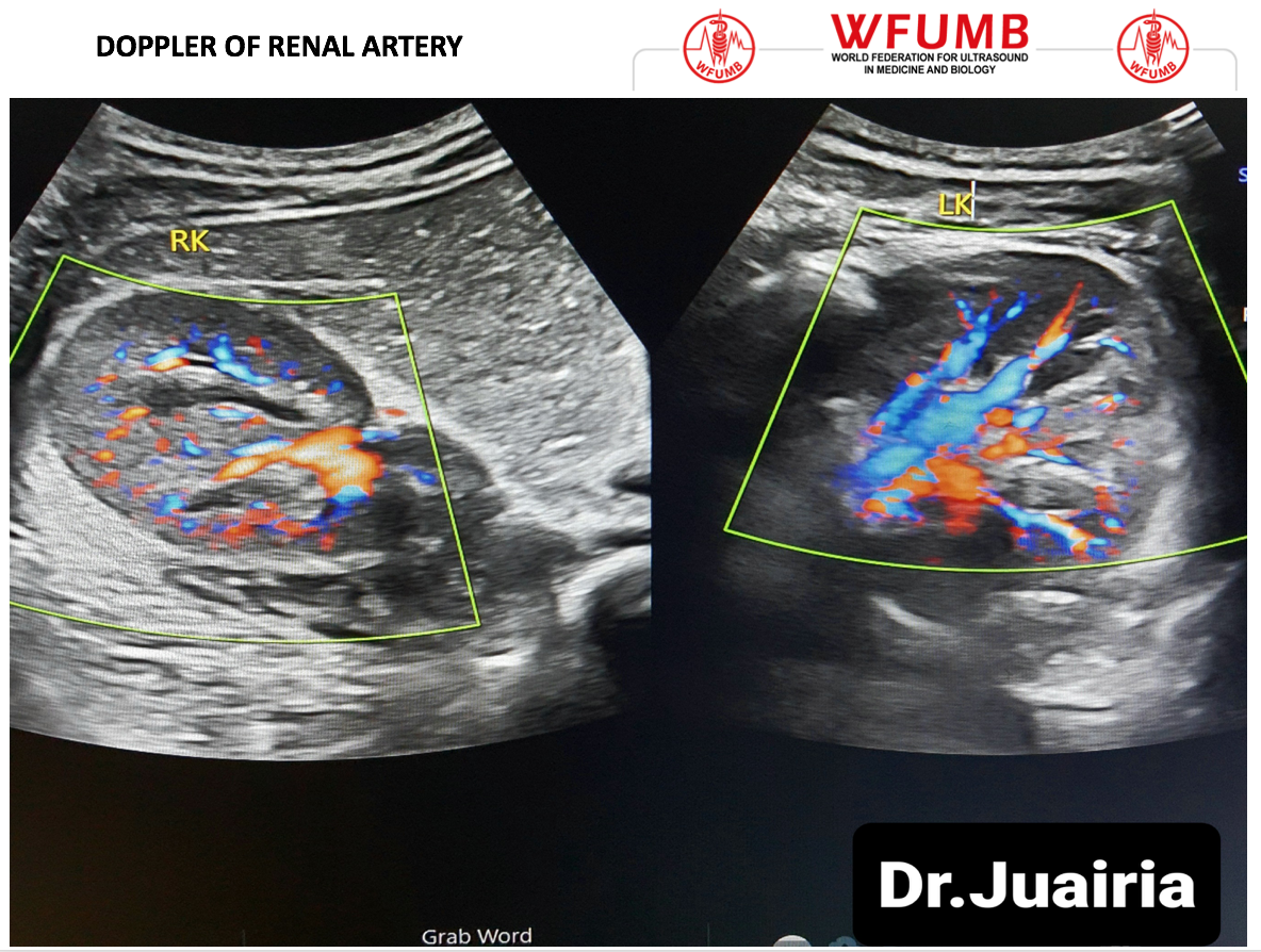
Ultrasound the Best #19: Doppler of Renal Artery
October 29, 2020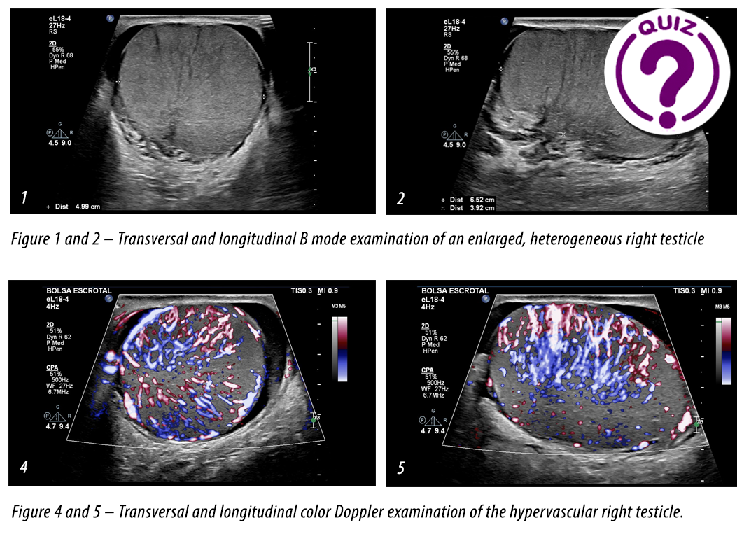
Case of the Month December 2020- Fast enlargement of the right testicle without trauma
December 2, 2020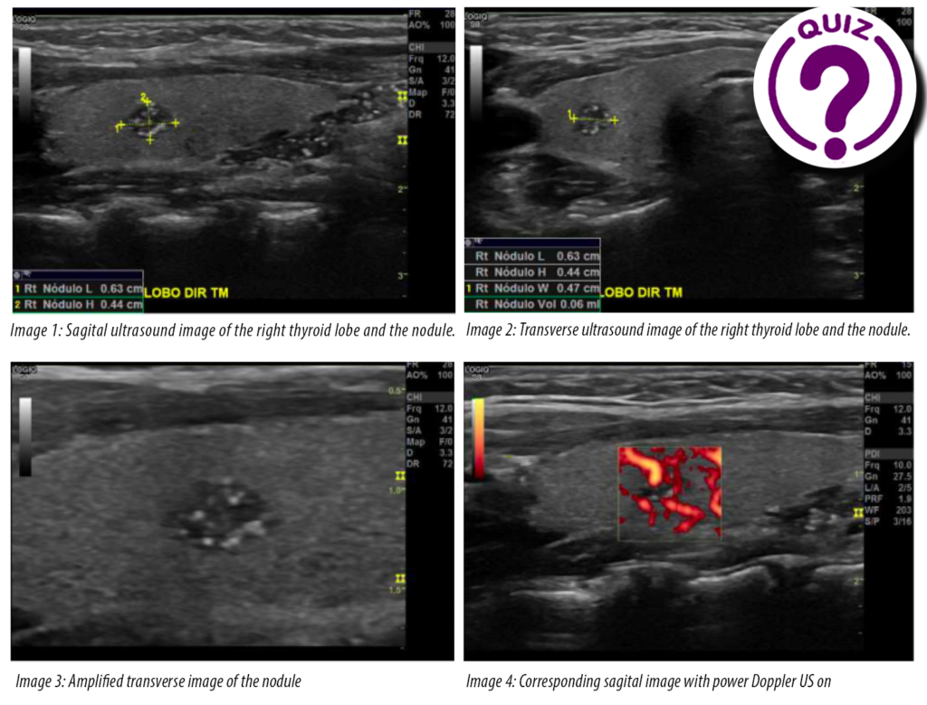
Alessandra Rodrigues Silva Chiovatto, Eduardo Davino Chiovatto, Julia Diva Zavariz, Maria Cristina Chammas.
Centro de Aperfeiçoamento e Pesquisa em Ultrassonografia Prof. Dr. Giovanni Guido Cerri, DASA, São Paulo, Brazil.
Clinical History:
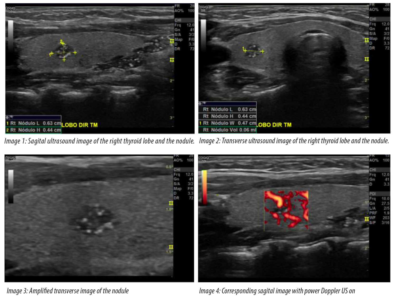
A 12-year-old female was referred to our service for a follow-up of suspected asymptomatic thyroid nodule found about 3 months ago. The patient was healthy, with no comorbidities.
Quiz-summary
0 of 1 questions completed
Questions:
- 1
Information
View the November Case below, answer the question and then click check >
You have already completed the quiz before. Hence you can not start it again.
Quiz is loading...
You must sign in or sign up to start the quiz.
You have to finish following quiz, to start this quiz:
Results
0 of 1 questions answered correctly
Your time:
Time has elapsed
You have reached 0 of 0 points, (0)
Categories
- Not categorized 0%
- 1
- Answered
- Review
-
Question 1 of 1
1. Question
Question: What is the most likely diagnosis?
Correct
CORRECT ANSWER EXPLAINED BELOW Correct answer is: Intrathyroidal thymic tissue
Explanation:
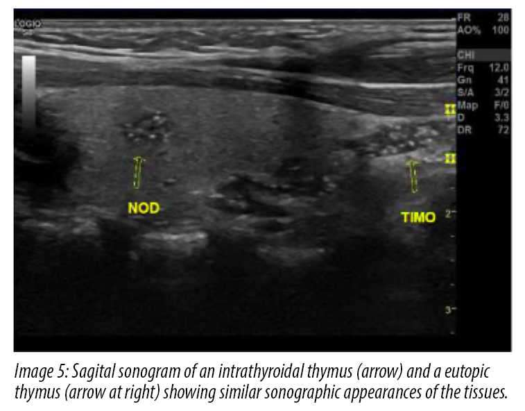
In the images, we see a nodule with ill-defined margins and multiple hyperechoic foci similar to microcalcifications, which matched the sonographic appearance of the normal eutopic thymus, in the mid-portion of the right thyroid lobe. Color Doppler sonography showed scanty blood flow within the nodule.
Intrathyroidal thymic tissue is a rare benign lesion found in the pediatric thyroid gland of nonthyroid origin and may be confused with a malignant thyroid nodule because of the hyperechoic dots mimicking microcalcifications (1-3).
Solitary thyroid nodules in children are present in 0.2%-1.5% (4) in comparison to the higher prevalence in adults. The probability of malignancy in children is higher than in adults (3). The accurate ultrasound diagnosis of such lesions is critical to avoid unnecessary biopsy or surgery. Most lesions identified within the thyroid gland in children are benign nodules of follicular cell origin or, rarely, differentiated thyroid cancers (6). The sonographic findings that suggest a malignant thyroid nodule are marked hypoechogenicity, irregular margins, microcalcifications, and a taller-than-wide shape.
Take Home Message
Solitary thyroid nodules in children are rare.
In ultrasound of the thyroid you must also assess the lymph nodes and the rest of the neck, especially the thymus in children. A good exam can avoid unnecessary biopsy or surgery.References
- BÜYÜKYAVUZ, I. et al. Ectopic thymic tissue as a rare and confusing entity. European journal of pediatric surgery, v. 12, n. 05, p. 327-329, 2002.
- http://www.pathologyoutlines.com/topic/thyroidthymictissue.html
- FRATES, Mary C. et al. Ectopic intrathyroidal thymic tissue mimicking thyroid nodules in children. Journal of Ultrasound in Medicine, v. 37, n. 3, p. 783-791, 2018.
- PARK, So Hyun et al. Intrathyroidal thymic tissue mimicking a malignant thyroid nodule in a 4-year-old child. Ultrasonography, v. 33, n. 1, p. 71, 2014.
- O’CONNOR, Kelly et al. An ectopic intrathyroidal thymic tissue and intrathymic parathyroid tissue in a patient with Graves disease. Gland surgery, v. 6, n. 6, p. 726, 2017.
- HUANG, Yanlei; ZHENG, Shan; XIAO, Xianmin. Ectopic intrathyroidal thymus in children: two case reports and review of the literature. Journal of Pediatric Surgery Case Reports, v. 1, n. 11, p. 386-390, 2013.
Incorrect
CORRECT ANSWER EXPLAINED BELOW Correct answer is: Intrathyroidal thymic tissue
Explanation:
In the images, we see a nodule with ill-defined margins and multiple hyperechoic foci similar to microcalcifications, which matched the sonographic appearance of the normal eutopic thymus, in the mid-portion of the right thyroid lobe. Color Doppler sonography showed scanty blood flow within the nodule.
Intrathyroidal thymic tissue is a rare benign lesion found in the pediatric thyroid gland of nonthyroid origin and may be confused with a malignant thyroid nodule because of the hyperechoic dots mimicking microcalcifications (1-3).
Solitary thyroid nodules in children are present in 0.2%-1.5% (4) in comparison to the higher prevalence in adults. The probability of malignancy in children is higher than in adults (3). The accurate ultrasound diagnosis of such lesions is critical to avoid unnecessary biopsy or surgery. Most lesions identified within the thyroid gland in children are benign nodules of follicular cell origin or, rarely, differentiated thyroid cancers (6). The sonographic findings that suggest a malignant thyroid nodule are marked hypoechogenicity, irregular margins, microcalcifications, and a taller-than-wide shape.
Take Home Message
Solitary thyroid nodules in children are rare.
In ultrasound of the thyroid you must also assess the lymph nodes and the rest of the neck, especially the thymus in children. A good exam can avoid unnecessary biopsy or surgery.References
- BÜYÜKYAVUZ, I. et al. Ectopic thymic tissue as a rare and confusing entity. European journal of pediatric surgery, v. 12, n. 05, p. 327-329, 2002.
- http://www.pathologyoutlines.com/topic/thyroidthymictissue.html
- FRATES, Mary C. et al. Ectopic intrathyroidal thymic tissue mimicking thyroid nodules in children. Journal of Ultrasound in Medicine, v. 37, n. 3, p. 783-791, 2018.
- PARK, So Hyun et al. Intrathyroidal thymic tissue mimicking a malignant thyroid nodule in a 4-year-old child. Ultrasonography, v. 33, n. 1, p. 71, 2014.
- O’CONNOR, Kelly et al. An ectopic intrathyroidal thymic tissue and intrathymic parathyroid tissue in a patient with Graves disease. Gland surgery, v. 6, n. 6, p. 726, 2017.
- HUANG, Yanlei; ZHENG, Shan; XIAO, Xianmin. Ectopic intrathyroidal thymus in children: two case reports and review of the literature. Journal of Pediatric Surgery Case Reports, v. 1, n. 11, p. 386-390, 2013.