Case of the Month December 2020- Fast enlargement of the right testicle without trauma
December 2, 2020Webinar: Hepatocellular Carcinoma (HCC)
December 14, 2020Sirine Dehmani 1, Christoph F Dietrich 2
- Kliniken Hirslanden Beau Site, Salem und Permanence, Bern, Switzerland, Sirine.Dehmani@hirslanden.ch
- *Kliniken Hirslanden Beau Site, Salem und Permanence, Bern, Switzerland, ChristophFrank.Dietrich@hirslanden.ch
* Correspondence: ChristophFrank.Dietrich@hirslanden.ch
Clinical History:
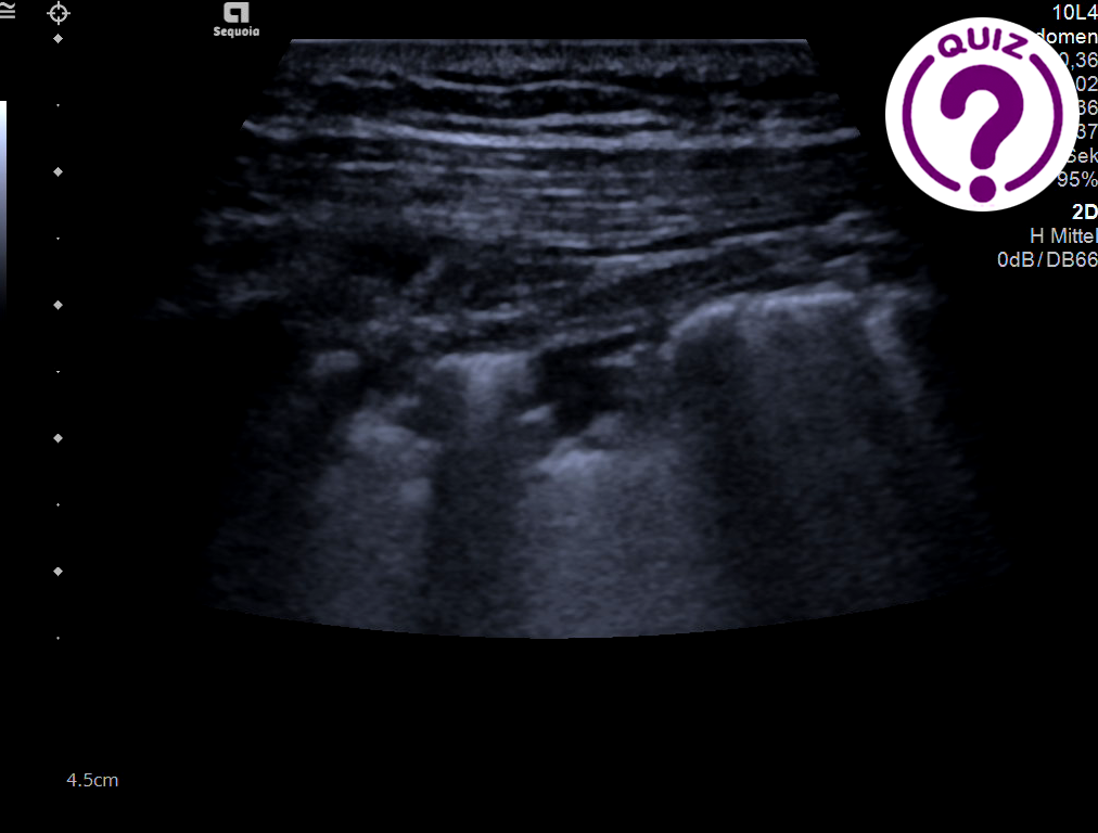
A 56-year-old-male with a history of previous cough and dyspnea on exercise was admitted to our clinic for evaluation and CoViD testing. At the time of the US examination he had no dyspnea or other symptoms.
Quiz-summary
0 of 2 questions completed
Questions:
- 1
- 2
Information
View the December Case below, answer the questions and then click check >
You have already completed the quiz before. Hence you can not start it again.
Quiz is loading...
You must sign in or sign up to start the quiz.
You have to finish following quiz, to start this quiz:
Results
0 of 2 questions answered correctly
Your time:
Time has elapsed
You have reached 0 of 0 points, (0)
Categories
- Not categorized 0%
-
CORRECT ANSWER EXPLAINED BELOW Correct answer to Q1 is:
- B-line artefacts
- Irregular pleural line and very small amount of free fluid located peripherally
- Multiple small consolidations in all lung regions
Correct answer to Q2 is:
- SARS-CoViD-19 infection
Discussion:
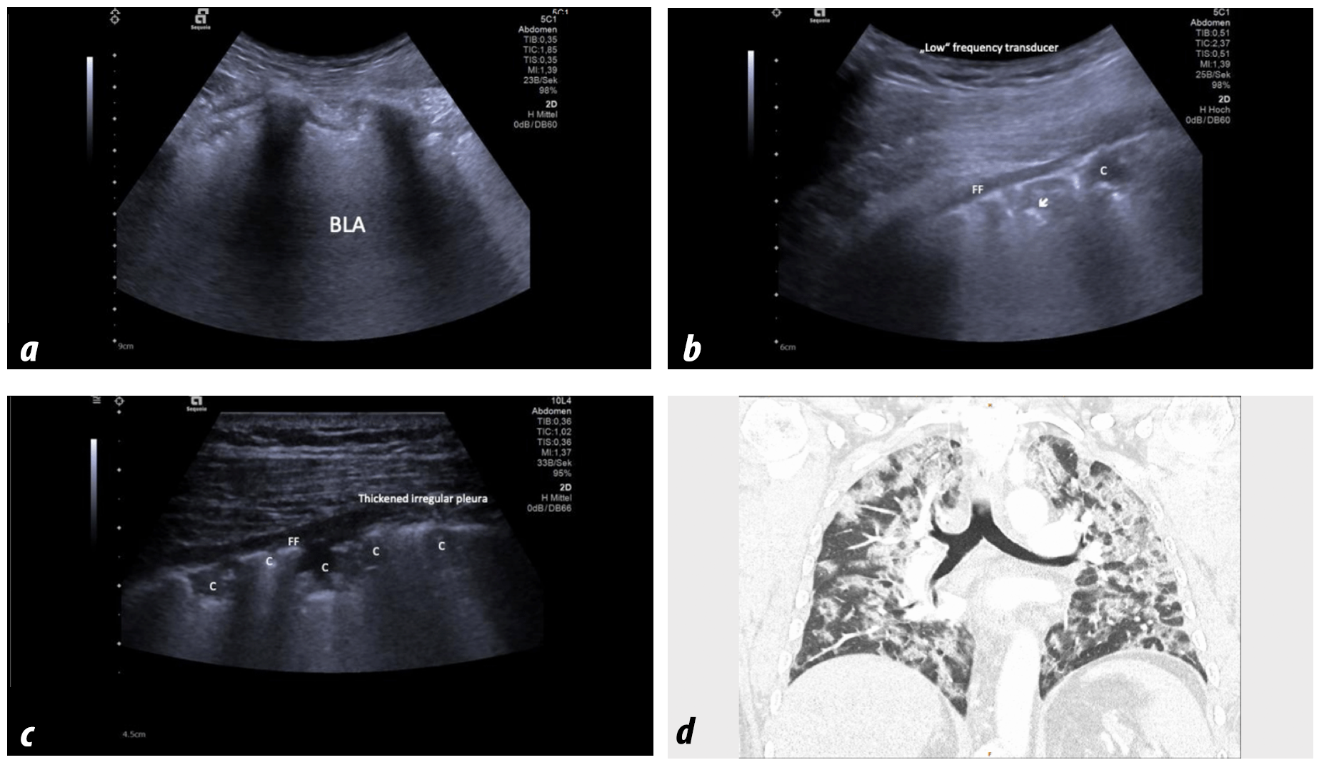
Since COVID-19 infection is primarily a respiratory disease the expectation would be that lung imaging would be essential for the diagnosis (1,2). The typical sonographic signs using lung US (LUS) identified in the course of COVID-19 infections include features of the pleura and pleural space (1), findings of interstitial pneumonia (2) and specific artefacts (3) (1,3-5). The signs are specific when there is a very high “a priori” probability of COVID-19 with cough and dyspnea (1). The initial lung involvement often posterior-basal may become extensive with involvement of the entire lung as shown in this patient. In later stages mixed and much less specific image patterns can be observed including the so-called white lung and bacterial pneumonia superinfection. US also allows detection of complications including pneumothorax, not only in ventilated patients, and pulmonary embolism (5).
Ultrasound signs and differential diagnoses
Pleura
- Thickened, irregular (coarse) and fragmented pleural line.
- Small amount of superficially located pleural fluid (sign of severity). More extensive pleural effusions are not typical.
Lung
- Consolidations, multiple. Very small consolidations often posterobasally, initially. Later in various distributions and possibly involving the entire lung.
Artefacts
- B-lines (initially discrete, later multifocal and confluent), alveolar-interstitial syndrome, so-called „white lung“ (ARDS).
- Respiratory dependent lung sliding (A-artefact), combined with B-line artefacts (mixing A and B patterns).
Differential diagnoses and complications
- Chronic heart insufficiency (BLA, larger pleural effusion than in CoViD-19).
- Bacterial pneumonia (typically larger consolidations than in CoViD-19).
- Pulmonary embolism (initially no vascularity)
Conflicts of interest
“The authors declare no conflict of interest.”
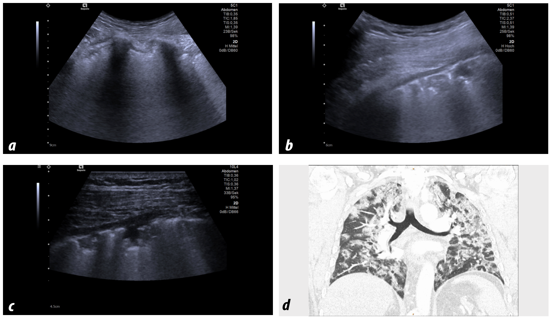
 Image(s): Confluent curtain like B-line artefacts (BLA) intercostally using 5 MHz curved array transducer (a). Thickened and irregular pleura line and tiny amount of pleural fluid using 5 MHz curved array transducer (b) and 10 MHz transducer revealing much more details (c). The corresponding computed tomography image is also shown (d). C: Consolidation. FF: free fluid. White arrow: Pneumo-alveolo-bronchogram.
References
- Piscaglia F, Stefanini F, Cantisani V, Sidhu PS, Barr R, Berzigotti A, Chammas MC, et al. Benefits, Open questions and Challenges of the use of Ultrasound in the COVID-19 pandemic era. The views of a panel of worldwide international experts. Ultraschall Med 2020;41:228-236.
- Zhu N, Zhang D, Wang W, Li X, Yang B, Song J, Zhao X, et al. A Novel Coronavirus from Patients with Pneumonia in China, 2019. N Engl J Med 2020;382:727-733.
- Dietrich CF, Mathis G, Cui XW, Ignee A, Hocke M, Hirche TO. Ultrasound of the pleurae and lungs. Ultrasound Med Biol 2015;41:351-365.
- Dietrich CF, Mathis G, Blaivas M, Volpicelli G, Seibel A, Atkinson NS, Cui XW, et al. Lung artefacts and their use. Med Ultrason 2016;18:488-499.
- Dietrich CF, Mathis G, Blaivas M, Volpicelli G, Seibel A, Wastl D, Atkinson NS, et al. Lung B-line artefacts and their use. J Thorac Dis 2016;8:1356-1365.
- 1
- 2
- Answered
- Review
-
Question 1 of 2
1. Question
Question 1: Which of the following US signs can you identify (more than one is possible)?
Correct
CORRECT ANSWER EXPLAINED BELOW Correct answer to Q1 is:
- B-line artefacts
- Irregular pleural line and very small amount of free fluid located peripherally
- Multiple small consolidations in all lung regions
Correct answer to Q2 is:
- SARS-CoViD-19 infection
Discussion:
Since COVID-19 infection is primarily a respiratory disease the expectation would be that lung imaging would be essential for the diagnosis (1,2). The typical sonographic signs using lung US (LUS) identified in the course of COVID-19 infections include features of the pleura and pleural space (1), findings of interstitial pneumonia (2) and specific artefacts (3) (1,3-5). The signs are specific when there is a very high “a priori” probability of COVID-19 with cough and dyspnea (1). The initial lung involvement often posterior-basal may become extensive with involvement of the entire lung as shown in this patient. In later stages mixed and much less specific image patterns can be observed including the so-called white lung and bacterial pneumonia superinfection. US also allows detection of complications including pneumothorax, not only in ventilated patients, and pulmonary embolism (5).
Ultrasound signs and differential diagnoses
Pleura
- Thickened, irregular (coarse) and fragmented pleural line.
- Small amount of superficially located pleural fluid (sign of severity). More extensive pleural effusions are not typical.
Lung
- Consolidations, multiple. Very small consolidations often posterobasally, initially. Later in various distributions and possibly involving the entire lung.
Artefacts
- B-lines (initially discrete, later multifocal and confluent), alveolar-interstitial syndrome, so-called „white lung“ (ARDS).
- Respiratory dependent lung sliding (A-artefact), combined with B-line artefacts (mixing A and B patterns).
Differential diagnoses and complications
- Chronic heart insufficiency (BLA, larger pleural effusion than in CoViD-19).
- Bacterial pneumonia (typically larger consolidations than in CoViD-19).
- Pulmonary embolism (initially no vascularity)
Conflicts of interest
“The authors declare no conflict of interest.”

Image(s): Confluent curtain like B-line artefacts (BLA) intercostally using 5 MHz curved array transducer (a). Thickened and irregular pleura line and tiny amount of pleural fluid using 5 MHz curved array transducer (b) and 10 MHz transducer revealing much more details (c). The corresponding computed tomography image is also shown (d). C: Consolidation. FF: free fluid. White arrow: Pneumo-alveolo-bronchogram.
References
- Piscaglia F, Stefanini F, Cantisani V, Sidhu PS, Barr R, Berzigotti A, Chammas MC, et al. Benefits, Open questions and Challenges of the use of Ultrasound in the COVID-19 pandemic era. The views of a panel of worldwide international experts. Ultraschall Med 2020;41:228-236.
- Zhu N, Zhang D, Wang W, Li X, Yang B, Song J, Zhao X, et al. A Novel Coronavirus from Patients with Pneumonia in China, 2019. N Engl J Med 2020;382:727-733.
- Dietrich CF, Mathis G, Cui XW, Ignee A, Hocke M, Hirche TO. Ultrasound of the pleurae and lungs. Ultrasound Med Biol 2015;41:351-365.
- Dietrich CF, Mathis G, Blaivas M, Volpicelli G, Seibel A, Atkinson NS, Cui XW, et al. Lung artefacts and their use. Med Ultrason 2016;18:488-499.
- Dietrich CF, Mathis G, Blaivas M, Volpicelli G, Seibel A, Wastl D, Atkinson NS, et al. Lung B-line artefacts and their use. J Thorac Dis 2016;8:1356-1365.
Incorrect
CORRECT ANSWER EXPLAINED BELOW Correct answer to Q1 is:
- B-line artefacts
- Irregular pleural line and very small amount of free fluid located peripherally
- Multiple small consolidations in all lung regions
Correct answer to Q2 is:
- SARS-CoViD-19 infection
Discussion:
Since COVID-19 infection is primarily a respiratory disease the expectation would be that lung imaging would be essential for the diagnosis (1,2). The typical sonographic signs using lung US (LUS) identified in the course of COVID-19 infections include features of the pleura and pleural space (1), findings of interstitial pneumonia (2) and specific artefacts (3) (1,3-5). The signs are specific when there is a very high “a priori” probability of COVID-19 with cough and dyspnea (1). The initial lung involvement often posterior-basal may become extensive with involvement of the entire lung as shown in this patient. In later stages mixed and much less specific image patterns can be observed including the so-called white lung and bacterial pneumonia superinfection. US also allows detection of complications including pneumothorax, not only in ventilated patients, and pulmonary embolism (5).
Ultrasound signs and differential diagnoses
Pleura
- Thickened, irregular (coarse) and fragmented pleural line.
- Small amount of superficially located pleural fluid (sign of severity). More extensive pleural effusions are not typical.
Lung
- Consolidations, multiple. Very small consolidations often posterobasally, initially. Later in various distributions and possibly involving the entire lung.
Artefacts
- B-lines (initially discrete, later multifocal and confluent), alveolar-interstitial syndrome, so-called „white lung“ (ARDS).
- Respiratory dependent lung sliding (A-artefact), combined with B-line artefacts (mixing A and B patterns).
Differential diagnoses and complications
- Chronic heart insufficiency (BLA, larger pleural effusion than in CoViD-19).
- Bacterial pneumonia (typically larger consolidations than in CoViD-19).
- Pulmonary embolism (initially no vascularity)
Conflicts of interest
“The authors declare no conflict of interest.”

Image(s): Confluent curtain like B-line artefacts (BLA) intercostally using 5 MHz curved array transducer (a). Thickened and irregular pleura line and tiny amount of pleural fluid using 5 MHz curved array transducer (b) and 10 MHz transducer revealing much more details (c). The corresponding computed tomography image is also shown (d). C: Consolidation. FF: free fluid. White arrow: Pneumo-alveolo-bronchogram.
References
- Piscaglia F, Stefanini F, Cantisani V, Sidhu PS, Barr R, Berzigotti A, Chammas MC, et al. Benefits, Open questions and Challenges of the use of Ultrasound in the COVID-19 pandemic era. The views of a panel of worldwide international experts. Ultraschall Med 2020;41:228-236.
- Zhu N, Zhang D, Wang W, Li X, Yang B, Song J, Zhao X, et al. A Novel Coronavirus from Patients with Pneumonia in China, 2019. N Engl J Med 2020;382:727-733.
- Dietrich CF, Mathis G, Cui XW, Ignee A, Hocke M, Hirche TO. Ultrasound of the pleurae and lungs. Ultrasound Med Biol 2015;41:351-365.
- Dietrich CF, Mathis G, Blaivas M, Volpicelli G, Seibel A, Atkinson NS, Cui XW, et al. Lung artefacts and their use. Med Ultrason 2016;18:488-499.
- Dietrich CF, Mathis G, Blaivas M, Volpicelli G, Seibel A, Wastl D, Atkinson NS, et al. Lung B-line artefacts and their use. J Thorac Dis 2016;8:1356-1365.
-
Question 2 of 2
2. Question
Question 2: Which of the following diseases is most probable (only one answer is correct)?
Correct
CORRECT ANSWER EXPLAINED BELOW Correct answer to Q1 is:
- B-line artefacts
- Irregular pleural line and very small amount of free fluid located peripherally
- Multiple small consolidations in all lung regions
Correct answer to Q2 is:
- SARS-CoViD-19 infection
Discussion:
Since COVID-19 infection is primarily a respiratory disease the expectation would be that lung imaging would be essential for the diagnosis (1,2). The typical sonographic signs using lung US (LUS) identified in the course of COVID-19 infections include features of the pleura and pleural space (1), findings of interstitial pneumonia (2) and specific artefacts (3) (1,3-5). The signs are specific when there is a very high “a priori” probability of COVID-19 with cough and dyspnea (1). The initial lung involvement often posterior-basal may become extensive with involvement of the entire lung as shown in this patient. In later stages mixed and much less specific image patterns can be observed including the so-called white lung and bacterial pneumonia superinfection. US also allows detection of complications including pneumothorax, not only in ventilated patients, and pulmonary embolism (5).
Ultrasound signs and differential diagnoses
Pleura
- Thickened, irregular (coarse) and fragmented pleural line.
- Small amount of superficially located pleural fluid (sign of severity). More extensive pleural effusions are not typical.
Lung
- Consolidations, multiple. Very small consolidations often posterobasally, initially. Later in various distributions and possibly involving the entire lung.
Artefacts
- B-lines (initially discrete, later multifocal and confluent), alveolar-interstitial syndrome, so-called „white lung“ (ARDS).
- Respiratory dependent lung sliding (A-artefact), combined with B-line artefacts (mixing A and B patterns).
Differential diagnoses and complications
- Chronic heart insufficiency (BLA, larger pleural effusion than in CoViD-19).
- Bacterial pneumonia (typically larger consolidations than in CoViD-19).
- Pulmonary embolism (initially no vascularity)
Conflicts of interest
“The authors declare no conflict of interest.”

Image(s): Confluent curtain like B-line artefacts (BLA) intercostally using 5 MHz curved array transducer (a). Thickened and irregular pleura line and tiny amount of pleural fluid using 5 MHz curved array transducer (b) and 10 MHz transducer revealing much more details (c). The corresponding computed tomography image is also shown (d). C: Consolidation. FF: free fluid. White arrow: Pneumo-alveolo-bronchogram.
References
- Piscaglia F, Stefanini F, Cantisani V, Sidhu PS, Barr R, Berzigotti A, Chammas MC, et al. Benefits, Open questions and Challenges of the use of Ultrasound in the COVID-19 pandemic era. The views of a panel of worldwide international experts. Ultraschall Med 2020;41:228-236.
- Zhu N, Zhang D, Wang W, Li X, Yang B, Song J, Zhao X, et al. A Novel Coronavirus from Patients with Pneumonia in China, 2019. N Engl J Med 2020;382:727-733.
- Dietrich CF, Mathis G, Cui XW, Ignee A, Hocke M, Hirche TO. Ultrasound of the pleurae and lungs. Ultrasound Med Biol 2015;41:351-365.
- Dietrich CF, Mathis G, Blaivas M, Volpicelli G, Seibel A, Atkinson NS, Cui XW, et al. Lung artefacts and their use. Med Ultrason 2016;18:488-499.
- Dietrich CF, Mathis G, Blaivas M, Volpicelli G, Seibel A, Wastl D, Atkinson NS, et al. Lung B-line artefacts and their use. J Thorac Dis 2016;8:1356-1365.
Incorrect
CORRECT ANSWER EXPLAINED BELOW Correct answer to Q1 is:
- B-line artefacts
- Irregular pleural line and very small amount of free fluid located peripherally
- Multiple small consolidations in all lung regions
Correct answer to Q2 is:
- SARS-CoViD-19 infection
Discussion:
Since COVID-19 infection is primarily a respiratory disease the expectation would be that lung imaging would be essential for the diagnosis (1,2). The typical sonographic signs using lung US (LUS) identified in the course of COVID-19 infections include features of the pleura and pleural space (1), findings of interstitial pneumonia (2) and specific artefacts (3) (1,3-5). The signs are specific when there is a very high “a priori” probability of COVID-19 with cough and dyspnea (1). The initial lung involvement often posterior-basal may become extensive with involvement of the entire lung as shown in this patient. In later stages mixed and much less specific image patterns can be observed including the so-called white lung and bacterial pneumonia superinfection. US also allows detection of complications including pneumothorax, not only in ventilated patients, and pulmonary embolism (5).
Ultrasound signs and differential diagnoses
Pleura
- Thickened, irregular (coarse) and fragmented pleural line.
- Small amount of superficially located pleural fluid (sign of severity). More extensive pleural effusions are not typical.
Lung
- Consolidations, multiple. Very small consolidations often posterobasally, initially. Later in various distributions and possibly involving the entire lung.
Artefacts
- B-lines (initially discrete, later multifocal and confluent), alveolar-interstitial syndrome, so-called „white lung“ (ARDS).
- Respiratory dependent lung sliding (A-artefact), combined with B-line artefacts (mixing A and B patterns).
Differential diagnoses and complications
- Chronic heart insufficiency (BLA, larger pleural effusion than in CoViD-19).
- Bacterial pneumonia (typically larger consolidations than in CoViD-19).
- Pulmonary embolism (initially no vascularity)
Conflicts of interest
“The authors declare no conflict of interest.”

Image(s): Confluent curtain like B-line artefacts (BLA) intercostally using 5 MHz curved array transducer (a). Thickened and irregular pleura line and tiny amount of pleural fluid using 5 MHz curved array transducer (b) and 10 MHz transducer revealing much more details (c). The corresponding computed tomography image is also shown (d). C: Consolidation. FF: free fluid. White arrow: Pneumo-alveolo-bronchogram.
References
- Piscaglia F, Stefanini F, Cantisani V, Sidhu PS, Barr R, Berzigotti A, Chammas MC, et al. Benefits, Open questions and Challenges of the use of Ultrasound in the COVID-19 pandemic era. The views of a panel of worldwide international experts. Ultraschall Med 2020;41:228-236.
- Zhu N, Zhang D, Wang W, Li X, Yang B, Song J, Zhao X, et al. A Novel Coronavirus from Patients with Pneumonia in China, 2019. N Engl J Med 2020;382:727-733.
- Dietrich CF, Mathis G, Cui XW, Ignee A, Hocke M, Hirche TO. Ultrasound of the pleurae and lungs. Ultrasound Med Biol 2015;41:351-365.
- Dietrich CF, Mathis G, Blaivas M, Volpicelli G, Seibel A, Atkinson NS, Cui XW, et al. Lung artefacts and their use. Med Ultrason 2016;18:488-499.
- Dietrich CF, Mathis G, Blaivas M, Volpicelli G, Seibel A, Wastl D, Atkinson NS, et al. Lung B-line artefacts and their use. J Thorac Dis 2016;8:1356-1365.

