Correct answer is: Know infant hip anatomy
Discussion
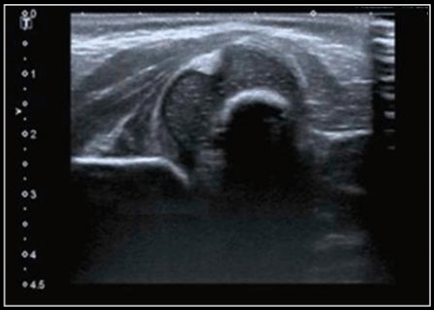
In this image, we can see a Type IV Graf dislocated hip with a poor bony roof, a flattened superior bony rim and with the cartilaginous roof pressed downwards. The angles are immeasurable in this case. By simply knowing the anatomy you are able to diagnose the hip dislocation.
We cannot measure the angles and draw the lines in this case because it is a decentered joint. Before a hip sonogram can be evaluated you must identify the anatomical structures in a methodical way (chondro-osseous junction, femoral head, synovial fold, joint capsule, acetabular labrum, cartilage acetabular roof, bony socket and osseous rim) (1). In order to reach a diagnostic and measurable sonogram the three landmarks must be present (the formed acetabular roof, bony part of the acetabular roof and bony rim: turning point from concavity to convexity).
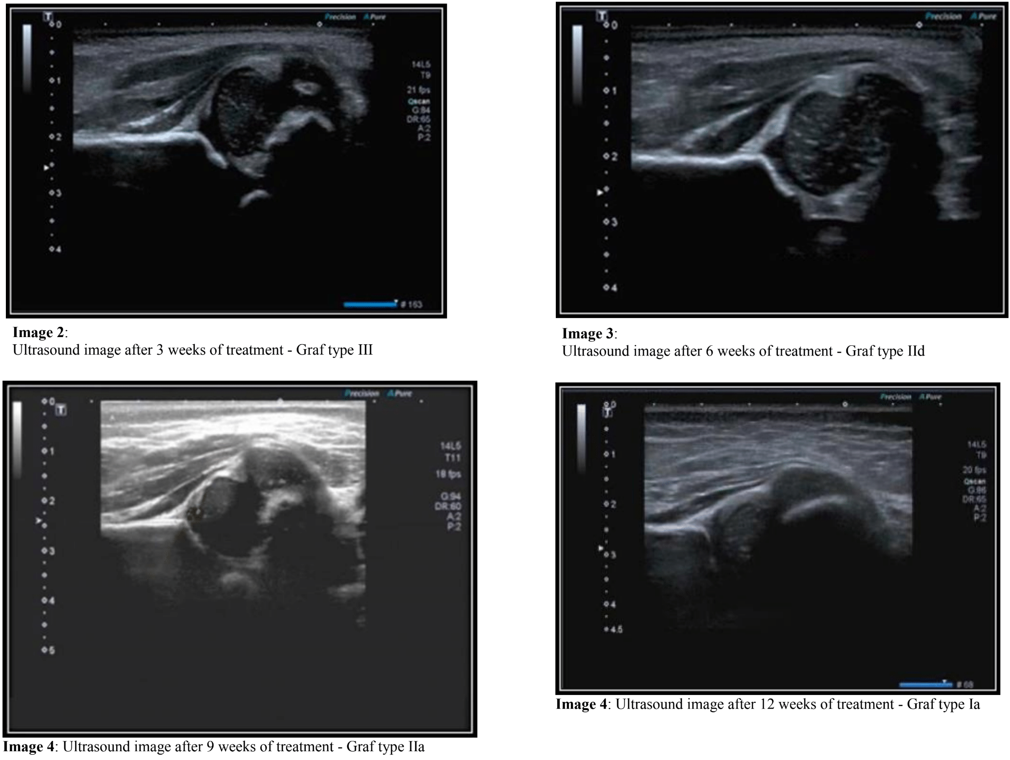
One of the main advantages of the Graf method is that it can be used as a criterion of improvement, stagnation or worsening of the condition during the follow-up of patients treated with Pavlik harness (2, 3). In this case, the infant had a dislocated hip, and after 3 weeks of treatment, this is the sonographic result:

In 6 weeks, 9 weeks and 12 weeks, the favorable evolution of the case can be observed, starting from a type IV to a type Ia hip, or from a dislocated hip to a mature hip.
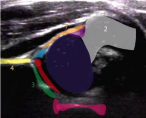
 Image 5: The first step: anatomical identification. Subtitles: Image 5: The first step: anatomical identification. Subtitles:
1-Femoral head
2-Large trochanter
3-Bony roof
4-Medial border of the Ilium
5-Labrum
6-Cartilaginous acetabular roof
7-Joint capsule
8-Synovial fold
9-Triradiate cartilage
Take home message
Through accurate analysis of the pathological changes in the cartilage and bony socket it is possible to state the severity of the pathology affecting the hip joint, even before the confirmation by the fourth step (the measurement with alpha and beta angles). Always remember the order of assessment: the first step is the anatomical identification, the second step is checking the three landmarks, the third step is the description of the structures (bony socket, bony rim and cartilaginous acetabular roof), and finally, the last one is the angles’ measurement that it must coincide (be congruent).
References
- GRAF, Reinhard.Hip sonography: diagnosis and management of infant hip dysplasia. Springer Science & Business Media, 2006.
- KOSAR, Pinar et al. Follow‐up SonographicResults for Graf Type 2a Hips: Association With Risk Factors for Developmental Dysplasia of the Hip and Instability.Journal of Ultrasound in Medicine, v. 30, n. 5, p. 677-683, 2011.
- JUDD, Julia; CLARKE, Nicholas MP. Treatment and prevention of hip dysplasia in infants and young children.Early human development, v. 90, n. 11, p. 731-734, 2014.
|


