Case of the Month October 2021- Preterm neonate with abdominal distention and clinical decompensation

Case of the Month September 2021- Intermittent upper abdominal pain with vomiting
September 7, 2021WFUMB / EFSUMB Students Webinar Series: 10 June 2021
October 26, 2021Martin Necas 1 & Holly Smith 1
1 Waikato Hospital, Hamilton, New Zealand; martin@antegrade.net
* Correspondence: martin@antegrade.net
Clinical History:
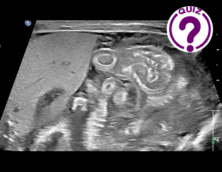
An 8-day-old female born at 23-weeks gestation was referred for abdominal ultrasound due to worsening metabolic acidosis, abdominal distension and discolouration. An X-ray performed prior to ultrasound showed paucity of bowel gas, a prominent fixed loop of small bowel centrally, with no definite signs of free gas. High-resolution abdominal ultrasound was performed using an ultra-broadband linear array transducer (eL18-4, Philips Epiq Elite ultrasound system).
Quiz-summary
0 of 1 questions completed
Questions:
- 1
Information
View the October Case below, answer the question and then click check >
You have already completed the quiz before. Hence you can not start it again.
Quiz is loading...
You must sign in or sign up to start the quiz.
You have to finish following quiz, to start this quiz:
Results
0 of 1 questions answered correctly
Your time:
Time has elapsed
You have reached 0 of 0 points, (0)
Categories
- Not categorized 0%
- 1
- Answered
- Review
-
Question 1 of 1
1. Question
Question: What is the diagnosis?
Correct
CORRECT ANSWER EXPLAINED BELOW Correct answer is: Necrotising enterocolitis with perforation
Discussion
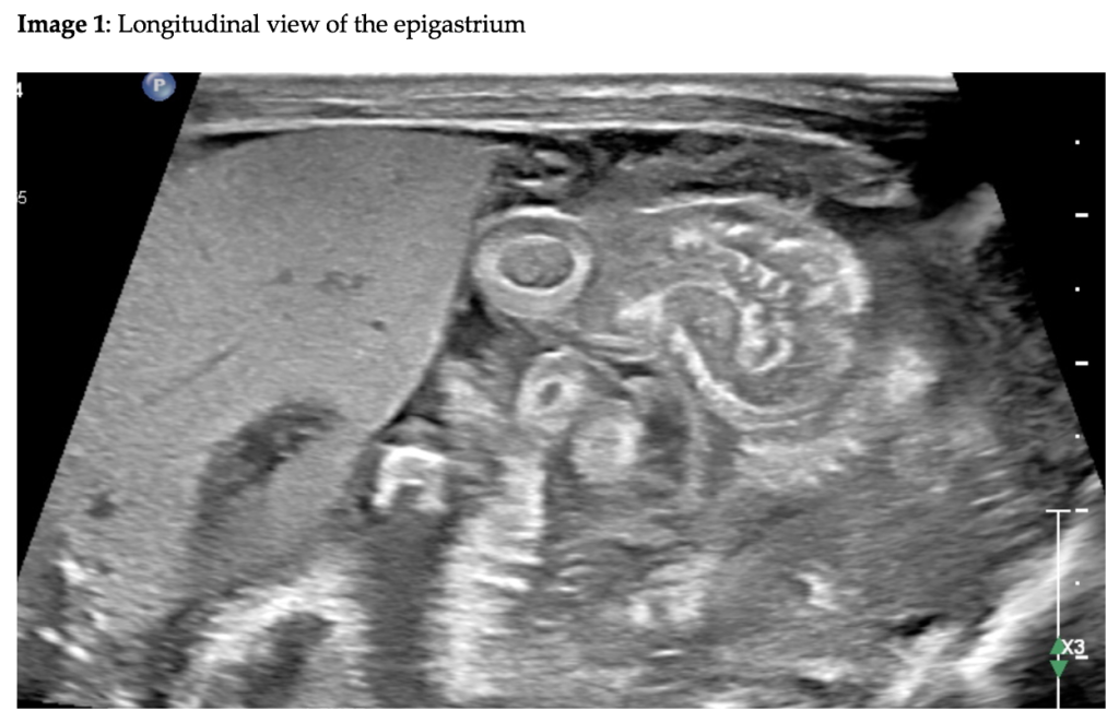
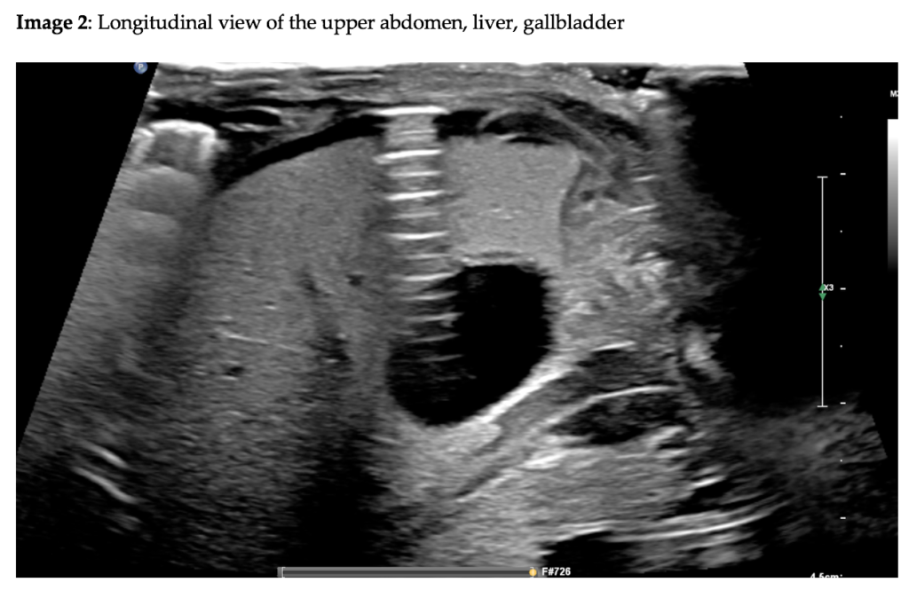
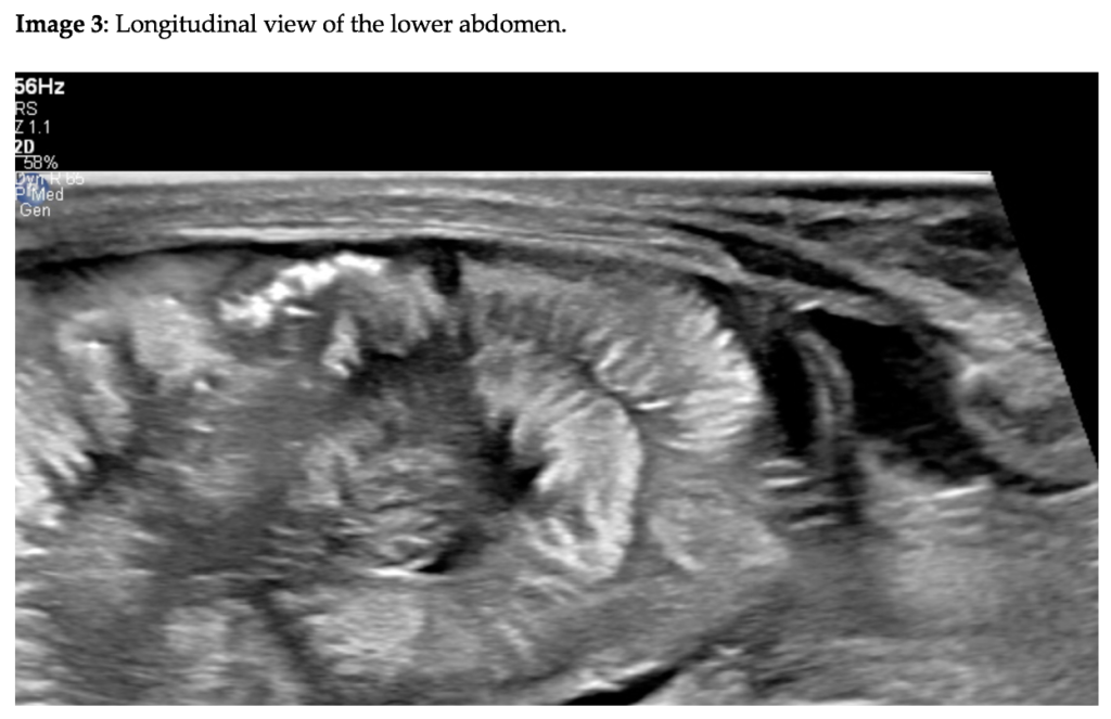
Image 1 and video 1 demonstrate thickened bowel loops and complex fluid throughout the abdomen. In video 1 and image 2, a bubble of free peritoneal gas is present anteriorly along the liver margin causing a strong reverberation artifact. In image 3, a cluster of echogenic foci representing gas is shown within the bowel wall, representing pneumatosis intestinalis. The overall appearance is that of necrotizing enterocolitis (NEC) with a perforation leading to the presence of intraperitoneal air.
NEC is a devastating intestinal disease of the neonate typically associated with prematurity. The condition is characterized by mucosal injury (due to severe prematurity, altered microbiome, food protein allergy, hypoxic injury and other causes) triggering a cascade of bacterial translocation (from the lumen into the bowel wall), acute inflammation, altered permeability, ischaemia, dysmotility, necrosis and perforation.
The clinical diagnosis of NEC is difficult. Abdominal X-ray and ultrasound aid in the identification of anatomical features of NEC. High-resolution ultrasound with the use of high-frequency ultra-broadband linear transducers provides unprecedented image resolution and allows the practitioner to distinguish minor gut changes in the inflammatory phase from more significant complications in the ischaemic phase and perforation. The typical features of NEC on ultrasound include bowel wall thickening, hyperemia (in the acute inflammatory phase), simple free fluid, bowel wall thinning (in the ischaemic phase), presence of gas within the bowel wall (pneumatosis), bowel dysmotility, portal venous gas, complex fluid and free peritoneal air (pneumoperitoneum). The presence of peritoneal air signals bowel perforation. It is important to keep in mind that more than one segment of the bowel may be affected by NEC and the involvement may vary in severity.
The neonate in this case study underwent laparotomy, confirming NEC with perforation.
Conflicts of Interest
The authors declare no conflict of interest
References
- Alexander KM, Chan SS, Opfer E, Cuna A, Fraser JD, Sharif S, et al. Implementation of bowel ultrasound practice for the diagnosis and management of necrotising enterocolitis. Archives of Disease in Childhood – Fetal and Neonatal Edition. 2021 Jan 1;106(1):96–103.
- Esposito F, Mamone R, Serafino MD, Mercogliano C, Vitale V, Vallone G, et al. Diagnostic imaging features of necrotizing enterocolitis: a narrative review. Quantitative Imaging in Medicine and Surgery. 2017 Jun;7(3):33644–33344.
- Chen S, Hu Y, Liu Q, Li X, Wang H, Wang K, et al. Application of abdominal sonography in diagnosis of infants with necrotizing enterocolitis. Medicine. 2019 Jul;98(28):e16202.
Incorrect
CORRECT ANSWER EXPLAINED BELOW Correct answer is: Necrotising enterocolitis with perforation
Discussion
Image 1 and video 1 demonstrate thickened bowel loops and complex fluid throughout the abdomen. In video 1 and image 2, a bubble of free peritoneal gas is present anteriorly along the liver margin causing a strong reverberation artifact. In image 3, a cluster of echogenic foci representing gas is shown within the bowel wall, representing pneumatosis intestinalis. The overall appearance is that of necrotizing enterocolitis (NEC) with a perforation leading to the presence of intraperitoneal air.
NEC is a devastating intestinal disease of the neonate typically associated with prematurity. The condition is characterized by mucosal injury (due to severe prematurity, altered microbiome, food protein allergy, hypoxic injury and other causes) triggering a cascade of bacterial translocation (from the lumen into the bowel wall), acute inflammation, altered permeability, ischaemia, dysmotility, necrosis and perforation.
The clinical diagnosis of NEC is difficult. Abdominal X-ray and ultrasound aid in the identification of anatomical features of NEC. High-resolution ultrasound with the use of high-frequency ultra-broadband linear transducers provides unprecedented image resolution and allows the practitioner to distinguish minor gut changes in the inflammatory phase from more significant complications in the ischaemic phase and perforation. The typical features of NEC on ultrasound include bowel wall thickening, hyperemia (in the acute inflammatory phase), simple free fluid, bowel wall thinning (in the ischaemic phase), presence of gas within the bowel wall (pneumatosis), bowel dysmotility, portal venous gas, complex fluid and free peritoneal air (pneumoperitoneum). The presence of peritoneal air signals bowel perforation. It is important to keep in mind that more than one segment of the bowel may be affected by NEC and the involvement may vary in severity.
The neonate in this case study underwent laparotomy, confirming NEC with perforation.
Conflicts of Interest
The authors declare no conflict of interest
References
- Alexander KM, Chan SS, Opfer E, Cuna A, Fraser JD, Sharif S, et al. Implementation of bowel ultrasound practice for the diagnosis and management of necrotising enterocolitis. Archives of Disease in Childhood – Fetal and Neonatal Edition. 2021 Jan 1;106(1):96–103.
- Esposito F, Mamone R, Serafino MD, Mercogliano C, Vitale V, Vallone G, et al. Diagnostic imaging features of necrotizing enterocolitis: a narrative review. Quantitative Imaging in Medicine and Surgery. 2017 Jun;7(3):33644–33344.
- Chen S, Hu Y, Liu Q, Li X, Wang H, Wang K, et al. Application of abdominal sonography in diagnosis of infants with necrotizing enterocolitis. Medicine. 2019 Jul;98(28):e16202.


