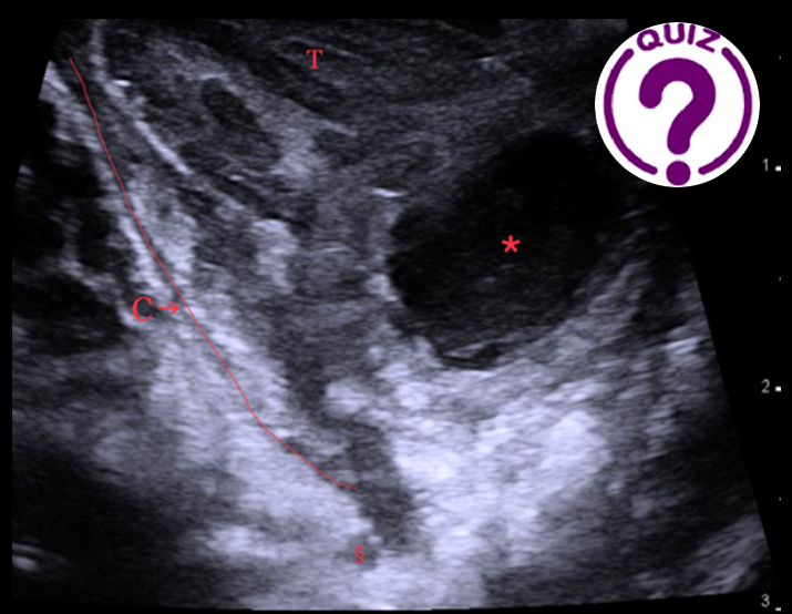
Correct answer is: Peritonsillar abscess
Discussion
A peritonsillar abscess (PTA) is a complication to an acute tonsillitis with accumulation of pus between the tonsil and the pharyngeal constrictor muscle. On transoral ultrasound, the image resembles an abscess in relation to the peritonsillar space (1).
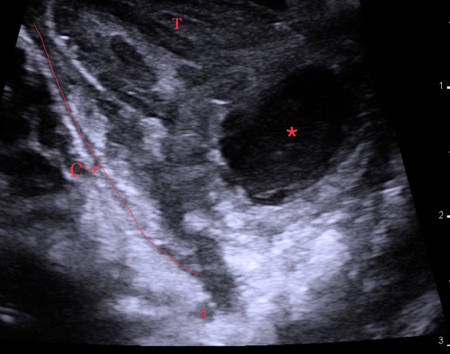
On the image shown, the abscess is easily recognized as an irregular hypoechoic area with posterior enhancement. By applying pressure to the abscess in pulses with the ultrasound probe, some fluctuation of the pus within the cavity may be seen.
In contrast to transcervical ultrasound, a transoral approach gives a much higher resolution and reduces the risk of air artefacts from the oral cavity because the probe is placed directly on the tonsil. Transoral ultrasound also allows for guided aspiration, but can be difficult due to the gag reflex and limited access due to trismus (2).
Acute tonsillitis with phlegmon of the peritonsillar tissue is the most common differential diagnosis to PTA. Tonsillitis is a common disease most commonly affecting children and young adults. This can progress to phlegmonous oedema of the peritonsillar tissue, mimicking PTA on appearance. On transoral ultrasonography, acute tonsillitis will reveal an enlarged inflamed tonsil with increased Doppler flow inside the tonsil. <<< See the image to the left of acute tonsillitis with increased flow on power doppler.
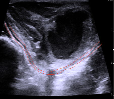
In case of phlegmon, a thickened submucosa surrounding the tonsil is seen. In contrast, a PTA requires a collection of pus located between the tonsil and pharyngeal constrictor muscle (see image above), and increased Doppler flow surrounding the lesion, but no Doppler flow inside the lesion (3).
Salivary retention cysts may present anywhere in the oral cavity and pharynx (e.g. from minor salivary glands). These may also be anechoic on ultrasound. They usually form slowly and without acute onset of pain and redness. Furthermore, the ultrasound appearance is usually with clearly defined margins. Cysts do not have increased Doppler flow surrounding the lesion, in contrast to a PTA (4).
A cancer in the tonsils may also present with pain and unilateral swelling. They usually present in older patients (40 years+). The swelling occurs more gradually and sub-acutely. There may be ulcers in the tonsillar tissue. On ultrasound, a tumour in the tonsil is hypoechoic, located within the tonsil and will have color flow inside the lesion (5).
Conflicts of Interest
The authors declare no conflict of interest
Ethics
The patient gave informed consent to publish this anonymized case.
References
- Todsen T, Stage MG, Holst Hahn C. A Novel Technique for Intraoral Ultrasound-Guided Aspiration of Peritonsillar Abscess. Diagnostics (Basel). 2018 Aug 2;8(3):50. doi: 10.3390/diagnostics8030050. PMID:30072648; PMCID: PMC6174334.
- Todsen T. Otorhinolaryngologist performed transcervical versus transoral ultrasonography in the management of peritonsillar abscess. Am J Otolaryngol. 2020 Oct 22:102768. doi: 10.1016/j.amjoto.2020.102768. Epub ahead of print. PMID: 33109415.
- Todsen T, Stage MG, Hahn CH. Surgeon-performed Transoral Ultrasound-Guided Aspiration of Peritonsillar Abscess. 2021 Mar 20. doi: 10.1002/lary.29525. Epub ahead of print. PMID: 33742694.
- Senthilkumar B, Mahabob MN. Mucocele: An unusual presentation of the minor salivary gland lesion. J Pharm Bioallied Sci. 2012 Aug;4(Suppl 2):S180-2. doi: 10.4103/0975-7406.100265. PMID:23066247; PMCID: PMC3467912.
- Fakhry C, Agrawal N, Califano J, Messing B, Liu J, Saunders J, Ha P, Coquia S, Hamper U, Gillison M, Blanco R. The use of ultrasound in the search for the primary site of unknown primary head and neck squamous cell cancers.
|