Case of the Month January 2022 – Superficial tumor in the abdominal wall
January 4, 2022
WFUMB / EFSUMB Students Webinar Series: 5 February 2022
February 8, 2022Holly Smith 1, Martin Necas 2 and Kara Prout3.
1 Waikato Hospital, Hamilton, New Zealand; h.milnesmith@gmail.com.
2 Waikato Hospital, Hamilton, New Zealand; martin@antegrade.net.
3 Waikato Hospital, Hamilton, New Zealand; kara.prout@waikatodhb.health.nz
* Correspondence: e-mail: h.milnesmith@gmail.com.
Clinical History:
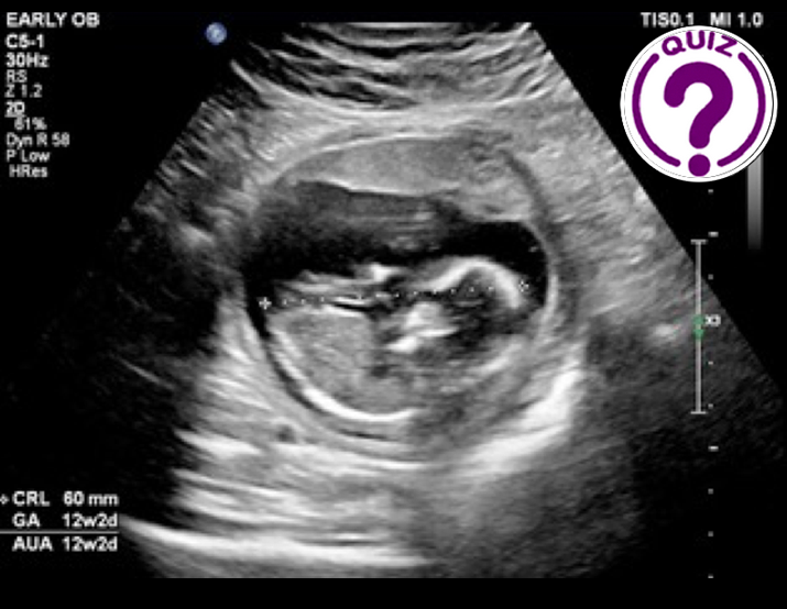
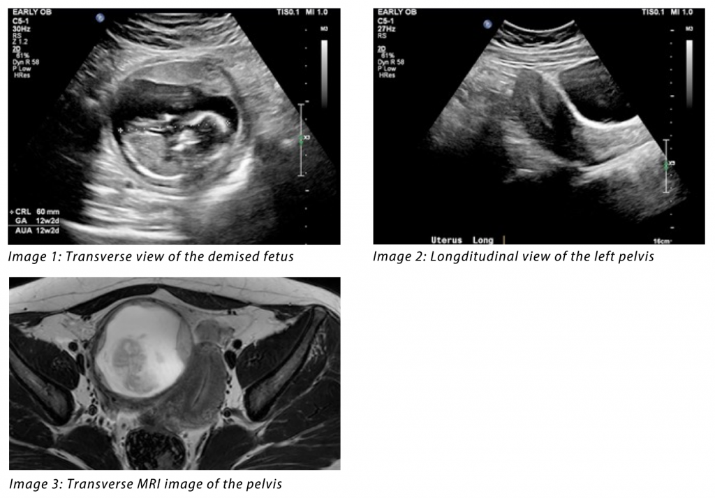
A 22-year-old woman, G2P0, presented for a nuchal translucency screening ultrasound. Unfortunately, the examination detected a non-viable fetus of 13 weeks’ gestation. The woman was subsequently given misoprostol to pass the products of conception, and experienced pain and cramping, however no tissue was passed. A repeat ultrasound was performed, followed by an MRI, which revealed the following images.
Video 1: Transverse sweep of the pelvis
Video 2: Transverse MRI sweep of the pelvis
Quiz-summary
0 of 1 questions completed
Questions:
- 1
Information
View the May Case below, answer the question and then click check >
You have already completed the quiz before. Hence you can not start it again.
Quiz is loading...
You must sign in or sign up to start the quiz.
You have to finish following quiz, to start this quiz:
Results
0 of 1 questions answered correctly
Your time:
Time has elapsed
You have reached 0 of 0 points, (0)
Categories
- Not categorized 0%
- 1
- Answered
- Review
-
Question 1 of 1
1. Question
Question: What is the most likely diagnosis?
Correct
CORRECT ANSWER EXPLAINED BELOW Correct answer is: Demised pregnancy in a rudimentary horn of a unicornuate uterus.
Discussion
Image 1 shows a gestational sac within the right horn of the uterus, with a non-viable fetus measuring 12w2d gestation. The placenta is visualised anteriorly, with a markedly thin myometrium surrounding the gestational sac. Image 2 demonstrates an empty left uterine horn, with a clearly visualised endometrium. In Video 1 we can see that there is no endometrial connection from the right uterine horn to the left horn, cervix, or vagina, and that the myometrium completely surrounds the gestational sac. These findings are suggestive of an ectopic pregnancy located within a non-communicating rudimentary horn of a unicornuate uterus.
An MRI was performed to confirm this diagnosis. Image 3 and Video 2 also show a gestational sac located within the right uterine horn, surrounded by myometrium. Again, there is no endometrial canal seen connecting the right horn to the left horn, cervix or vaginal canal. The diagnosis was confirmed, and the patient was subsequently referred for surgical excision of the pregnancy and the rudimentary horn.
Pregnancy within a rudimentary horn is extremely rare, with an incidence of 1 in 76,000-150,000 pregnancies (1). If undetected, 80-90% of these pregnancies will result in uterine wall rupture by the third trimester, due to the poorly developed myometrium (2,3). The sensitivity of ultrasound in the diagnosis of a rudimentary horn pregnancy has been reported as low as 26% (2). However, this should be considered as a differential diagnosis in ectopic pregnancies, cornual pregnancies, and intrauterine pregnancies in a bicornuate or didelphic uterus (3). Ultrasound criteria for the diagnosis of this condition include the following: appearances of asymmetrical bicornuate/didelphic uterus, absence of continuation of the endometrium of the pregnant horn to the contralateral horn, cervix or vagina, and myometrial tissue surrounding the gestational sac (1–3).
Conflicts of Interest
The authors declare no conflict of interest
References
- Moawad GN, Abi Khalil ED. A Case of Recurrent Rudimentary Horn Ectopic Pregnancies Managed by Methotrexate Therapy and Laparoscopic Excision of the Rudimentary Horn. Case Rep Obstet Gynecol. 2016;2016:5747524.
- Thurber BW, Fleischer AC. Ultrasound Features of Rudimentary Horn Ectopic Pregnancies. J Ultrasound Med. 2019;38(6):1643–7.
- Lai Y-J, Lin C-H, Hou W-C, Hwang K-S, Yu M-H, Su H-Y. Pregnancy in a Noncommunicating Rudimentary Horn of a Unicornuate Uterus: Prerupture Diagnosis and Management. Taiwan J Obstet Gynecol. 2016 Aug 1;55(4):604–6.
Incorrect
CORRECT ANSWER EXPLAINED BELOW Correct answer is: Demised pregnancy in a rudimentary horn of a unicornuate uterus.
Discussion
Image 1 shows a gestational sac within the right horn of the uterus, with a non-viable fetus measuring 12w2d gestation. The placenta is visualised anteriorly, with a markedly thin myometrium surrounding the gestational sac. Image 2 demonstrates an empty left uterine horn, with a clearly visualised endometrium. In Video 1 we can see that there is no endometrial connection from the right uterine horn to the left horn, cervix, or vagina, and that the myometrium completely surrounds the gestational sac. These findings are suggestive of an ectopic pregnancy located within a non-communicating rudimentary horn of a unicornuate uterus.
An MRI was performed to confirm this diagnosis. Image 3 and Video 2 also show a gestational sac located within the right uterine horn, surrounded by myometrium. Again, there is no endometrial canal seen connecting the right horn to the left horn, cervix or vaginal canal. The diagnosis was confirmed, and the patient was subsequently referred for surgical excision of the pregnancy and the rudimentary horn.
Pregnancy within a rudimentary horn is extremely rare, with an incidence of 1 in 76,000-150,000 pregnancies (1). If undetected, 80-90% of these pregnancies will result in uterine wall rupture by the third trimester, due to the poorly developed myometrium (2,3). The sensitivity of ultrasound in the diagnosis of a rudimentary horn pregnancy has been reported as low as 26% (2). However, this should be considered as a differential diagnosis in ectopic pregnancies, cornual pregnancies, and intrauterine pregnancies in a bicornuate or didelphic uterus (3). Ultrasound criteria for the diagnosis of this condition include the following: appearances of asymmetrical bicornuate/didelphic uterus, absence of continuation of the endometrium of the pregnant horn to the contralateral horn, cervix or vagina, and myometrial tissue surrounding the gestational sac (1–3).
Conflicts of Interest
The authors declare no conflict of interest
References
- Moawad GN, Abi Khalil ED. A Case of Recurrent Rudimentary Horn Ectopic Pregnancies Managed by Methotrexate Therapy and Laparoscopic Excision of the Rudimentary Horn. Case Rep Obstet Gynecol. 2016;2016:5747524.
- Thurber BW, Fleischer AC. Ultrasound Features of Rudimentary Horn Ectopic Pregnancies. J Ultrasound Med. 2019;38(6):1643–7.
- Lai Y-J, Lin C-H, Hou W-C, Hwang K-S, Yu M-H, Su H-Y. Pregnancy in a Noncommunicating Rudimentary Horn of a Unicornuate Uterus: Prerupture Diagnosis and Management. Taiwan J Obstet Gynecol. 2016 Aug 1;55(4):604–6.


