Atlas of Thyroid Ultrasound Images
May 30, 2022
Congress Committee FLAUSCristina Chammas
June 9, 2022Bezzina Atef, Lahmar Lilia, Moalla Salma, Douira-Khomsi Wièm *
Department of Paediatric radiology, Béchir Hamza Children’s Hospital, Tunis, Tunisia
* Correspondence: Khomsiwiem@yahoo.fr
Clinical History:
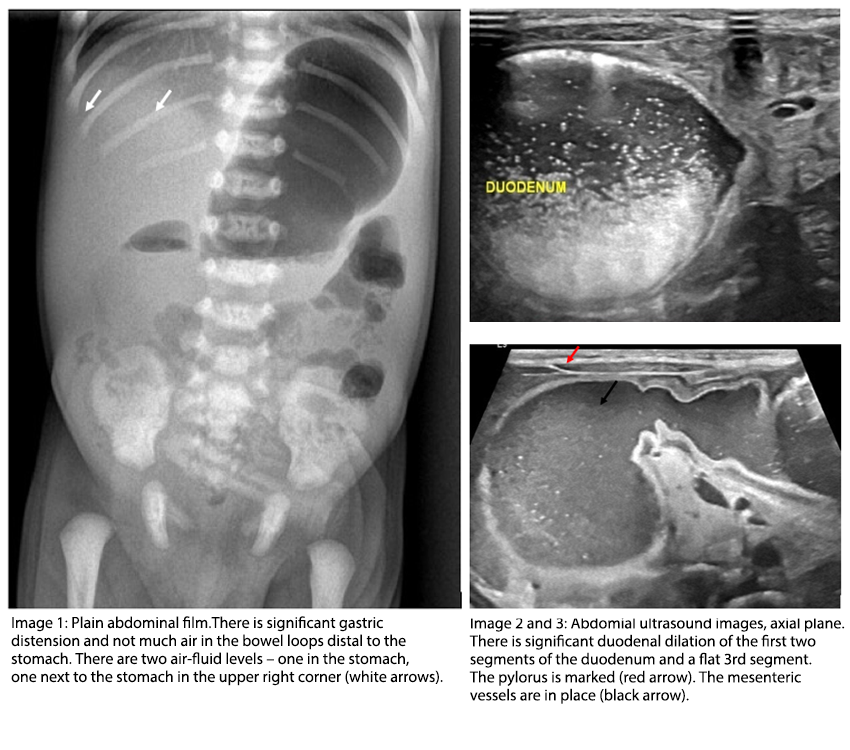
A 5-days-old boy born at term was admitted to our hospital after 3 days of alimentary and later biliary vomiting. There was no failure of passaging air and stool. The pregnancy had been normal and the Apgar Score at birth was 9/10. The meconium had passed within the first 24 hours. The abdomen was soft without distension on palpation. A plain abdominal film was obtained at admission as well as abdominal ultrasound.
Video 1-4: Abdominal ultrasound, axial plane
Quiz-summary
0 of 2 questions completed
Questions:
- 1
- 2
Information
View the June Case below, answer the question and then click check >
You have already completed the quiz before. Hence you can not start it again.
Quiz is loading...
You must sign in or sign up to start the quiz.
You have to finish following quiz, to start this quiz:
Results
0 of 2 questions answered correctly
Your time:
Time has elapsed
You have reached 0 of 0 points, (0)
Categories
- Not categorized 0%
- 1
- 2
- Answered
- Review
-
Question 1 of 2
1. Question
Question 1: Which diagnosis do you consider?
Correct
CORRECT ANSWER EXPLAINED BELOW Correct answer to Q1 is: Incomplete obstruction of the 2nd segment of duodenum
Incorrect
CORRECT ANSWER EXPLAINED BELOW Correct answer to Q1 is: Incomplete obstruction of the 2nd segment of duodenum
-
Question 2 of 2
2. Question
The mesenteric vessels are wellpositioned and there is air in the bowel loops in the upper left quadrant indicating an incomplete obstruction in the 2nd segment of duodenum. Gastroenteritis would not cause dilatation of the stomach especially not after excessive vomiting. There is no stenosis or enlargement of the stomach wall at the level of the pylorus. As the fluid and air filled structure is identified as the stomach, caecal volvulus is not likely.
Question 2: What would be your next step?
Correct
CORRECT ANSWER EXPLAINED BELOW Correct answer to Q2 is: Small bowel follow through.
Additional Images:
Discussion:
In a small bowel follow through examination the baby drinks water soluble oral contrast and fluoroscopy images are obtained consequtively. This examination shows dilated stomach and proximal duodenum, but slight, delayed passage. There is no stenosis at the level of the pylorus. The proximal jejunal bowel loops are located in the correct anatomical position (upper left quadrant). The examination indicates a partial obstruction (a web) in the 2nd part of the duodenum. The patient underwent surgery which confirmed the finding of a duodenal web.
Ultrasound is a non-invasive examination, which is readily available also in the acute setting. It is often a supplement to plain abdominal film in the acute pediatric setting. It is helpful to determine the level of obstruction and may indicate the cause.
Conclusion
Vomiting in neonates or small infants may indicate obstruction.
Conflicts of interest
“The authors declare no conflict of interest.”
References
- Weerakkody, Y., Glick, Y. Duodenal web. Reference article, Radiopaedia.org. (accessed on 16 May 2022) https://doi.org/10.53347/rID-7630
- Poddar U, Jain V, Yachha SK, Srivastava A. Congenital duodenal web: successful management with endoscopic dilatation. Endosc Int Open. 2016;4(3):E238-E241. doi:10.1055/s-0041-110955
Incorrect
CORRECT ANSWER EXPLAINED BELOW Correct answer to Q2 is: Small bowel follow through.
Additional Images:
Discussion:
In a small bowel follow through examination the baby drinks water soluble oral contrast and fluoroscopy images are obtained consequtively. This examination shows dilated stomach and proximal duodenum, but slight, delayed passage. There is no stenosis at the level of the pylorus. The proximal jejunal bowel loops are located in the correct anatomical position (upper left quadrant). The examination indicates a partial obstruction (a web) in the 2nd part of the duodenum. The patient underwent surgery which confirmed the finding of a duodenal web.
Ultrasound is a non-invasive examination, which is readily available also in the acute setting. It is often a supplement to plain abdominal film in the acute pediatric setting. It is helpful to determine the level of obstruction and may indicate the cause.
Conclusion
Vomiting in neonates or small infants may indicate obstruction.
Conflicts of interest
“The authors declare no conflict of interest.”
References
- Weerakkody, Y., Glick, Y. Duodenal web. Reference article, Radiopaedia.org. (accessed on 16 May 2022) https://doi.org/10.53347/rID-7630
- Poddar U, Jain V, Yachha SK, Srivastava A. Congenital duodenal web: successful management with endoscopic dilatation. Endosc Int Open. 2016;4(3):E238-E241. doi:10.1055/s-0041-110955


