E-learning & Student Education Task Force ASUMAlison Deslandes
June 16, 2022
26th Workshop of WFUMB COE ~ Bangladesh
July 5, 2022Shakiba Mosadegh(1),*, Jonathan Cohen(1)
1. Department of Radiology, Copenhagen University Hospital, Rigshospitalet, Copenhagen, Denmark; shakibamosadegh@gmail.com
* Correspondence: shakibamosadegh@gmail.com
Quiz-summary
0 of 3 questions completed
Questions:
- 1
- 2
- 3
Information
View the July Case below, answer the question and then click check >
You have already completed the quiz before. Hence you can not start it again.
Quiz is loading...
You must sign in or sign up to start the quiz.
You have to finish following quiz, to start this quiz:
Results
0 of 3 questions answered correctly
Your time:
Time has elapsed
You have reached 0 of 0 points, (0)
Categories
- Not categorized 0%
- 1
- 2
- 3
- Answered
- Review
-
Question 1 of 3
1. Question
Clinical History:
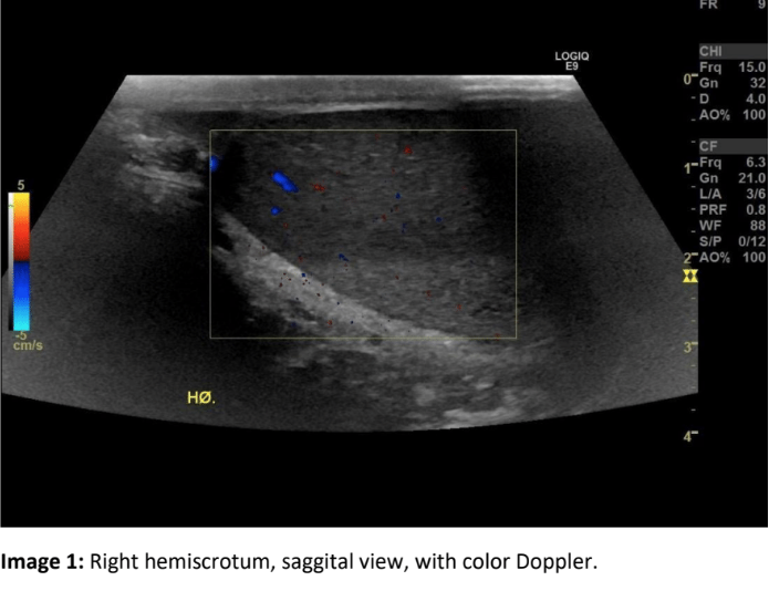
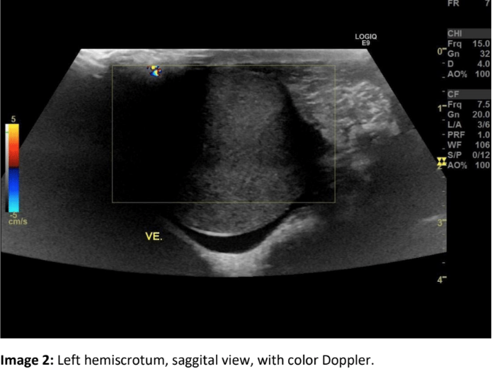
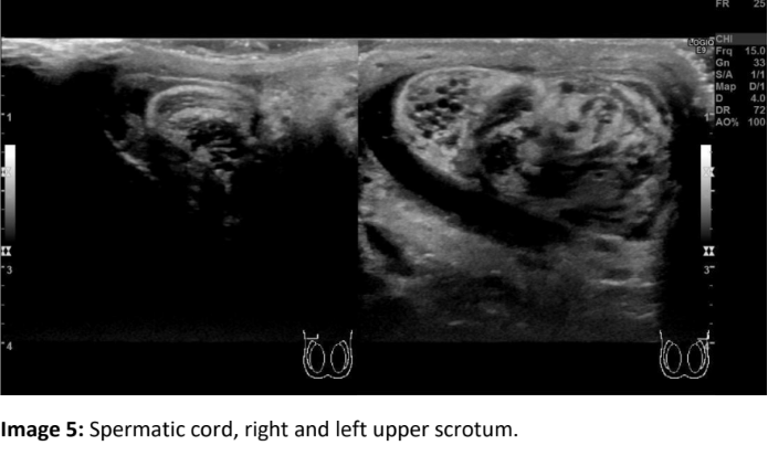
A 14-year-old boy with no former medical or surgical history presented to the Emergency Department with sudden onset severe testicular pain on the left side, radiating to the groin and lower abdomen. He had gastrointestinal upset with nausea. No history of trauma. Physical examination revealed a firm, swollen left hemiscrotum. Ultrasound (US) examination was performed.
Question 1: Which of the following diagnoses needs to be considered first?
Correct
CORRECT ANSWER EXPLAINED BELOW Correct answer to Q1 is:Testicular torsion
Incorrect
CORRECT ANSWER EXPLAINED BELOW Correct answer to Q1 is:Testicular torsion
-
Question 2 of 3
2. Question
Additional history and images:
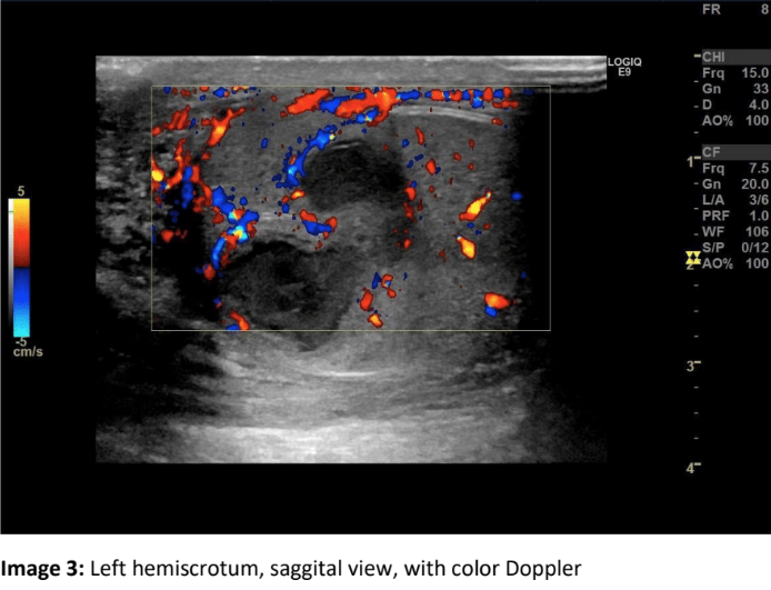
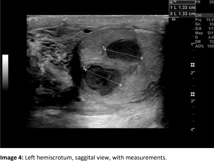
Ultrasound images revealed left-sided testicular torsion with a raised left testis with no apparent blood flow. The spermatic cord was visibly twisted. Surgery confirmed torsion of the testis and spermatic cord. Bilateral orchiopexy was performed with no immediate complications. After one month, the patient was brought to the emergency department again because of testicular pain. Physical examination revealed a warm, erythematous, swollen scrotum on the left side, which was tender to palpation. US was performed again.
Question 2: What is the most likely diagnosis?
Correct
CORRECT ANSWER EXPLAINED BELOW Correct answer to Q2 is: Testicular abscesses
Incorrect
CORRECT ANSWER EXPLAINED BELOW Correct answer to Q2 is: Testicular abscesses
-
Question 3 of 3
3. Question
Question 3: What is the most common cause of testicular abscess?
Correct
CORRECT ANSWER EXPLAINED BELOW Correct answer to Q3 is: Untreated or severe epididymo-orchitis
Discussion:
The acute scrotum is a syndrome characterised by acute onset painful hemiscrotumwhich can be accompanied by abdominal pain, fever or gastrointestinal upset such as nausea or vomiting. It is most commonly caused by torsion of the testis, torsion of epididymal appendage, epididymitis or epididymo-orchitis.Patients presenting with sudden painful hemiscrotum with vomiting is typical for testicular torsion or torsion of epididymal appendage.Duration of symptoms is shorter in testicular torsion (69% present within twelve hours) and torsion of the appendix testes (62%) compared to epididymitis (31%). In the early phase, location of the pain can lead to diagnosis. Patients with acute epididymitis experience a tender epididymis, whereas patients with testicular torsion are more likely to have a tender testis, and patients with torsion of the appendix testis feel isolated tenderness of the superior pole of the testis (1).Apart from pain and hemiscrotalswelling, physical examination in patients with testicular torsion may reveal elevation of the scrotum without cremasteric reflex, and a testis sometimes in horizontal lie. The affected testis may be larger than the asymptomatic testis.Doppler US is the modality of choice for evaluating testicular torsion. Ultrasound of the testis may demonstrate that the affected testis is enlarged, heterogeneous in echotexture, and without evidence of any blood flow. It is important to compare the affected testis with the normal side (2). Treatment is bilateral orchiopexy.An important differential diagnosis is epididymitis, which is inflammation of the epididymis and may be extended to the testis, in which case it is called epididymo-orchitis.Epididymitis and epididymo-orchitis are usually caused by a bacterial infection, often due to surgery, bladder catheter, urinary tract infection or sexually transmitted disease. The clinical spectrum ranges from mild tenderness to a severe febrile process with acute unilateral scrotal pain. US often shows that epididymis is swollen and heterogeneous, with hydrocele and scrotal wall thickening. Doppler US may demonstrate hyperaemia.A complication of severe epididymo-orchitis is abscess formation. Doppler ultrasound shows a focal hypoechoic area with a surrounding hyperaemic rim. Treatment may include prolonged antibiotics, surgical debridement or even orchiectomy (3).Conclusion
In conclusion, US is an important imaging tool in patients with acute scrotal pain.
Conflicts of interest
“The authors declare no conflict of interest.”
References
- C. Radmayr (Chair), G. Bogaert, H.S. Dogan, R. Koc ̆vara, J.M. Nijman (Vice-chair), R. Stein, S. Tekgül Guidelines Associates: L.A. ‘t Hoen, J. Quaedackers, M.S. Silay, S. Undre. EAU Guidelines on Paediatric Urology. https://uroweb.org/wp-content/uploads/EAU-Guidelines-on-Paediatric-Urology-2019.pdf
- Testicular torsion| Radiology Reference Article | Radiopaedia.org. Available online: https://radiopaedia.org/articles/testicular-torsion?lang=us (accessed on 18.01.2022).
- Testicular abscess| Radiology Reference Article | Radiopaedia.org. Available online: https://radiopaedia.org/articles/testicular-abscess (accessed 07. 05.2018)
Incorrect
CORRECT ANSWER EXPLAINED BELOW Correct answer to Q3 is: Untreated or severe epididymo-orchitis
Discussion:
The acute scrotum is a syndrome characterised by acute onset painful hemiscrotumwhich can be accompanied by abdominal pain, fever or gastrointestinal upset such as nausea or vomiting. It is most commonly caused by torsion of the testis, torsion of epididymal appendage, epididymitis or epididymo-orchitis.Patients presenting with sudden painful hemiscrotum with vomiting is typical for testicular torsion or torsion of epididymal appendage.Duration of symptoms is shorter in testicular torsion (69% present within twelve hours) and torsion of the appendix testes (62%) compared to epididymitis (31%). In the early phase, location of the pain can lead to diagnosis. Patients with acute epididymitis experience a tender epididymis, whereas patients with testicular torsion are more likely to have a tender testis, and patients with torsion of the appendix testis feel isolated tenderness of the superior pole of the testis (1).Apart from pain and hemiscrotalswelling, physical examination in patients with testicular torsion may reveal elevation of the scrotum without cremasteric reflex, and a testis sometimes in horizontal lie. The affected testis may be larger than the asymptomatic testis.Doppler US is the modality of choice for evaluating testicular torsion. Ultrasound of the testis may demonstrate that the affected testis is enlarged, heterogeneous in echotexture, and without evidence of any blood flow. It is important to compare the affected testis with the normal side (2). Treatment is bilateral orchiopexy.An important differential diagnosis is epididymitis, which is inflammation of the epididymis and may be extended to the testis, in which case it is called epididymo-orchitis.Epididymitis and epididymo-orchitis are usually caused by a bacterial infection, often due to surgery, bladder catheter, urinary tract infection or sexually transmitted disease. The clinical spectrum ranges from mild tenderness to a severe febrile process with acute unilateral scrotal pain. US often shows that epididymis is swollen and heterogeneous, with hydrocele and scrotal wall thickening. Doppler US may demonstrate hyperaemia.A complication of severe epididymo-orchitis is abscess formation. Doppler ultrasound shows a focal hypoechoic area with a surrounding hyperaemic rim. Treatment may include prolonged antibiotics, surgical debridement or even orchiectomy (3).Conclusion
In conclusion, US is an important imaging tool in patients with acute scrotal pain.
Conflicts of interest
“The authors declare no conflict of interest.”
References
- C. Radmayr (Chair), G. Bogaert, H.S. Dogan, R. Koc ̆vara, J.M. Nijman (Vice-chair), R. Stein, S. Tekgül Guidelines Associates: L.A. ‘t Hoen, J. Quaedackers, M.S. Silay, S. Undre. EAU Guidelines on Paediatric Urology. https://uroweb.org/wp-content/uploads/EAU-Guidelines-on-Paediatric-Urology-2019.pdf
- Testicular torsion| Radiology Reference Article | Radiopaedia.org. Available online: https://radiopaedia.org/articles/testicular-torsion?lang=us (accessed on 18.01.2022).
- Testicular abscess| Radiology Reference Article | Radiopaedia.org. Available online: https://radiopaedia.org/articles/testicular-abscess (accessed 07. 05.2018)


