Echoes Issue No. 28 [July 2022]
September 30, 2022
Epicondyle pain ultrasound ~ Fernando Jimenez (Spain)
October 19, 2022Won Jae Lee
Department of Radiology, Samsung Medical Center, Sungkyunkwan University School of Medicine, Seoul, Korea; wjlee@skku.du
* Correspondence: wjlee@skku.edu
Clinical history
A 42-year-old man was referred to my hospital for further evaluation of an incidentally found hepatic tumor during a routine check at another hospital. He was previously healthy, except a history of surgery for duodenal perforation. His laboratory findings were all in normal ranges including liver function tests and tumor markers.
Contrast enhanced ultrasound (CEUS) was performed.
Images / Videos

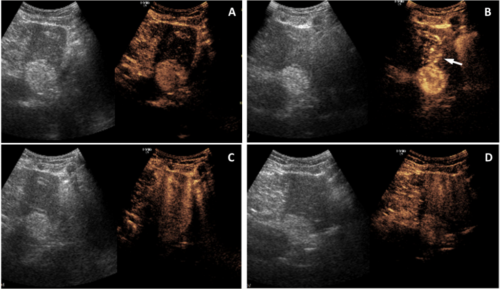
Image 1: Sonazoid-enhanced ultrasound images of an incidentally found hepatic tumor in the lateral segment. A 3.7 cm, hyperechoic lesion on the pre-contrast image (A) shows the same hyperechoic mass with heterogeneous enhancement in the arterial phase (B), which turns isoechoic in the late phase (C) due to surrounding parenchymal enhancement, and finally hyperechoic in the post-vascular phase (D) due to decreased enhancement of the liver parenchyma. Note the early draining vein (arrow).
Video 1: The arterial phase video of the same tumor as Image 1 obtained around 20 seconds after the contrast injection shows a hyperechoic lesion with heterogeneous enhancement. Early draining venous flow from the lesion is noted during the arterial phase.
Video 2: The late phase video of the same tumor as Image 1 obtained around 3 minutes after the contrast injection shows that the same lesion becomes isoechoic due to surrounding parenchymal enhancement.
Quiz-summary
0 of 2 questions completed
Questions:
- 1
- 2
Information
View the October Case below, answer the question and then click check >
You have already completed the quiz before. Hence you can not start it again.
Quiz is loading...
You must sign in or sign up to start the quiz.
You have to finish following quiz, to start this quiz:
Results
0 of 2 questions answered correctly
Your time:
Time has elapsed
You have reached 0 of 0 points, (0)
Categories
- Not categorized 0%
- 1
- 2
- Answered
- Review
-
Question 1 of 2
1. Question
Question 1: Which of the following diagnoses needs to be considered first?
Correct
CORRECT ANSWER EXPLAINED BELOW Correct answer to Q1 is: Angiomyolipoma
Discussion
The patient later underwent laparoscopic lateral segmentectomy for this tumor, and pathological analyses revealed the tumor to be an angiomyolipoma.
Typical ultrasound findings of hepatic angiomyolipoma are homogeneous and strong hyperechogenicity in most cases (1). Therefore, angiomyolipoma should be included in the differential diagnosis of hyperechoic hepatic tumors. Contrast-enhanced ultrasound findings show hyperenhancement in the arterial phase, and contrast defect in the post-vascular phase, which may resemble hypervascular malignancies such as hepatocellular carcinoma (2). Angiomyolipoma can be isoechoic during the late phase, which may be a differential point from hepatocellular carcinoma, which often exhibit late-phase washout. Early draining vein shown in image B is also an important differential point from other hyperechoic hepatic tumors, which will be discussed later.
Incorrect
CORRECT ANSWER EXPLAINED BELOW Correct answer to Q1 is: Angiomyolipoma
Discussion
The patient later underwent laparoscopic lateral segmentectomy for this tumor, and pathological analyses revealed the tumor to be an angiomyolipoma.
Typical ultrasound findings of hepatic angiomyolipoma are homogeneous and strong hyperechogenicity in most cases (1). Therefore, angiomyolipoma should be included in the differential diagnosis of hyperechoic hepatic tumors. Contrast-enhanced ultrasound findings show hyperenhancement in the arterial phase, and contrast defect in the post-vascular phase, which may resemble hypervascular malignancies such as hepatocellular carcinoma (2). Angiomyolipoma can be isoechoic during the late phase, which may be a differential point from hepatocellular carcinoma, which often exhibit late-phase washout. Early draining vein shown in image B is also an important differential point from other hyperechoic hepatic tumors, which will be discussed later.
-
Question 2 of 2
2. Question
Additional Images
Image 2. Three-phase CT images of another incidentally found hepatic tumor in segment 5. The arterial phase images (A and D) show a 4.8 cm, hyperenhancing lesion with tortuous vascularity in the segment 5. Note the early draining portal vein (arrow) from the lesion. The portal and equilibrium phase images (B and C) show the same hypoenhancing lesion, i.e., delayed with washout.
Question 2: What is the most likely diagnosis?
Correct
CORRECT ANSWER EXPLAINED BELOW Correct answer to Q2 is: Fat-deficient angiomyolipoma
Additional discussion
Three-phase CT findings of hepatic angiomyolipoma show arterial hyperenhancement and delayed washout. Therefore it is difficult to differentiate angiomyolipoma from hypervascular malignancies such as hepatocellular carcinoma, especially when it is fat-deficient and the patient has chronic liver disease. Some additional CT findings are helpful and can be summarized as central and peripheral distribution of tortuous vascularity and early draining vein connected with the portal vein and hepatic vein (3).
Conclusion
As mentioned above, detailed knowledge on ultrasound and CT findings of hepatic angiomyolipoma are important in the differentiantion from hyperechoic and hypervascular hepatic tumors, especially from hepatocellular carcinoma.
Conflicts of Interest:
No conflict of interest.
References
- Dietrich CF, Nolsoe CP, et al. Guidelines and good clinical practice recommendations for contrast-enhanced ultrasound (CEUS) in the liver – update 2020 WFUMB in cooperation with EFSUMB, AFSUMB, AIUM, and FLAUS. Ultrasound Med Biol. 2020;46(10):2579-2604.
- Lee JY, Minami Y, et al. The AFSUMB consensus statements and recommendations for the clinical practice of contrast-enhanced ultrasound using Sonazoid. Ultrasonography. 2020;39:191-220.
- Jeon TY, Kim SH, et al. Assessment of triple-phase CT findings for the differentiation of fat-deficient hepatic angiomyolipoma from hepatocellular carcinoma in non-cirrhotic liver. Eur J Radiol. 2010;73(3):601-606.
Incorrect
CORRECT ANSWER EXPLAINED BELOW Correct answer to Q2 is: Fat-deficient angiomyolipoma
Additional discussion
Three-phase CT findings of hepatic angiomyolipoma show arterial hyperenhancement and delayed washout. Therefore it is difficult to differentiate angiomyolipoma from hypervascular malignancies such as hepatocellular carcinoma, especially when it is fat-deficient and the patient has chronic liver disease. Some additional CT findings are helpful and can be summarized as central and peripheral distribution of tortuous vascularity and early draining vein connected with the portal vein and hepatic vein (3).
Conclusion
As mentioned above, detailed knowledge on ultrasound and CT findings of hepatic angiomyolipoma are important in the differentiantion from hyperechoic and hypervascular hepatic tumors, especially from hepatocellular carcinoma.
Conflicts of Interest:
No conflict of interest.
References
- Dietrich CF, Nolsoe CP, et al. Guidelines and good clinical practice recommendations for contrast-enhanced ultrasound (CEUS) in the liver – update 2020 WFUMB in cooperation with EFSUMB, AFSUMB, AIUM, and FLAUS. Ultrasound Med Biol. 2020;46(10):2579-2604.
- Lee JY, Minami Y, et al. The AFSUMB consensus statements and recommendations for the clinical practice of contrast-enhanced ultrasound using Sonazoid. Ultrasonography. 2020;39:191-220.
- Jeon TY, Kim SH, et al. Assessment of triple-phase CT findings for the differentiation of fat-deficient hepatic angiomyolipoma from hepatocellular carcinoma in non-cirrhotic liver. Eur J Radiol. 2010;73(3):601-606.


