MV-Flow imaging: New Doppler technology to investigate breast tumors ~ Bao Quan Nguyen Phuoc (Vietnam)
November 3, 2022
WFUMB / EFSUMB Students Webinar Series: 26 November 2022
November 28, 2022Susan Campbell Westerway
1 Womens Imaging Group – NSW, Australia scwus@hotmail.com
* Correspondence: scwus@hotmail.com
Clinical history
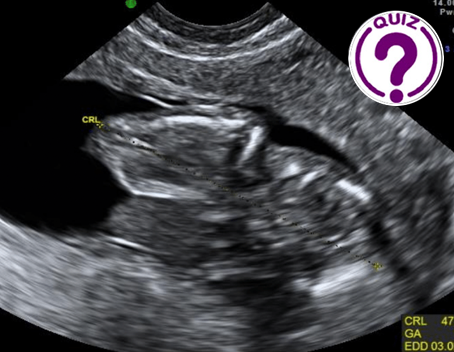
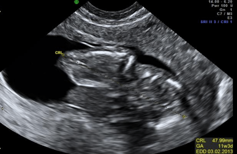
25 yr old G2P1 for routine 1st trimester ultrasound at 12 weeks 3 days to confirm dates of pregnancy by measuring the crown rump length (CRL) and head circumference (HC). There was a one week discrepancy between the CRL & HC (images 1,2).
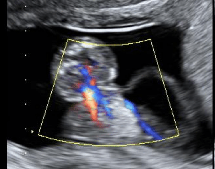
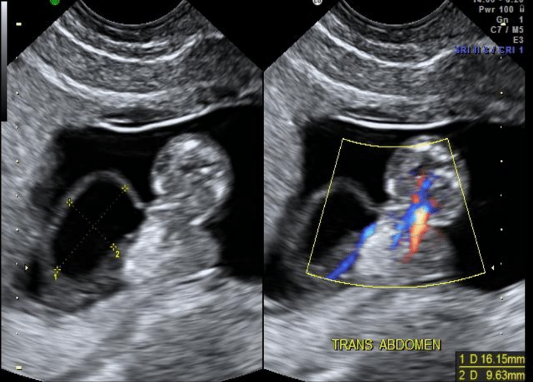
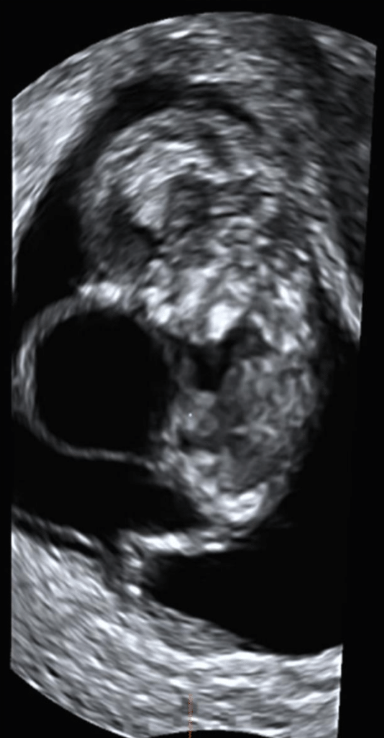
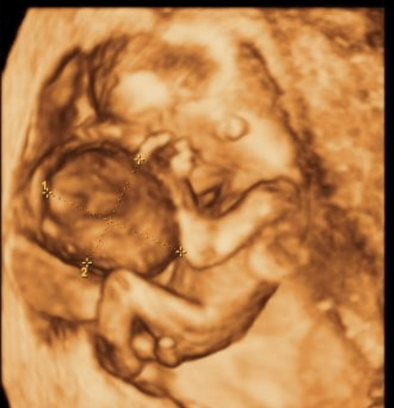
Further scanning revealed a midline anterior abdominal wall defect, a herniated sac with visceral contents (image 3). The abdominal wall defect was attached to the posterior uterine wall with a separate large cyst on the lateral segment of the defect (image 4).
Images / Videos
Quiz-summary
0 of 1 questions completed
Questions:
- 1
Information
View the Second November Case below, answer the question and then click check >
You have already completed the quiz before. Hence you can not start it again.
Quiz is loading...
You must sign in or sign up to start the quiz.
You have to finish following quiz, to start this quiz:
Results
0 of 1 questions answered correctly
Your time:
Time has elapsed
You have reached 0 of 0 points, (0)
Categories
- Not categorized 0%
- 1
- Answered
- Review
-
Question 1 of 1
1. Question
Question: What could this abdominal wall defect represent?
Correct
CORRECT ANSWER EXPLAINED BELOW Correct answer is: Limb body wall variant
Discussion
This case highlights the importance of scanning both in sagittal & transverse image planes.The abdominal wall defect and cyst cannot be appreciated in the long axis view of the fetus. The addition of colour Doppler imaging in the transverse plane confirms the herniated sac with visceral content in the defect which is consistent with an omphalocele. However, the adherence to the placenta changes the diagnosis to a limb body wall variant.
Limb body wall complex (LBWC), or body stalk syndrome is characterized by abdominoschisis, limb defects, neural tube defects, possible facial clefts and either a placentocranial or placentoabdominal adhesion. LBWC tends to occur between the 3rd – 6th week of embryonic development. With an incidence of ~ 1:14000 (1) there is a raised maternal alpha foeto protein (MAFP) and although the karyotype is usually normal, the affected fetus is not compatible with life (2,3).
Additional Discussion
The alternative diagnoses were:
- Gastroschisis is when bowel loops herniate through an abdominal wall defect located lateral & right of the umbilical cord insertion. On ultrasound it appears as herniated bowel loops floating in amniotic fluid.
- An omphalocele is a midline anterior abdominal wall defect, herniated sac with visceral contents and umbilical cord insertion at apex of sac.
- TRAP – Twin Reversed Arterial Perfusion Sequence is the most severe case of twin-to-twin transfusion syndrome. With an incidence of < 1% of monozygotic / monochorionic twins there is an acardiac twin with absent / mishaped upper body/limbs. With colour Doppler imaging there is flow from the normal (pump) twin to acadiac. The perfusion of the acardiac twin is via umbilical artery to artery and vein to vein anastomoses between twins leading to an inbalance of fetal circulation resulting in reversed flow in the umbilical artery of acardiac twin. The pump twin is at risk of polyhydramnios, hydrops fetalis, congestive cardiac failure with 50% having a chromosomal abnormality.
Conclusion
The key learning points in this case were:
- Multiple imaging planes are essential when scanning any pregnancy
- The addition of colour Doppler imaging assists in defining the diagnosis
- As LBWC is not compatible with life, early diagnosis will assist with management
Conflicts of Interest
The authors declare no conflict of interest
References
- Mann L, Ferguson-Smith MA, Desai M, Gibson AA, Raine PA. Prenatal assessment of anterior abdominal wall defects and their prognosis. Prenat Diagn 1984;4:427-35.
- Van Allen MI, Curry C, Gallagher L. Limb body wall complex: I. Pathogenesis. Am J Med Genet 1987;28:529-48.
- Okposio MM, Ugwueke TN, Alekwe L. Limb body wall complex: Case report and review of literature. Trop J Med Res 2015;18:113-5.
Incorrect
CORRECT ANSWER EXPLAINED BELOW Correct answer is: Limb body wall variant
Discussion
This case highlights the importance of scanning both in sagittal & transverse image planes.The abdominal wall defect and cyst cannot be appreciated in the long axis view of the fetus. The addition of colour Doppler imaging in the transverse plane confirms the herniated sac with visceral content in the defect which is consistent with an omphalocele. However, the adherence to the placenta changes the diagnosis to a limb body wall variant.
Limb body wall complex (LBWC), or body stalk syndrome is characterized by abdominoschisis, limb defects, neural tube defects, possible facial clefts and either a placentocranial or placentoabdominal adhesion. LBWC tends to occur between the 3rd – 6th week of embryonic development. With an incidence of ~ 1:14000 (1) there is a raised maternal alpha foeto protein (MAFP) and although the karyotype is usually normal, the affected fetus is not compatible with life (2,3).
Additional Discussion
The alternative diagnoses were:
- Gastroschisis is when bowel loops herniate through an abdominal wall defect located lateral & right of the umbilical cord insertion. On ultrasound it appears as herniated bowel loops floating in amniotic fluid.
- An omphalocele is a midline anterior abdominal wall defect, herniated sac with visceral contents and umbilical cord insertion at apex of sac.
- TRAP – Twin Reversed Arterial Perfusion Sequence is the most severe case of twin-to-twin transfusion syndrome. With an incidence of < 1% of monozygotic / monochorionic twins there is an acardiac twin with absent / mishaped upper body/limbs. With colour Doppler imaging there is flow from the normal (pump) twin to acadiac. The perfusion of the acardiac twin is via umbilical artery to artery and vein to vein anastomoses between twins leading to an inbalance of fetal circulation resulting in reversed flow in the umbilical artery of acardiac twin. The pump twin is at risk of polyhydramnios, hydrops fetalis, congestive cardiac failure with 50% having a chromosomal abnormality.
Conclusion
The key learning points in this case were:
- Multiple imaging planes are essential when scanning any pregnancy
- The addition of colour Doppler imaging assists in defining the diagnosis
- As LBWC is not compatible with life, early diagnosis will assist with management
Conflicts of Interest
The authors declare no conflict of interest
References
- Mann L, Ferguson-Smith MA, Desai M, Gibson AA, Raine PA. Prenatal assessment of anterior abdominal wall defects and their prognosis. Prenat Diagn 1984;4:427-35.
- Van Allen MI, Curry C, Gallagher L. Limb body wall complex: I. Pathogenesis. Am J Med Genet 1987;28:529-48.
- Okposio MM, Ugwueke TN, Alekwe L. Limb body wall complex: Case report and review of literature. Trop J Med Res 2015;18:113-5.

