Echoes Issue No. 30 [December 2022]
March 31, 2023
Real-time ultrasound in jaundice – tips and tricks ~ Adrian Saftoiu (Romania)
April 19, 2023Stian Solumsmoen 1, Ditte Dencker 1 and Mia Ostergaard 1
1 Dept. of Radiology, Copenhagen University Hospital – Rigshospitalet, Denmark
* Correspondence: stian.kinder.solumsmoen@regionh.dk
Clinical history
A 48-year-old female who recently underwent liver resection due a Klatskin type 3A tumour was referred to our department for an ultrasound-guided pleural drainage. The tumor had invaded and occluded the entire right portal vein, both the right liver vein and artery, the right intrahepatic ducts, the left hepatic duct and the proximal common hepatic duct. After resection, segments 2 and 3 were left, and a hepaticojejunal anastomosis was established. The patient had subsequent leakage from the hepaticojejunal anastomosis, and developed pleural effusion on the right side two months after primary surgery.
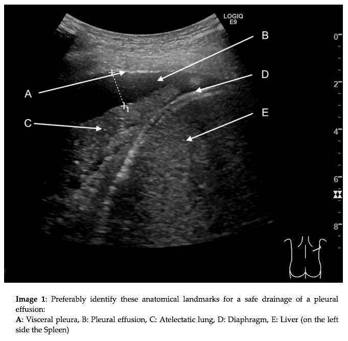
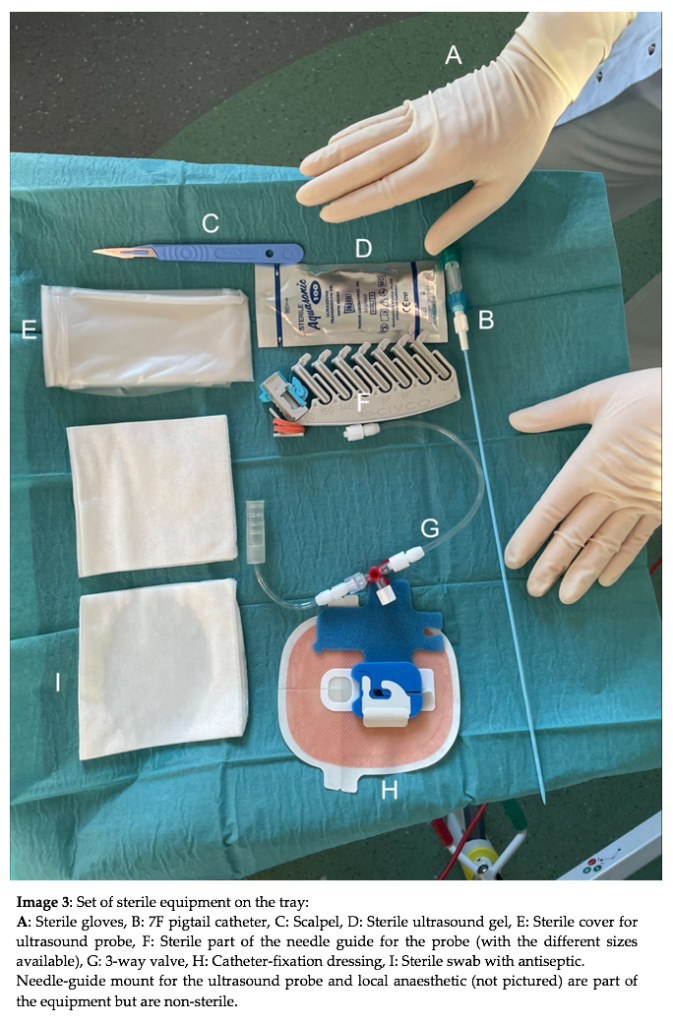
This case will provide an overview of the anatomical structures used to identify and plan your procedure as well as the equipment needed to perform an ultrasound guided drainage of a pleural effusion.
Video 1: Showing the penetration of the chest wall and visceral pleura by 7F pigtail catheter in the upper right corner of the video along the dotted needle-guide trajectory.
Quiz-summary
0 of 1 questions completed
Questions:
- 1
Information
View the April Case below, answer the question and then click check >
You have already completed the quiz before. Hence you can not start it again.
Quiz is loading...
You must sign in or sign up to start the quiz.
You have to finish following quiz, to start this quiz:
Results
0 of 1 questions answered correctly
Your time:
Time has elapsed
You have reached 0 of 0 points, (0)
Categories
- Not categorized 0%
- 1
- Answered
- Review
-
Question 1 of 1
1. Question
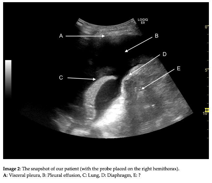
Question: What is the arrow “E” pointing at in image 2?
Correct
CORRECT ANSWER EXPLAINED BELOW Correct answer is: Bowel
Discussion
As mentioned in the clinical history the patient underwent a liver resection leaving only segment 2 and 3. This created a potential space to be filled. The primary retroperitoneal organs such as the kidneys are immobile and would therefore not be able to move and fill this potential space. Secondary retroperitoneal organs such as the pancreas (where the head and the body are retroperitoneal but not the tail) are also less mobile. This leaves the intraperitoneal organs, which are mobile within the peritoneum, such as the small bowel or the transverse colon, as the most obvious structures to fill the potential space.
Conclusion
Knowing your anatomical landmarks and familiarizing yourself with your equipment are the two key points in performing a successful ultrasound guided drainage of a pleural effusion.
Conflicts of Interest
The authors declare no conflict of interest
Incorrect
CORRECT ANSWER EXPLAINED BELOW Correct answer is: Bowel
Discussion
As mentioned in the clinical history the patient underwent a liver resection leaving only segment 2 and 3. This created a potential space to be filled. The primary retroperitoneal organs such as the kidneys are immobile and would therefore not be able to move and fill this potential space. Secondary retroperitoneal organs such as the pancreas (where the head and the body are retroperitoneal but not the tail) are also less mobile. This leaves the intraperitoneal organs, which are mobile within the peritoneum, such as the small bowel or the transverse colon, as the most obvious structures to fill the potential space.
Conclusion
Knowing your anatomical landmarks and familiarizing yourself with your equipment are the two key points in performing a successful ultrasound guided drainage of a pleural effusion.
Conflicts of Interest
The authors declare no conflict of interest


