Case of the Month August 2023 – Importance of contrast enhanced ultrasound in assessment of the uterus

Case of the Month July 2023 – Postprandial vomiting in a newborn
July 4, 2023
Case of the Month September 2023 – Flow in a AAA sac following EVAR. Is it an endoleak?
September 1, 2023Nidhi Leekha1, * and Dayib Buthul1
1 Department of Radiology & Imaging, Aga Khan University Hospital Nairobi; nidhi.leekha@aku.edu
1 Department of Radiology & Imaging, Aga Khan University Hospital Nairobi; dayib.buthul@aku.edu
* Correspondence: nidhi.leekha@aku.edu
Clinical history
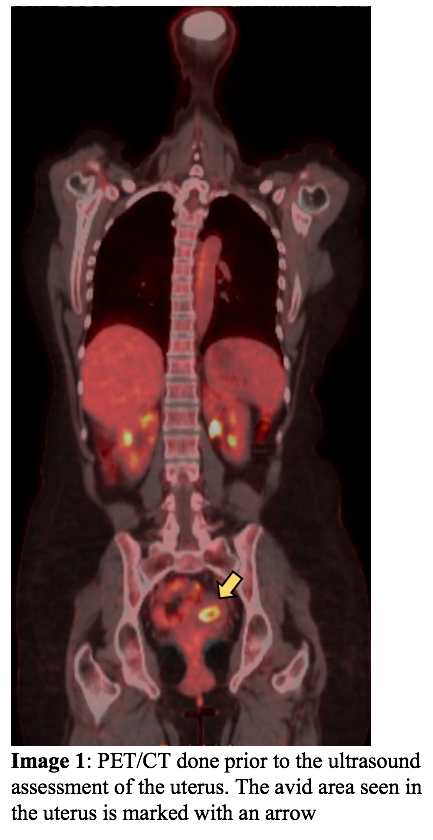
A 53-year-old-female who underwent surgery for cholangiocarcinoma with hepaticojejunostomy 3 years ago with 4 cycles of chemo and 25 sessions of radiotherapy, presented for a routine FDG-PET/CT follow up which showed diffuse increased endometrial uptake. She was referred for a gynecological review, but was lost to follow up. She presented 5 months later with worsening symptoms secondary to a known biliary stricture. A new PET/CT revealed an increase in the FDG avid endometrial lining with increased endometrial distention. CA125 levels were elevated at 286 U/mL.
Images
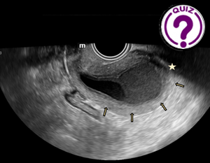
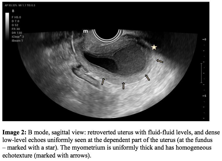
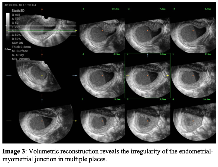
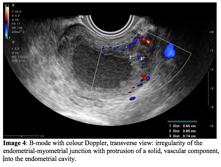
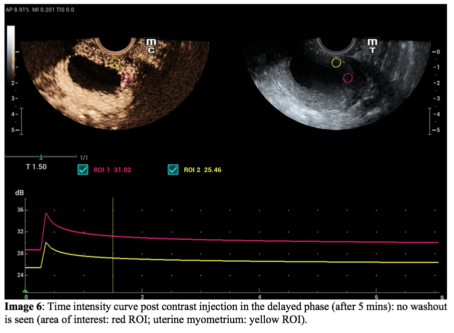
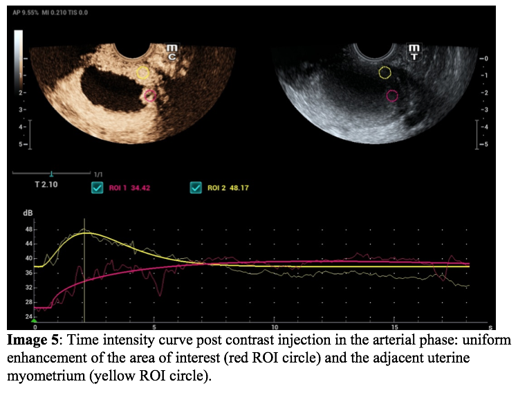
Pelvic ultrasound (US) examination revealed a retroverted uterus with a fluid-fluid level, with dense low-level echoes in the dependent part of the uterus at the fundus. Assessment of the endometrial lining revealed irregularities with solid components projecting into the endometrial cavity (corresponding to the increased metabolic activity detected on PET/CT). Color Doppler assessment revealed internal vascularity. A contrast enhanced ultrasound was also performed and showed uniform enhancement of this irregular region at the endometrial-myometrial junction with no washout in the delayed phase. The endometrial contents did not enhance.
Video 1: B-mode, transverse and sagittal view: Uterus, depicted from fundus to cervix, demonstrating the quality of the endometrial contents and demonstrating irregularity of the endometrial lining.
Video 3: CEUS, 25 seconds post-injection: uniform enhancement of the region of interest as compared to the adjacent myometrium in the arterial phase.
Quiz-summary
0 of 1 questions completed
Questions:
- 1
Information
View the August Case below, answer the question and then click check >
You have already completed the quiz before. Hence you can not start it again.
Quiz is loading...
You must sign in or sign up to start the quiz.
You have to finish following quiz, to start this quiz:
Results
0 of 1 questions answered correctly
Your time:
Time has elapsed
You have reached 0 of 0 points, (0)
Categories
- Not categorized 0%
- 1
- Answered
- Review
-
Question 1 of 1
1. Question
Question: What is the most likely diagnosis?
Correct
CORRECT ANSWER EXPLAINED BELOW Correct answer is: Endometritis
Discussion
As there was still a slight suspicion of malignancy and the patient wanted a hysterectomy, the patient went on to have a total laparoscopic hysterectomy, bilateral salpingo-oopherectomy and pelvic lymphadenectomy. Histopathology confirmed a diagnosis of extensive necrotizing granulomatous endometritis, adenomyosis, and bilateral chronic necrotizing granulomatous salpingo-oophoritis.
Necrotizing granulomatous endometritis (NGE) is a rare but serious inflammatory condition of the uterine lining that can cause significant pelvic pain and infertility. Differentiating NGE from other conditions, such as hematometra, endometrial cancer, and endometriosis, is important for proper diagnosis and treatment. Ultrasound is a commonly used imaging modality that can help differentiate these conditions.
Hematometra is a condition where blood accumulates in the uterus due to an obstruction in the cervical or uterine canal. On ultrasound, hematometra appears as a fluid-filled cavity in the uterus with a smooth, echogenic lining and uniformly homogeneous myometrium.
Endometrial carcinoma is a malignant growth of the endometrial tissue that can cause abnormal vaginal bleeding and pelvic pain. On ultrasound, endometrial carcinoma appears as a thickening of the endometrial lining, often with irregular borders and heterogeneous echogenicity. There may also be masses or nodules within the endometrial cavity or myometrium. Unlike endometritis, endometrial carcinoma demonstrates washout in the delayed phase on CEUS examination.
Endometriosis is a condition where endometrial tissue grows outside the uterus, often on the ovaries or other organs within the pelvis. On ultrasound, endometriosis appears as well-defined masses that are hypoechoic or cystic in nature with diffuse low-level echoes within. Endometriosis and adenomyosis can also be a cause of elevated CA125. Adenomyosis can also cause irregularity of the endometrial lining with uniform appearance of the myometrium in early stages.
NGE, on the other hand, appears as multiple, small cystic spaces within the endometrial cavity(image 6) often with thickening and irregularity of the surrounding endometrium. These cystic spaces may contain fluid, debris, or necrotic tissue. On ultrasound, NGE can appear similar to endometriosis or endometrial carcinoma on B-mode imaging, however CEUS reveals no delayed washout. Endometritis may also show FDG uptake on PET/CT, where the degree of uptake varies by the severity and chronicity of the inflammation.
Conclusion
Contrast enhanced ultrasound is a safe and inexpensive imaging modality providing bed side diagnosis when used, and can aid in planning further management.
Conflicts of Interest
The authors declare no conflict of interest
References
- Geng J, Tang J. Contrast-enhanced ultrasound in the diagnosis of endometrial carcinoma: A meta-analysis. Exp Ther Med. 2018 Dec;16(6):5310-5314. doi: 10.3892/etm.2018.6889. Epub 2018 Oct 22. PMID: 30542488; PMCID: PMC6257399.
- Merviel, P, James, P, Carlier, M, et al. Xanthogranulomatous endometritis: A case report and literature review. Clin Case Rep. 2021; 9:e04299.
- Pop CM, Mihu D, Badea R. Role of contrast-enhanced ultrasound (CEUS) in the diagnosis of endometrial pathology. Clujul Med. 2015;88(4):433-7. doi: 10.15386/cjmed-499. Epub 2015 Nov 15. PMID: 26733740; PMCID: PMC4689232.
Incorrect
CORRECT ANSWER EXPLAINED BELOW Correct answer is: Endometritis
Discussion
As there was still a slight suspicion of malignancy and the patient wanted a hysterectomy, the patient went on to have a total laparoscopic hysterectomy, bilateral salpingo-oopherectomy and pelvic lymphadenectomy. Histopathology confirmed a diagnosis of extensive necrotizing granulomatous endometritis, adenomyosis, and bilateral chronic necrotizing granulomatous salpingo-oophoritis.
Necrotizing granulomatous endometritis (NGE) is a rare but serious inflammatory condition of the uterine lining that can cause significant pelvic pain and infertility. Differentiating NGE from other conditions, such as hematometra, endometrial cancer, and endometriosis, is important for proper diagnosis and treatment. Ultrasound is a commonly used imaging modality that can help differentiate these conditions.
Hematometra is a condition where blood accumulates in the uterus due to an obstruction in the cervical or uterine canal. On ultrasound, hematometra appears as a fluid-filled cavity in the uterus with a smooth, echogenic lining and uniformly homogeneous myometrium.
Endometrial carcinoma is a malignant growth of the endometrial tissue that can cause abnormal vaginal bleeding and pelvic pain. On ultrasound, endometrial carcinoma appears as a thickening of the endometrial lining, often with irregular borders and heterogeneous echogenicity. There may also be masses or nodules within the endometrial cavity or myometrium. Unlike endometritis, endometrial carcinoma demonstrates washout in the delayed phase on CEUS examination.
Endometriosis is a condition where endometrial tissue grows outside the uterus, often on the ovaries or other organs within the pelvis. On ultrasound, endometriosis appears as well-defined masses that are hypoechoic or cystic in nature with diffuse low-level echoes within. Endometriosis and adenomyosis can also be a cause of elevated CA125. Adenomyosis can also cause irregularity of the endometrial lining with uniform appearance of the myometrium in early stages.
NGE, on the other hand, appears as multiple, small cystic spaces within the endometrial cavity(image 6) often with thickening and irregularity of the surrounding endometrium. These cystic spaces may contain fluid, debris, or necrotic tissue. On ultrasound, NGE can appear similar to endometriosis or endometrial carcinoma on B-mode imaging, however CEUS reveals no delayed washout. Endometritis may also show FDG uptake on PET/CT, where the degree of uptake varies by the severity and chronicity of the inflammation.
Conclusion
Contrast enhanced ultrasound is a safe and inexpensive imaging modality providing bed side diagnosis when used, and can aid in planning further management.
Conflicts of Interest
The authors declare no conflict of interest
References
- Geng J, Tang J. Contrast-enhanced ultrasound in the diagnosis of endometrial carcinoma: A meta-analysis. Exp Ther Med. 2018 Dec;16(6):5310-5314. doi: 10.3892/etm.2018.6889. Epub 2018 Oct 22. PMID: 30542488; PMCID: PMC6257399.
- Merviel, P, James, P, Carlier, M, et al. Xanthogranulomatous endometritis: A case report and literature review. Clin Case Rep. 2021; 9:e04299.
- Pop CM, Mihu D, Badea R. Role of contrast-enhanced ultrasound (CEUS) in the diagnosis of endometrial pathology. Clujul Med. 2015;88(4):433-7. doi: 10.15386/cjmed-499. Epub 2015 Nov 15. PMID: 26733740; PMCID: PMC4689232.

