Beijing, China WFUMB COE
November 15, 2023
Webinar: Gastrointestinal ultrasound (GIUS) in IBD – 11-03-2024
January 4, 2024Qingli Zhu1, Yuanjing Gao1, Yi-Hong Chou*2
1 Peking Union Medical College Hospital, Chinese Academy of Medical Sciences and Peking Union Medical College, Beijing, China
2 Department of Radiology, Taipei Veterans General Hospital, Taipei, Taiwan; School of Medicine, National Yang Ming Chiao Tung University, Taipei, Taiwan
* Correspondence: yhchou7@gmail.com
Clinical history
A 57-year-old woman was admitted to the hospital with dyspnea caused by an increasingly swollen right neck for the past three months. She denied fever or heart palpitations but had had a weight loss of 5 kg in the past three months.
Laboratory tests showed a normal thyroid function and elevated thyroid antibodies (T3 0.94 (0.66-1.92) ng/ml; T4: 7.80 (4.30-12.50) μg/dl; anti-thyroglobulin antibodies: 522 (<115) IU/ml; Anti-thyroid peroxidase antibodies: 432 (<34) IU/ml).
White blood cell count, C-reactive protein, and erythrocyte sedimentation rate were within normal range.
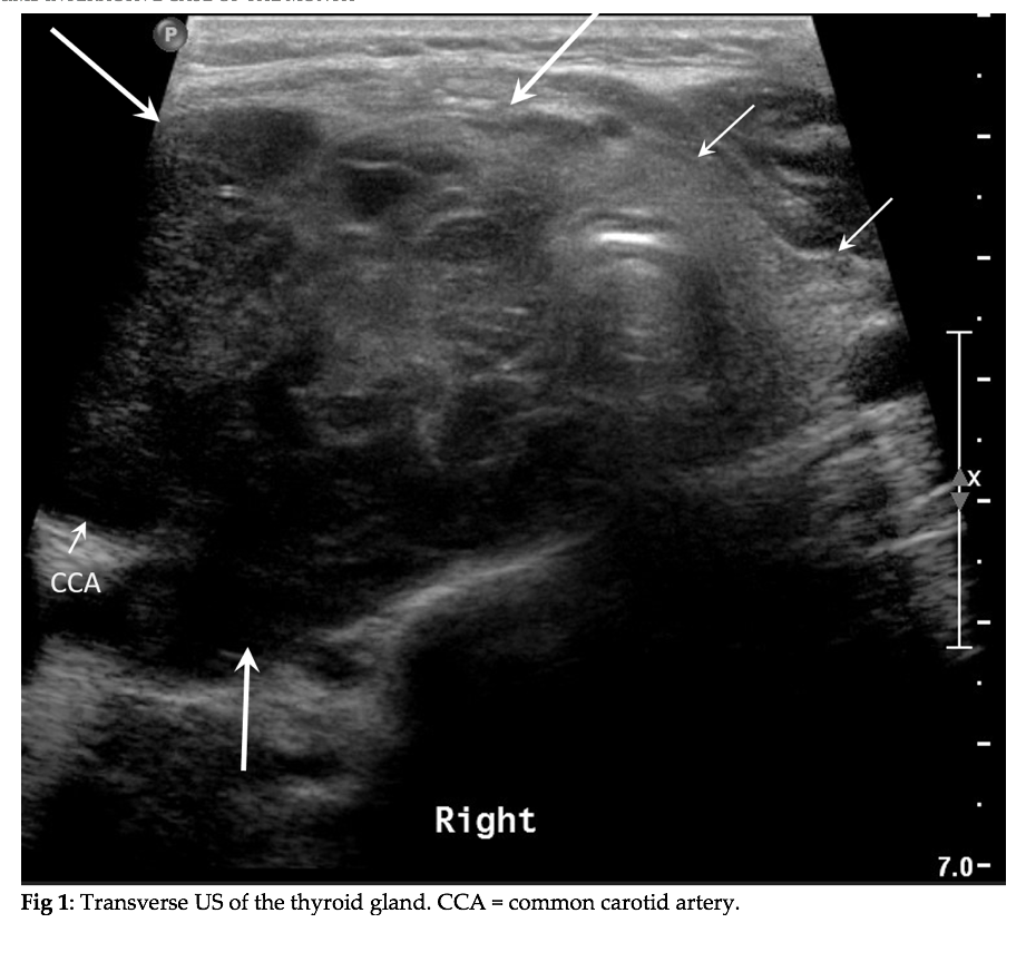
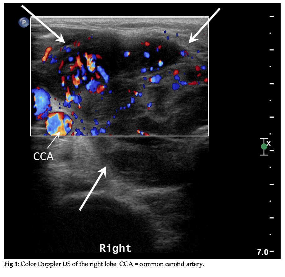
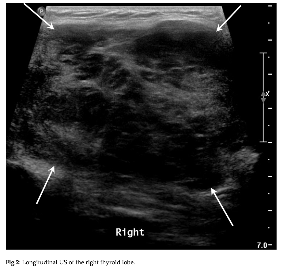
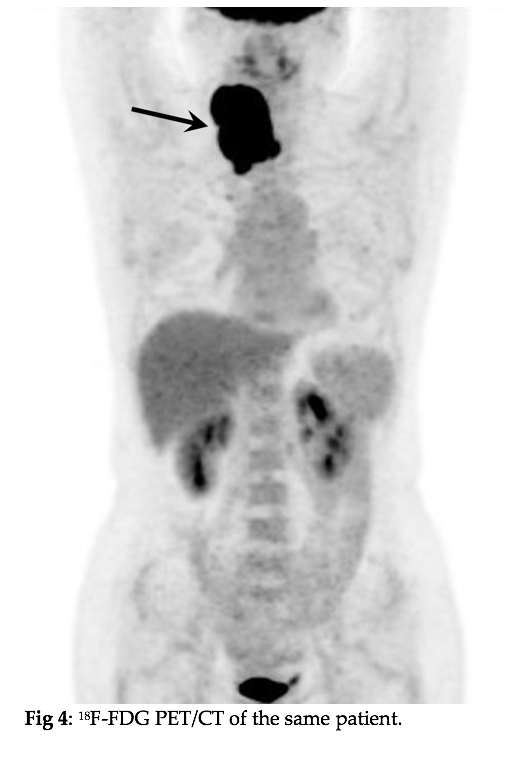
An ultrasound examination of the neck was first performed (Fig 1-3). After a US-guided biopsy, 18F-FDG PET/CT was performed (Fig 4).
Images
Quiz-summary
0 of 1 questions completed
Questions:
- 1
Information
View the December Case below, answer the question and then click check >
You have already completed the quiz before. Hence you can not start it again.
Quiz is loading...
You must sign in or sign up to start the quiz.
You have to finish following quiz, to start this quiz:
Results
0 of 1 questions answered correctly
Your time:
Time has elapsed
You have reached 0 of 0 points, (0)
Categories
- Not categorized 0%
- 1
- Answered
- Review
-
Question 1 of 1
1. Question
Question: What is the most likely diagnosis?
Correct
CORRECT ANSWER EXPLAINED BELOW Correct answer is: Primary thyroid lymphoma.
Review of the imaging studies
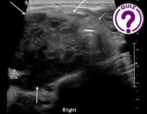
A transverse US scan of the thyroid (Fig 1) shows a large, lobulated hypoechoic mass in the right thyroid lobe (large arrows). The mass invades the soft tissues surrounding the common carotid artery (CCA). The isthmus and left thyroid lobe are essentially of normal appearance (small arrows). A longitudinal US scan of the right thyroid lobe (Fig 2) shows a diffusely enlarged right lobe which is totally infiltrated by a large heterogeneous hypoechoic mass (arrows). Color Doppler US of the right lobe (Fig 3) demonstrates abundant color flow signals in the mass (arrows). Note that the mass invades the thyroid capsule. 18F-FDG PET/CT of the same patient (Fig 4) shows a significantly increased metabolic activity in the right lobe of the thyroid (arrow) (SUVmax = 46.1), which is highly suggestive of malignancy. No other sites showed distinct areas of increased FDG-metabolism.
Sonographic Appearance of Thyroid Lymphoma
Thyroid lymphoma is most commonly described as bilateral or unilateral asymmetric goiter. The lesions typically show heterogeneous echotexture and marked hypoechogenicity with posterior acoustic enhancement and may invade the thyroid capsule. There is increased chaotic vascularity which is shown on the color Doppler flow image.
Clinical Course of the Patient
The patient received a US-guided core needle biopsy 1 day after the ultrasound examination, and the pathological study revealed that the tumor was consistent with large B cell lymphoma. Since there is no evidence of lymphomatous lesions in other parts of the body and bone marrow biopsy showed no abnormalities, the patient was diagnosed as a case of primary thyroid lymphoma. She subsequently received chemotherapy and total remission was achieved. She was doing well one year after chemotherapy.
Differential Diagnoses
A goiter with markedly hypoechoic, and heterogeneous internal echoes could also be observed in Hashimoto’s thyroiditis. However, rapid growth and pressure symptoms are rare in diffuse Hashimoto’s thyroiditis.
Nodular goiter may present with multiple solid nodules with large size (5 cm in greatest diameter). However, there will not be thyroid capsule invasion or increased chaotic vascularity.
Goiter and rapid progression are also observed in undifferentiated thyroid carcinoma. Undifferentiated thyroid carcinoma often presents with microcalcifications and cystic changes on sonograms.
Conclusion
The clinical presentation with rapid thyroid enlargement and breathing difficulties, may indicate PTL. The US features of PTL include asymmetric goiter, heterogeneous echotexture, and marked hypoechogenicity with posterior acoustic enhancement. The thyroid capsule invasion and the focal chaotic vascularity further raise suspicion for lymphoma.
Conflicts of Interest
The authors declare no conflict of interest
References
- Xia Y, Wang L, et al. Sonographic appearance of primary thyroid lymphoma-preliminary experience. PLoS One.2014,9(12):e114080.
- Peng C, Chen Yang C, et al. Multimodal Sonographic Appearance and Survival Outcomes of 69 Cases of Primary Thyroid Lymphoma Over 10 Years. J Ultrasound Med. 2022, 41(12):3031-3040.
Incorrect
CORRECT ANSWER EXPLAINED BELOW Correct answer is: Primary thyroid lymphoma.
Review of the imaging studies
A transverse US scan of the thyroid (Fig 1) shows a large, lobulated hypoechoic mass in the right thyroid lobe (large arrows). The mass invades the soft tissues surrounding the common carotid artery (CCA). The isthmus and left thyroid lobe are essentially of normal appearance (small arrows). A longitudinal US scan of the right thyroid lobe (Fig 2) shows a diffusely enlarged right lobe which is totally infiltrated by a large heterogeneous hypoechoic mass (arrows). Color Doppler US of the right lobe (Fig 3) demonstrates abundant color flow signals in the mass (arrows). Note that the mass invades the thyroid capsule. 18F-FDG PET/CT of the same patient (Fig 4) shows a significantly increased metabolic activity in the right lobe of the thyroid (arrow) (SUVmax = 46.1), which is highly suggestive of malignancy. No other sites showed distinct areas of increased FDG-metabolism.
Sonographic Appearance of Thyroid Lymphoma
Thyroid lymphoma is most commonly described as bilateral or unilateral asymmetric goiter. The lesions typically show heterogeneous echotexture and marked hypoechogenicity with posterior acoustic enhancement and may invade the thyroid capsule. There is increased chaotic vascularity which is shown on the color Doppler flow image.
Clinical Course of the Patient
The patient received a US-guided core needle biopsy 1 day after the ultrasound examination, and the pathological study revealed that the tumor was consistent with large B cell lymphoma. Since there is no evidence of lymphomatous lesions in other parts of the body and bone marrow biopsy showed no abnormalities, the patient was diagnosed as a case of primary thyroid lymphoma. She subsequently received chemotherapy and total remission was achieved. She was doing well one year after chemotherapy.
Differential Diagnoses
A goiter with markedly hypoechoic, and heterogeneous internal echoes could also be observed in Hashimoto’s thyroiditis. However, rapid growth and pressure symptoms are rare in diffuse Hashimoto’s thyroiditis.
Nodular goiter may present with multiple solid nodules with large size (5 cm in greatest diameter). However, there will not be thyroid capsule invasion or increased chaotic vascularity.
Goiter and rapid progression are also observed in undifferentiated thyroid carcinoma. Undifferentiated thyroid carcinoma often presents with microcalcifications and cystic changes on sonograms.
Conclusion
The clinical presentation with rapid thyroid enlargement and breathing difficulties, may indicate PTL. The US features of PTL include asymmetric goiter, heterogeneous echotexture, and marked hypoechogenicity with posterior acoustic enhancement. The thyroid capsule invasion and the focal chaotic vascularity further raise suspicion for lymphoma.
Conflicts of Interest
The authors declare no conflict of interest
References
- Xia Y, Wang L, et al. Sonographic appearance of primary thyroid lymphoma-preliminary experience. PLoS One.2014,9(12):e114080.
- Peng C, Chen Yang C, et al. Multimodal Sonographic Appearance and Survival Outcomes of 69 Cases of Primary Thyroid Lymphoma Over 10 Years. J Ultrasound Med. 2022, 41(12):3031-3040.

