VICE PRESIDENT 1 Acting President Ioan Sporea – EFSUMB
January 31, 2024
EUROSON SCHOOL: 6th International Paediatric Advanced Ultrasound Course
February 19, 2024Adrian Goudie 1
1 Fiona Stanley Hospital, Perth, Australia.
* Correspondence: adrian.goudie@health.wa.gov.au
Clinical history
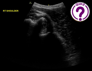
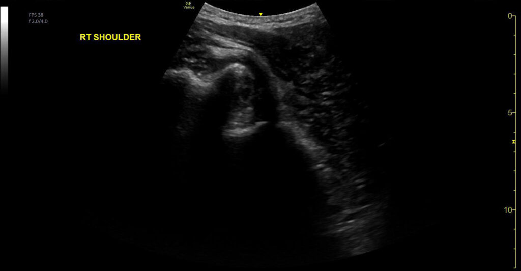
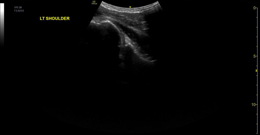
A middle aged man presented with a painful right shoulder after falling off his bike. Examination revealed loss of the normal shoulder contour and markedly restricted movement due to pain. After confirmation of the diagnosis a procedure was performed.
Images
Quiz-summary
0 of 1 questions completed
Questions:
- 1
Information
View the May Case below, answer the question and then click check >
You have already completed the quiz before. Hence you can not start it again.
Quiz is loading...
You must sign in or sign up to start the quiz.
You have to finish following quiz, to start this quiz:
Results
0 of 1 questions answered correctly
Your time:
Time has elapsed
You have reached 0 of 0 points, (0)
Categories
- Not categorized 0%
- 1
- Answered
- Review
-
Question 1 of 1
1. Question
Question: What is the diagnosis?
Correct
CORRECT ANSWER EXPLAINED BELOW Correct answer is: Glenohumeral joint dislocation
Discussion
The initial image shows the glenoid fossa is ‘empty’ with the humeral head displaced away from the transducer, diagnostic of an anterior glenohumeral joint dislocation. The video demonstrates the humeral head being maneuvered during reduction, with the head finally returning to the glenoid fossa.
Additional Discussion
Whilst plain radiography is the most common imaging modality to diagnose shoulder dislocation and confirm reduction, ultrasound has high sensitivity and specificity(1, 2). Whilst shoulder dislocation and reduction is often clinically evident, it may not be if there is significant swelling or joint instability. In these cases, the portability and immediacy of ultrasound allows rapid confirmation, and if necessary, further reduction attempts (for example whilst the patient is still sedated, if that has been required).
Conclusion
Shoulder ultrasound is a rapid and useful technique for the diagnosis of shoulder dislocation and reduction.
Conflicts of Interest
The authors declare no conflict of interest
References
- Attard Biancardi MA, Jarman RD, Cardona T. Diagnostic accuracy of point-of-care ultrasound (PoCUS) for shoulder dislocations and reductions in the emergency department: a diagnostic randomised control trial (RCT). Emerg Med J. 2022;39(9):655-61.
- Gottlieb M, Patel D, Marks A, Peksa GD. Ultrasound for the diagnosis of shoulder dislocation and reduction: A systematic review and meta-analysis. Acad Emerg Med. 2022;29(8):999-1007.
Incorrect
CORRECT ANSWER EXPLAINED BELOW Correct answer is: Glenohumeral joint dislocation
Discussion
The initial image shows the glenoid fossa is ‘empty’ with the humeral head displaced away from the transducer, diagnostic of an anterior glenohumeral joint dislocation. The video demonstrates the humeral head being maneuvered during reduction, with the head finally returning to the glenoid fossa.
Additional Discussion
Whilst plain radiography is the most common imaging modality to diagnose shoulder dislocation and confirm reduction, ultrasound has high sensitivity and specificity(1, 2). Whilst shoulder dislocation and reduction is often clinically evident, it may not be if there is significant swelling or joint instability. In these cases, the portability and immediacy of ultrasound allows rapid confirmation, and if necessary, further reduction attempts (for example whilst the patient is still sedated, if that has been required).
Conclusion
Shoulder ultrasound is a rapid and useful technique for the diagnosis of shoulder dislocation and reduction.
Conflicts of Interest
The authors declare no conflict of interest
References
- Attard Biancardi MA, Jarman RD, Cardona T. Diagnostic accuracy of point-of-care ultrasound (PoCUS) for shoulder dislocations and reductions in the emergency department: a diagnostic randomised control trial (RCT). Emerg Med J. 2022;39(9):655-61.
- Gottlieb M, Patel D, Marks A, Peksa GD. Ultrasound for the diagnosis of shoulder dislocation and reduction: A systematic review and meta-analysis. Acad Emerg Med. 2022;29(8):999-1007.


