Webinar: Update to WFUMB 2018 Guidelines on Liver Ultrasound Elastography – 15-04-2024
March 4, 2024
Case of the Month April 2024 – Bilateral fetal adrenal calcifications
March 30, 2024Maria Franca Meloni1 *, Debora Cidoni2 , Giacomo Gazzano1, and Sandro Sironi2 ,
1 Casa di Cura Igea: Department of Radiology, Milano, Italy
2 Department of Radiology, University of Milano-Bicocca, Papa Giovanni XXIII Hospital Bergamo- Italy
2 Instituto Auxologico Italiano, Department of Pathology, Milano, Italy
* Correspondence: meloni.mariafranca@gmail.com
Clinical history
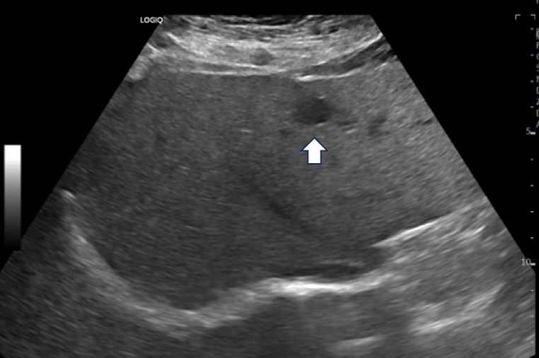
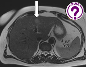
A 55-year-old healthy female, without a history of cirrhosis or cancer had undergone a follow-up MRI for renal angiomyolipoma with contrast agent and diffusion weighted sequences at a different hospital. She had not taken any estrogen or progesterone drugs in the past. The MRI incidentally identified a focal lesion in liver segment III measuring 16 mm with intense diffusion restriction.
Images


Video 1 – CEUS shows transient arterial enhancement and early wash-out 22 seconds after injection.

Quiz-summary
0 of 2 questions completed
Questions:
- 1
- 2
Information
View the May Case below, answer the question and then click check >
You have already completed the quiz before. Hence you can not start it again.
Quiz is loading...
You must sign in or sign up to start the quiz.
You have to finish following quiz, to start this quiz:
Results
0 of 2 questions answered correctly
Your time:
Time has elapsed
You have reached 0 of 0 points, (0)
Categories
- Not categorized 0%
- 1
- 2
- Answered
- Review
-
Question 1 of 2
1. Question
Question 1: What is the most likely diagnosis?
Correct
CORRECT ANSWER EXPLAINED BELOW Correct answer to Q1 is: Other
Discussion
The vascular behavior on CEUS could indicate a malignant lesion, but the absence of symptoms or a history of cancer along with negative laboratory tests including tumor markers lead us to continue the clinical investigations.
Incorrect
CORRECT ANSWER EXPLAINED BELOW Correct answer to Q1 is: Other
Discussion
The vascular behavior on CEUS could indicate a malignant lesion, but the absence of symptoms or a history of cancer along with negative laboratory tests including tumor markers lead us to continue the clinical investigations.
-
Question 2 of 2
2. Question
Question 2: Is the information we have enough for a diagnosis?
Correct
CORRECT ANSWER EXPLAINED BELOW Correct answer to Q2 is: Biopsy is strongly recommended.
Additional Imaging
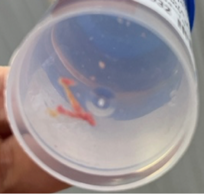
Video 2 – Ultrasound guided biopsy performed with an 18 G semi-automatic needle of the liver lesion in segment III.

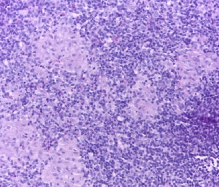
Image 5 – Hematoxylin-Eosin staining. Epitheloid microgranuloma and lymphocytic infiltration. The histological diagnosis was compatible with atypical mycobacteriosis.
Additional discussion
Hepatic granulomas are rare benign lesions. They are found in approximately 2-15% of liver biopsies. The causes can be infectious and non-infectious. Infectious granulomas are linked to primary biliary cirrhosis, tuberculosis, viral hepatitis B and C, bacterial, fungal, and parasitic infections, while non-infectious ones are usually linked to drugs or sarcoidosis (1).
Hepatic granulomas may be found incidentally in asymptomatic patients or patients with vague or non-specific symptoms. The do not have a specific enhancement pattern in CT or MRI, and can in fact present without enhancement, with hyper-enhancement or with peripheral rim-like enhancement (2).
The CEUS behavior of granuloma is described as highly variable, making the differential diagnosis with malignant neoplasms difficult (3-5).
In our case the patient was asymptomatic, and the detection of the liver lesion was incidental. CEUS, performed after B-mode ultrasonography, showed a rapid and fleeting arterial enhancement followed by an early (22 sec) and progressively intense wash-out. The CEUS behavior was compatible with a malignant lesion, but in the absence of a clinical history of cancer, a biopsy was necessary, which correctly led to the benign diagnosis.
Conclusion
Hepatic granuloma are rare lesions in the liver, but they should be considered in the differential diagnosis of focal liver lesions. CEUS cannot differentiate granulomatous lesions from focal malignant lesions such as metastases or intrahepatic cholcangiocarcinoma. Biopsy plays a fundamental role in the diagnosis as it excludes malignant pathology.
Conflicts of Interest:
The author declares no conflict of interest.
References
- Choi EK, Lamps LW. Granulomas in the liver with a Focus on Infectious Causes Surg Pathol Clin. 2018 Jun;11(2):231-250. doi: 10.1016/j.path.2018.02.008
- Mortelé KJ, Segatto E, Ros PR. The infected liver: radiologic-pathologic correlation. Radiographics. 2004 Jul-Aug;24(4):937-55. doi: 10.1148/rg.244035719.
- Dietrich CF, Nolsøe CP, Barr RG, et al., Guidelines and Good Clinical Practice Recommendations for Contrast Enhanced Ultra- sound (CEUS) in the Liver – Update 2020 – WFUMB in Cooper- ation with EFSUMB, AFSUMB, AIUM, and FLAUS Ultraschall Med 2020; 39:187–210.
- Kong WT, Wang WP, Cai H, Huang BJ, Ding H, Mao F. The analysis of enhancement pattern of hepatic inflamma- tory pseudotumor on contrast-enhanced ultrasound. Abdom Imaging 2014;39:168-174.
- Kong WT, Wang WP, Shen HY, Xue HY, Liu CR, Huang DQ, Wu M. Hepatic inflammatory pseudotumor mimicking malignancy: the value of differential diagnosis on contrast enhanced ultrasound.. Med Ultrason. 2021 Feb 18;23(1):15-21. doi: 10.11152/mu-2542.
Incorrect
CORRECT ANSWER EXPLAINED BELOW Correct answer to Q2 is: Biopsy is strongly recommended.
Additional Imaging
Video 2 – Ultrasound guided biopsy performed with an 18 G semi-automatic needle of the liver lesion in segment III.

Image 5 – Hematoxylin-Eosin staining. Epitheloid microgranuloma and lymphocytic infiltration. The histological diagnosis was compatible with atypical mycobacteriosis.
Additional discussion
Hepatic granulomas are rare benign lesions. They are found in approximately 2-15% of liver biopsies. The causes can be infectious and non-infectious. Infectious granulomas are linked to primary biliary cirrhosis, tuberculosis, viral hepatitis B and C, bacterial, fungal, and parasitic infections, while non-infectious ones are usually linked to drugs or sarcoidosis (1).
Hepatic granulomas may be found incidentally in asymptomatic patients or patients with vague or non-specific symptoms. The do not have a specific enhancement pattern in CT or MRI, and can in fact present without enhancement, with hyper-enhancement or with peripheral rim-like enhancement (2).
The CEUS behavior of granuloma is described as highly variable, making the differential diagnosis with malignant neoplasms difficult (3-5).
In our case the patient was asymptomatic, and the detection of the liver lesion was incidental. CEUS, performed after B-mode ultrasonography, showed a rapid and fleeting arterial enhancement followed by an early (22 sec) and progressively intense wash-out. The CEUS behavior was compatible with a malignant lesion, but in the absence of a clinical history of cancer, a biopsy was necessary, which correctly led to the benign diagnosis.
Conclusion
Hepatic granuloma are rare lesions in the liver, but they should be considered in the differential diagnosis of focal liver lesions. CEUS cannot differentiate granulomatous lesions from focal malignant lesions such as metastases or intrahepatic cholcangiocarcinoma. Biopsy plays a fundamental role in the diagnosis as it excludes malignant pathology.
Conflicts of Interest:
The author declares no conflict of interest.
References
- Choi EK, Lamps LW. Granulomas in the liver with a Focus on Infectious Causes Surg Pathol Clin. 2018 Jun;11(2):231-250. doi: 10.1016/j.path.2018.02.008
- Mortelé KJ, Segatto E, Ros PR. The infected liver: radiologic-pathologic correlation. Radiographics. 2004 Jul-Aug;24(4):937-55. doi: 10.1148/rg.244035719.
- Dietrich CF, Nolsøe CP, Barr RG, et al., Guidelines and Good Clinical Practice Recommendations for Contrast Enhanced Ultra- sound (CEUS) in the Liver – Update 2020 – WFUMB in Cooper- ation with EFSUMB, AFSUMB, AIUM, and FLAUS Ultraschall Med 2020; 39:187–210.
- Kong WT, Wang WP, Cai H, Huang BJ, Ding H, Mao F. The analysis of enhancement pattern of hepatic inflamma- tory pseudotumor on contrast-enhanced ultrasound. Abdom Imaging 2014;39:168-174.
- Kong WT, Wang WP, Shen HY, Xue HY, Liu CR, Huang DQ, Wu M. Hepatic inflammatory pseudotumor mimicking malignancy: the value of differential diagnosis on contrast enhanced ultrasound.. Med Ultrason. 2021 Feb 18;23(1):15-21. doi: 10.11152/mu-2542.


