ASUM New Zealand 2024 Annual Conference
May 30, 2024
Case of the month July 2024 – Swelling of the cheeks
July 5, 2024Rune Fuglø-Mortensen1*, Jonathan Frydenlund Cohen2
- Department of Radiology, Herlev and Gentofte Hospital, Denmark;
- Department of Radiology, Rigshospitalet, Copenhagen University Hospital, Denmark
* Correspondences: rune.fugloe.mortensen@regionh.dk
Clinical history
A 14-year-old boy was referred to the pediatric department due to several hours of constant right testicular pain. He had not experienced any nausea or vomiting. No history of recent infections. The physical examination showed no redness or swelling of the scrotum, no size difference of the testes, but a diffuse soreness of the right testis. An ultrasound examination was performed.
Images


Quiz-summary
0 of 2 questions completed
Questions:
- 1
- 2
Information
View the June Case below, answer the question and then click check >
You have already completed the quiz before. Hence you can not start it again.
Quiz is loading...
You must sign in or sign up to start the quiz.
You have to finish following quiz, to start this quiz:
Results
0 of 2 questions answered correctly
Your time:
Time has elapsed
You have reached 0 of 0 points, (0)
Categories
- Not categorized 0%
- 1
- 2
- Answered
- Review
-
Question 1 of 2
1. Question
Question 1: What is the most likely diagnosis?
Correct
CORRECT ANSWER EXPLAINED BELOW Correct answer to Q1 is: Testicular epidermoid cysts
Incorrect
CORRECT ANSWER EXPLAINED BELOW Correct answer to Q1 is: Testicular epidermoid cysts
-
Question 2 of 2
2. Question
Discussion
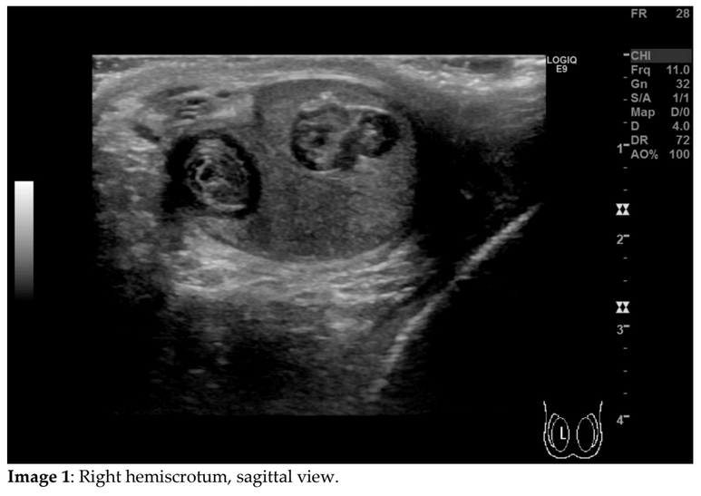
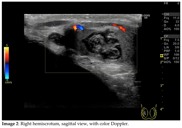
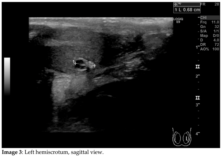
In the right testis, there are three well circumscribed, round tumors, some with calcifications. The tumors measure about 1.2 cm at largest and show no vascularity using colour Doppler. They all have the classic “onion-peel” appearance of epidermoid cysts. In the left testis, there is a similar, but smaller tumor. No signs of testicular torsion.
Epidermoid cysts are rare benign testicular tumors, which account for around 1-2% of focal testicular lesions. Testicular epidermoid cysts usually present in the second to fourth decades, and often present as a painless enlargement of the testis [1-3].
Epidermoid cysts are solid tumors located beneath the tunica albuginea and they are typically surrounded by normal testis parenchyma. They have fibrous walls and the lumen contains keratin deposits [1]. Sonographically, the appearance of epidermoid cysts has classically been described as an “onion peel” appearance of alternating rings of hyper-and hypoechogenicity. At least three other appearances have been described, including a densely calcified mass, a cyst with peripheral rim or central calcifications, and a mixed-pattern cyst [4]. On elastography, epidermoid cysts appear hard [5].Unlike most testicular neoplasms, epidermoid cysts are avascular [5, 6], and for this reason the use of colour Doppler is important in the differentiation from testicular neoplasms.
Unlike teratomas or dermoid cysts, epidermoid cysts do not contain any other teratomatous contents such as teeth, sebaceous glands or hair follicles [1, 3, 6]. It is important to recognize the appearance of testicular epidermoid cysts, as it enables treatment by enucleation. The patient underwent enucleation instead of orchiectomy and frozen sections were obtained during surgery confirming the benign diagnosis.
Testicular infarction would not show as focal well circumscribed lesions, but more as wedge shaped hypoechoic areas.In the present case, the histopathological diagnosis of epidermoid cysts was indeed confirmed intraoperatively, and the four tumors (three from the right testis and one from the left testis) were enucleated while the rest of the testis parenchyma was left intact. Outpatient follow-up ultrasound showed no signs of recurrence.
Question 2: Given the fact that the patient had no other testicular lesions, and was otherwise well with an unremarkable medical history, what do you expect the blood work-up to show?
Correct
CORRECT ANSWER EXPLAINED BELOW Correct answer to Q2 is: Normal alpha-fetoprotein, normal beta human chorionic gonadotropin.
Additional Discussion
Testicular epidermoid cysts are typically not associated with increase in alpha-fetoprotein or beta human chorionic gonadotropin, which is more characteristic of malignant germ cell tumors [3].
Conclusion
Epidermoid cysts are rare benign testicular tumors that classically have a well-circumscribed sonographic appearance with alternating hyperechogenicities and hypochogenicites giving an “onion peel” appearance.
Conflicts of Interest:
The author declares no conflict of interest with respect to this case.
References
- Moghe PK, Brady AP. Ultrasound of testicular epidermoid cysts. Br J Radiol. 1999 Oct;72(862):942-5. doi: 10.1259/bjr.72.862.10673943. PMID: 10673943.
- Radiopaedia.org. Available online: https://radiopaedia.org/articles/testicular-epidermoid-cyst?lang=us (accessed on 24.04.24)
- Ashouri KB, Heiman JM, Kelly EF, Manganiotis AN. Testicular epidermoid cyst: A rare case. Urol Ann. 2017 Jul-Sep;9(3):296-298. doi: 10.4103/UA.UA_37_17. PMID: 28794603; PMCID: PMC5532904.
- Atchley JT, Dewbury KC. Ultrasound appearances of testicular epidermoid cysts. Clin Radiol. 2000 Jul;55(7):493-502. doi: 10.1053/crad.1999.0472. PMID: 10924372.
- EFSUMB Course Book, 2nd Edition, Editor: Christoph F. Dietrich, Ultrasound of the scrotum (Available online: https://efsumb.org/wp-content/uploads/2023/07/ECB2nd_Chapter_Scrotum_FULL.pdf). [pp. 24-24] (accessed on 24.04.24)
- Saouli, A., Karmouni, T., El Khader, K. et al. Testicular epidermoid cyst: about a case report and a review of the literature. Afr J Urol 27, 30 (2021). https://doi.org/10.1186/s12301-021-00135-z
- DMCG.dk. Available online: https://www.dmcg.dk/siteassets/kliniske-retningslinjer—skabeloner-og-vejledninger/kliniske-retningslinjer-opdelt-pa-dmcg/dateca/dateca_testikel-kraft_admgodk150720.pdf (accessed on22.05.24)
Incorrect
CORRECT ANSWER EXPLAINED BELOW Correct answer to Q2 is: Normal alpha-fetoprotein, normal beta human chorionic gonadotropin.
Additional Discussion
Testicular epidermoid cysts are typically not associated with increase in alpha-fetoprotein or beta human chorionic gonadotropin, which is more characteristic of malignant germ cell tumors [3].
Conclusion
Epidermoid cysts are rare benign testicular tumors that classically have a well-circumscribed sonographic appearance with alternating hyperechogenicities and hypochogenicites giving an “onion peel” appearance.
Conflicts of Interest:
The author declares no conflict of interest with respect to this case.
References
- Moghe PK, Brady AP. Ultrasound of testicular epidermoid cysts. Br J Radiol. 1999 Oct;72(862):942-5. doi: 10.1259/bjr.72.862.10673943. PMID: 10673943.
- Radiopaedia.org. Available online: https://radiopaedia.org/articles/testicular-epidermoid-cyst?lang=us (accessed on 24.04.24)
- Ashouri KB, Heiman JM, Kelly EF, Manganiotis AN. Testicular epidermoid cyst: A rare case. Urol Ann. 2017 Jul-Sep;9(3):296-298. doi: 10.4103/UA.UA_37_17. PMID: 28794603; PMCID: PMC5532904.
- Atchley JT, Dewbury KC. Ultrasound appearances of testicular epidermoid cysts. Clin Radiol. 2000 Jul;55(7):493-502. doi: 10.1053/crad.1999.0472. PMID: 10924372.
- EFSUMB Course Book, 2nd Edition, Editor: Christoph F. Dietrich, Ultrasound of the scrotum (Available online: https://efsumb.org/wp-content/uploads/2023/07/ECB2nd_Chapter_Scrotum_FULL.pdf). [pp. 24-24] (accessed on 24.04.24)
- Saouli, A., Karmouni, T., El Khader, K. et al. Testicular epidermoid cyst: about a case report and a review of the literature. Afr J Urol 27, 30 (2021). https://doi.org/10.1186/s12301-021-00135-z
- DMCG.dk. Available online: https://www.dmcg.dk/siteassets/kliniske-retningslinjer—skabeloner-og-vejledninger/kliniske-retningslinjer-opdelt-pa-dmcg/dateca/dateca_testikel-kraft_admgodk150720.pdf (accessed on22.05.24)

