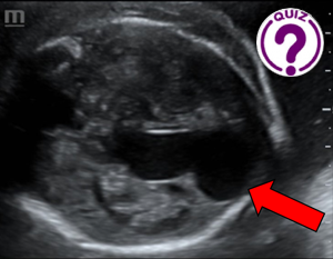
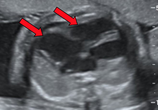
Case of the month September 2024 – 26-week prenatal ultrasound: anechoic, cystic structure next to the midline of the brain. What do you think?

COP29 Special Report consultation (online, 4 September, 14:00 CET):
August 15, 2024
Case of the month October 2024: Unusual neck lesion – CEUS as an aid to diagnosis
November 19, 2024Nadhem KAMMOUN, Dora KHOMSI, Wièm DOUIRA-KHOMSI
Department of Paediatric Radiology, Béchir Hamza Children’s Hospital, Tunis, Tunisia *
Correspondences: Nadhem.kammoun@medecinesfax.org; dora.khomsi@gmail.com; khomsiwiem@yahoo.fr
Clinical history
A 22-year-old patient in her first pregnancy was referred to our department for prenatal ultrasound at 26 weeks of gestation.
Ultrasound revealed an anechoic, cystic structure above the cerebellum and below the thalamus with blood flow on Colour Doppler
(images 1 and 2).
Image


Quiz-summary
0 of 2 questions completed
Questions:
- 1
- 2
Information
View the May Case below, answer the question and then click check >
You have already completed the quiz before. Hence you can not start it again.
Quiz is loading...
You must sign in or sign up to start the quiz.
You have to finish following quiz, to start this quiz:
Results
0 of 2 questions answered correctly
Your time:
Time has elapsed
You have reached 0 of 0 points, (0)
Categories
- Not categorized 0%
- 1
- 2
- Answered
- Review
-
Question 1 of 2
1. Question
Question 1: Findings included a central anechoic structure next to the midline of the brain with blood flow in colour Doppler images. What diagnosis do you propose and what anatomical structure does it involve?
Correct
CORRECT ANSWER EXPLAINED BELOW Correct answer to Q1 is: Vein of galen aneurysmal malformation.
Incorrect
CORRECT ANSWER EXPLAINED BELOW Correct answer to Q1 is: Vein of galen aneurysmal malformation.
-
Question 2 of 2
2. Question
Discussion
Ultrasound revealed an anechoic, interhemispheric structure posterior to the 3rd ventricle, with vascular flow on color Doppler. There were no other cerebral abnormalities. The diagnosis of aneurysm of the vein of Galen was retained.
Classically, it has a “racket” or “keyhole” shape, as the oval lumen of the malformation communicates posteriorly with a tubular structure corresponding to a venous drainage sinus.
Color Doppler confirms the vascular nature of the lesion, demonstrating bidirectional turbulent flow (1,2,3). It enables differential diagnosis with other midline fluid lesions of the brain with different prognoses (4).
Additional Images
Image 3: B-mode ultrasound shows increased cardiothoracic ratio and dilatation of the right heart cavities (red arrows).
Image 4: Colour Doppler shows tricuspid regurgitation and dilatation of the jugular vessels
Image 5: B-mode ultrasound (left: transverse; right: sagittal) shows a peritoneal effusion (white arrow). VB: gallbladder
Question 2: What is the most likely explanation for the thoracoabdominal findings?
Correct
CORRECT ANSWER EXPLAINED BELOW Correct answer to Q2 is: Foetal heart failure caused by the cerebral vascular malformation
Additional discussion
Patients with aneurysmal malformation of the vein of Galen have an underlying arteriovenous shunting of blood in the cerebral circulation. Ultrasound assessment therefore also includes evaluation of the hemodynamic impact of the intracranial shunt. The cardiac outcome is characterized by dilatation of the right cavities (image 3). Colour Doppler may reveal tricuspid regurgitation and dilatation of the jugular vessels (image 4). These signs can occur early, sometimes as early as the second trimester, and are pathognomonic of intracranial arteriovenous fistula (5).
Other signs of right heart failure, such as hepatomegaly, serous intraperitoneal effusion (image 5) and hydramnios, are more rare and appear later. The final stage is fetoplacental hydrops (6).
Conclusion
Vein of Galen aneurysm is a rare, intracranial, vascular, congenital malformation. We report an antenatal diagnosis by ultrasound at 26 weeks gestation. The neonatal prognosis is poor if fetal cardiac insufficiency or cerebral lesions (hydrocephaly, porencephaly, intracerebral hemorrhage) are present antenatally.
Conflicts of Interest:
“The authors declare no conflict of interest.”
References
- Li TG, Zhang YY, Nie F, Peng MJ, Li YZ, Li PL. Diagnosis of foetal vein of galen aneurysmal malformation by ultrasound combined with magnetic resonance imaging: a case series. BMC Med Imaging. 2020 Jun 12;20(1):63.
- Michaels AY, Sood S, Frates MC. Vein of Galen aneurysmal malformation [J] Ultrasound Q. 2016;32(4):366–369.
- Hartung J, Heling KS, Rake A, Zimmer C, Chaoui R. Detection of an aneurysm of the vein of Galen following signs of cardiac overload in a 22-week old fetus. Prenat Diagn 2003; 23: 901-3.
- Pilu G, Falco P, Perolo A, Sandri F, Cocchi G, Ancora G et al. Differential diagnosis and outcome of fetal intracranial hypoechoic lesions: report of 21 cases. Ultrasound Obstet Gynecol 1997; 9: 229-36.
- Doren M, Tercanli S, Holzgreve W. Prenatal sonographic diagnosis of a vein of Galen aneurysm: relevance of associated malformations for timing and mode of delivery. Ultrasound Obstet Gynecol 1995; 6: 287-9.
- Sepulveda W, Platt CC, Fisk NM. Prenatal diagnosis of cerebral arteriovenous malformation using color Doppler ultrasonography: case report and review of the literature. Ultrasound Obstet Gynecol 1995; 6: 282-6.
Incorrect
CORRECT ANSWER EXPLAINED BELOW Correct answer to Q2 is: Foetal heart failure caused by the cerebral vascular malformation
Additional discussion
Patients with aneurysmal malformation of the vein of Galen have an underlying arteriovenous shunting of blood in the cerebral circulation. Ultrasound assessment therefore also includes evaluation of the hemodynamic impact of the intracranial shunt. The cardiac outcome is characterized by dilatation of the right cavities (image 3). Colour Doppler may reveal tricuspid regurgitation and dilatation of the jugular vessels (image 4). These signs can occur early, sometimes as early as the second trimester, and are pathognomonic of intracranial arteriovenous fistula (5).
Other signs of right heart failure, such as hepatomegaly, serous intraperitoneal effusion (image 5) and hydramnios, are more rare and appear later. The final stage is fetoplacental hydrops (6).
Conclusion
Vein of Galen aneurysm is a rare, intracranial, vascular, congenital malformation. We report an antenatal diagnosis by ultrasound at 26 weeks gestation. The neonatal prognosis is poor if fetal cardiac insufficiency or cerebral lesions (hydrocephaly, porencephaly, intracerebral hemorrhage) are present antenatally.
Conflicts of Interest:
“The authors declare no conflict of interest.”
References
- Li TG, Zhang YY, Nie F, Peng MJ, Li YZ, Li PL. Diagnosis of foetal vein of galen aneurysmal malformation by ultrasound combined with magnetic resonance imaging: a case series. BMC Med Imaging. 2020 Jun 12;20(1):63.
- Michaels AY, Sood S, Frates MC. Vein of Galen aneurysmal malformation [J] Ultrasound Q. 2016;32(4):366–369.
- Hartung J, Heling KS, Rake A, Zimmer C, Chaoui R. Detection of an aneurysm of the vein of Galen following signs of cardiac overload in a 22-week old fetus. Prenat Diagn 2003; 23: 901-3.
- Pilu G, Falco P, Perolo A, Sandri F, Cocchi G, Ancora G et al. Differential diagnosis and outcome of fetal intracranial hypoechoic lesions: report of 21 cases. Ultrasound Obstet Gynecol 1997; 9: 229-36.
- Doren M, Tercanli S, Holzgreve W. Prenatal sonographic diagnosis of a vein of Galen aneurysm: relevance of associated malformations for timing and mode of delivery. Ultrasound Obstet Gynecol 1995; 6: 287-9.
- Sepulveda W, Platt CC, Fisk NM. Prenatal diagnosis of cerebral arteriovenous malformation using color Doppler ultrasonography: case report and review of the literature. Ultrasound Obstet Gynecol 1995; 6: 282-6.


