

Case of the month March 2025: Acute pediatric groin bulge and pain
March 3, 2025
Case of the month May 2025: Primary or secondary liver lesion – the lifelong question
May 1, 2025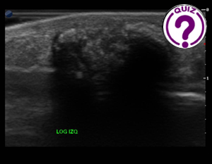
Edda Chaves 1*
1 Hospital Regional Nicolas Solano La Chorrera. Panamá
*correspondence: echavess@minsa.gob.pa
Clinical history
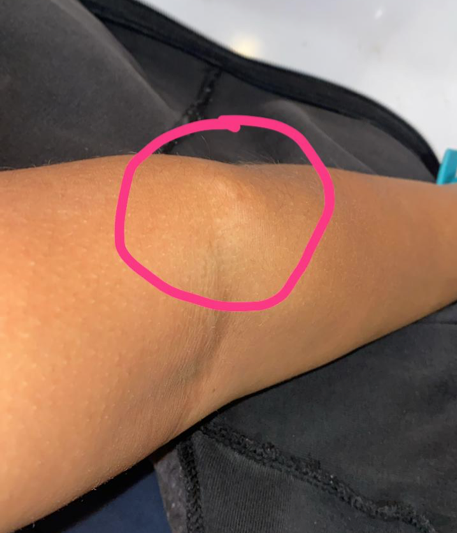
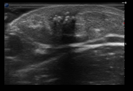
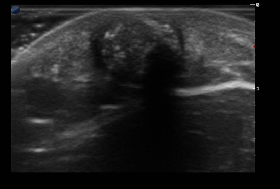
A 25-year-old female patient consulted a dermatologist due to the presence of a mobile, well-defined mass in the lateral anterior surface of her left forearm. The mass was painful on palpation, but otherwise not. It had grown slowly for several years. There was no prior trauma to the area, and no previous history of cancer. She was referred to an ultrasound examination.
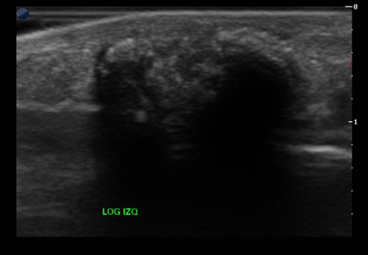
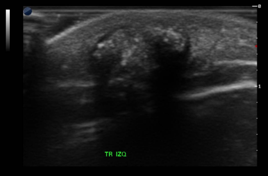
An ultrasound examination was performed with a high frequency linear array transducer.
Images
Quiz-summary
0 of 3 questions completed
Questions:
- 1
- 2
- 3
Information
View the April Case below, answer the question and then click check >
You have already completed the quiz before. Hence you can not start it again.
Quiz is loading...
You must sign in or sign up to start the quiz.
You have to finish following quiz, to start this quiz:
Results
0 of 3 questions answered correctly
Your time:
Time has elapsed
You have reached 0 of 0 points, (0)
Categories
- Not categorized 0%
- 1
- 2
- 3
- Answered
- Review
-
Question 1 of 3
1. Question
Question 1: What are your diagnostic considerations?
Correct
CORRECT ANSWER EXPLAINED BELOW Correct answer to Q1 is: Calcified solid mass
Incorrect
CORRECT ANSWER EXPLAINED BELOW Correct answer to Q1 is: Calcified solid mass
-
Question 2 of 3
2. Question
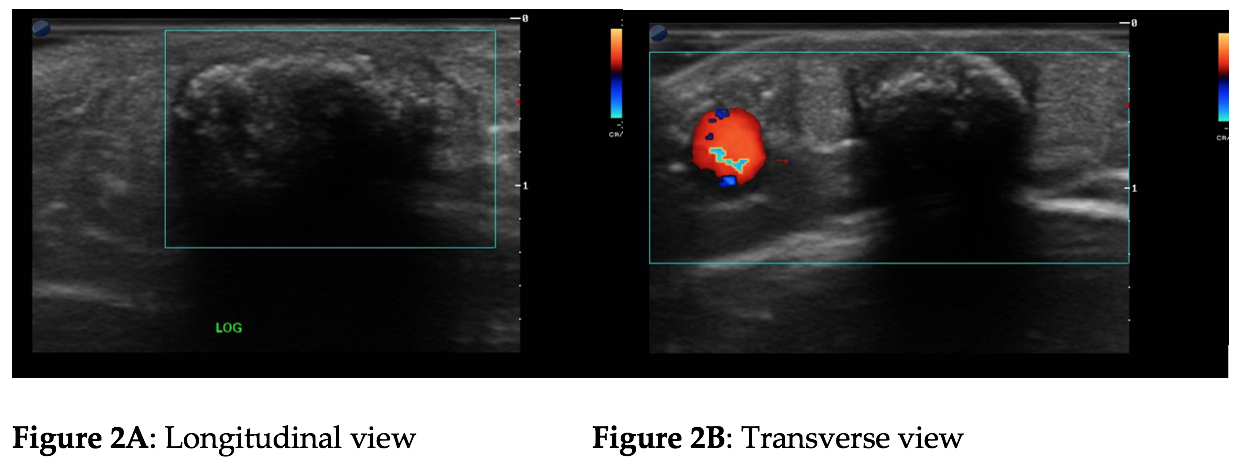
Supplemental images with colour Doppler are shown below:
Question 2: What is your impression now?
Correct
CORRECT ANSWER EXPLAINED BELOW Correct answer to Q2 is: Benign avascular mass in the subcutaneous fat.
Incorrect
CORRECT ANSWER EXPLAINED BELOW Correct answer to Q2 is: Benign avascular mass in the subcutaneous fat.
-
Question 3 of 3
3. Question
Question 3: Considering all the findings, what is the most likely diagnosis?
Correct
CORRECT ANSWER EXPLAINED BELOW Correct answer to Q3 is: Pilomatricoma
The patient underwent surgery with the final histopathological result of pilomatricoma.
Discussion:
Pilomatricoma or Malherbe calcifying epithelioma, is a benign tumour of the germinal matrix of the hair follicle. The most common locations are the head, neck and upper limbs, less frequently the lower limbs. They are generally solitary, but can be multiple. 60% of cases occur before the age of 20 and 40% under the age of 10 mainly in women (1-3).
Five different patterns of pilomatricoma have been described according to their characteristics; type I, completely calcified; type 2, partially calcified with hypoechoic peripheral halo and avascular; type 3, complex lesion with undefined edges; type 4, pseudocystic lesion; type 5, pseudoneoplastic, with irregular contours and evident vascularization. Type 2 is the most common (1-3).
In the differential diagnosis, an epidermoid cyst should be considered. These present as hypoechoic avascular masses usually not with as many calcifications as pilomaticomas. They can be heterogeneous and sometimes the connection to the epidermis is visible. Usually they appear in the 3rd to 4th decade of life and are more common in males than in females (4). Haemangiomas are vascular lesions that may present with calcifications, but usually scarce. In calcified hematomas the patient has a history of trauma and the ultrasound findings vary according to the time since the trauma.
Conclusion
This case has shown a pilomatricoma, corresponding to type 2. The diagnosis of pilomatricoma should be considered when ultrasound shows a well-defined mass with hypoechoic halo and calcifications, located in the subcutaneous tissue in the head, neck or limbs.
The use of high-frequency transducers and the knowledge of the pathology are very important to obtain an accurate diagnosis.
Conflicts of interest
The authors declare no conflict of interest.
References
- Radiopaedia.org. Available online: https://doi.org/10.53347/rID-19245 (Accessed on 21 Oct 2024)
- Garioni E, Danesino GM, Madonia L. Pilomatricoma: Sonographic features. J Ultrasound 2008; 11(2): 76–78.
- Hwang JY, Lee SW, Lee SM. The common ultrasonographic features of pilomatricoma. J Ultrasound Med 2005; 24(10): 1397-402.
- National Library of Medicine. Available online: https://www.ncbi.nlm.nih.gov/books/NBK499974/ (Accessed on 19 Nov 2024)
Incorrect
CORRECT ANSWER EXPLAINED BELOW Correct answer to Q3 is: Pilomatricoma
The patient underwent surgery with the final histopathological result of pilomatricoma.
Discussion:
Pilomatricoma or Malherbe calcifying epithelioma, is a benign tumour of the germinal matrix of the hair follicle. The most common locations are the head, neck and upper limbs, less frequently the lower limbs. They are generally solitary, but can be multiple. 60% of cases occur before the age of 20 and 40% under the age of 10 mainly in women (1-3).
Five different patterns of pilomatricoma have been described according to their characteristics; type I, completely calcified; type 2, partially calcified with hypoechoic peripheral halo and avascular; type 3, complex lesion with undefined edges; type 4, pseudocystic lesion; type 5, pseudoneoplastic, with irregular contours and evident vascularization. Type 2 is the most common (1-3).
In the differential diagnosis, an epidermoid cyst should be considered. These present as hypoechoic avascular masses usually not with as many calcifications as pilomaticomas. They can be heterogeneous and sometimes the connection to the epidermis is visible. Usually they appear in the 3rd to 4th decade of life and are more common in males than in females (4). Haemangiomas are vascular lesions that may present with calcifications, but usually scarce. In calcified hematomas the patient has a history of trauma and the ultrasound findings vary according to the time since the trauma.
Conclusion
This case has shown a pilomatricoma, corresponding to type 2. The diagnosis of pilomatricoma should be considered when ultrasound shows a well-defined mass with hypoechoic halo and calcifications, located in the subcutaneous tissue in the head, neck or limbs.
The use of high-frequency transducers and the knowledge of the pathology are very important to obtain an accurate diagnosis.
Conflicts of interest
The authors declare no conflict of interest.
References
- Radiopaedia.org. Available online: https://doi.org/10.53347/rID-19245 (Accessed on 21 Oct 2024)
- Garioni E, Danesino GM, Madonia L. Pilomatricoma: Sonographic features. J Ultrasound 2008; 11(2): 76–78.
- Hwang JY, Lee SW, Lee SM. The common ultrasonographic features of pilomatricoma. J Ultrasound Med 2005; 24(10): 1397-402.
- National Library of Medicine. Available online: https://www.ncbi.nlm.nih.gov/books/NBK499974/ (Accessed on 19 Nov 2024)