

Case of the month July 2025: A case of neonatal vomiting
July 1, 2025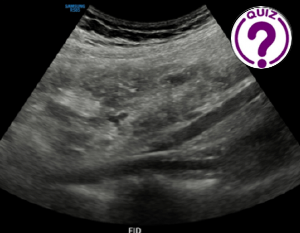
Edda Leonor Chaves S MD *
Servicio de Radiología Hospital Nicolas Solano La Chorrera Panamá
Correspondence: echavess@minsa.gob.pa
Clinical history
A 17-year-old female patient is referred from the emergency room for ultrasound, with the aim of ruling out appendicitis; she reports abdominal pain of approximately 14 hours duration. In the last few hours the pain has settled in the lower right quadrant. The only significant finding is a value of 17.560 white cell count, 89.3% neutrophils. Ultrasound is performed with emphasis on the right iliac fossa with the following findings:
Images
Quiz-summary
0 of 4 questions completed
Questions:
- 1
- 2
- 3
- 4
Information
View the August Case below, answer the question and then click check >
You have already completed the quiz before. Hence you can not start it again.
Quiz is loading...
You must sign in or sign up to start the quiz.
You have to finish following quiz, to start this quiz:
Results
0 of 4 questions answered correctly
Your time:
Time has elapsed
You have reached 0 of 0 points, (0)
Categories
- Not categorized 0%
- 1
- 2
- 3
- 4
- Answered
- Review
-
Question 1 of 4
1. Question
Question 1: What is your diagnostic impression:
Correct
CORRECT ANSWER EXPLAINED BELOW Correct answer to Q1 is: Dilated bowel loops
Incorrect
CORRECT ANSWER EXPLAINED BELOW Correct answer to Q1 is: Dilated bowel loops
-
Question 2 of 4
2. Question
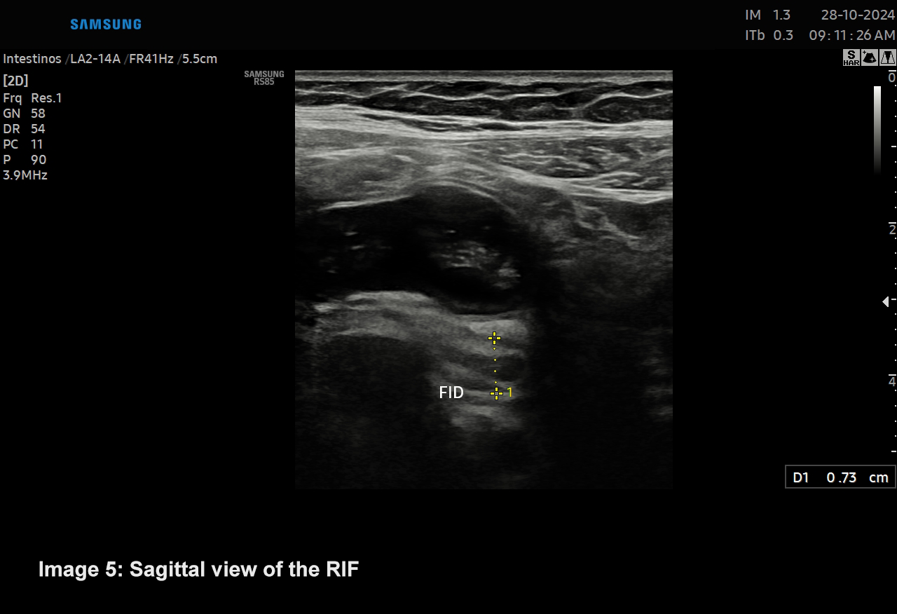
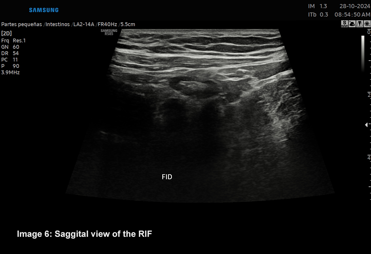
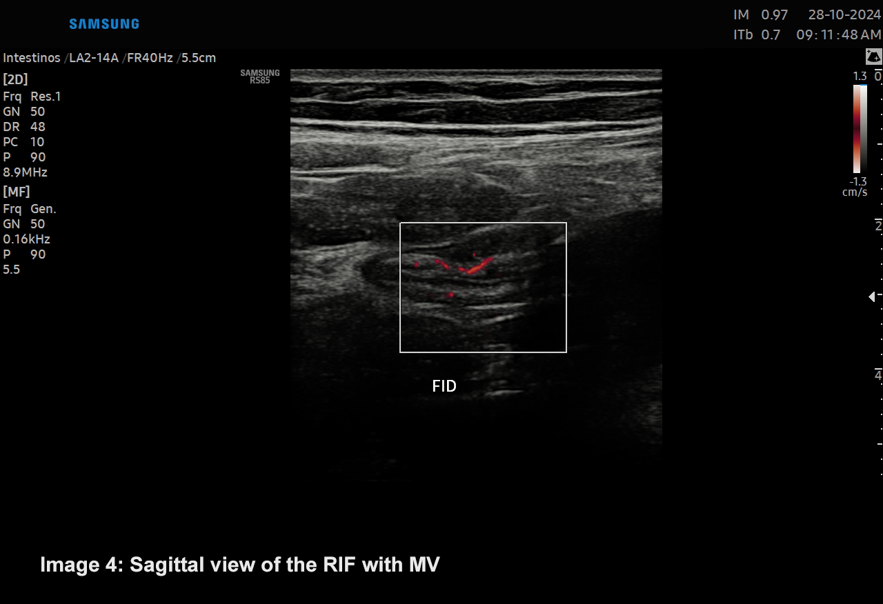
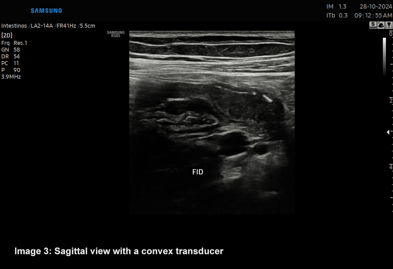
A second look was performed since there were no significant findings and the patient was very symptomatic with the following findings:
Question 2: Please select a potential answer
Correct
CORRECT ANSWER EXPLAINED BELOW Correct answer to Q2 is: Acute retrocecal appendicitis and a mesenteric node
Incorrect
CORRECT ANSWER EXPLAINED BELOW Correct answer to Q2 is: Acute retrocecal appendicitis and a mesenteric node
-
Question 3 of 4
3. Question
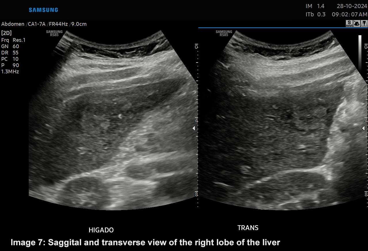
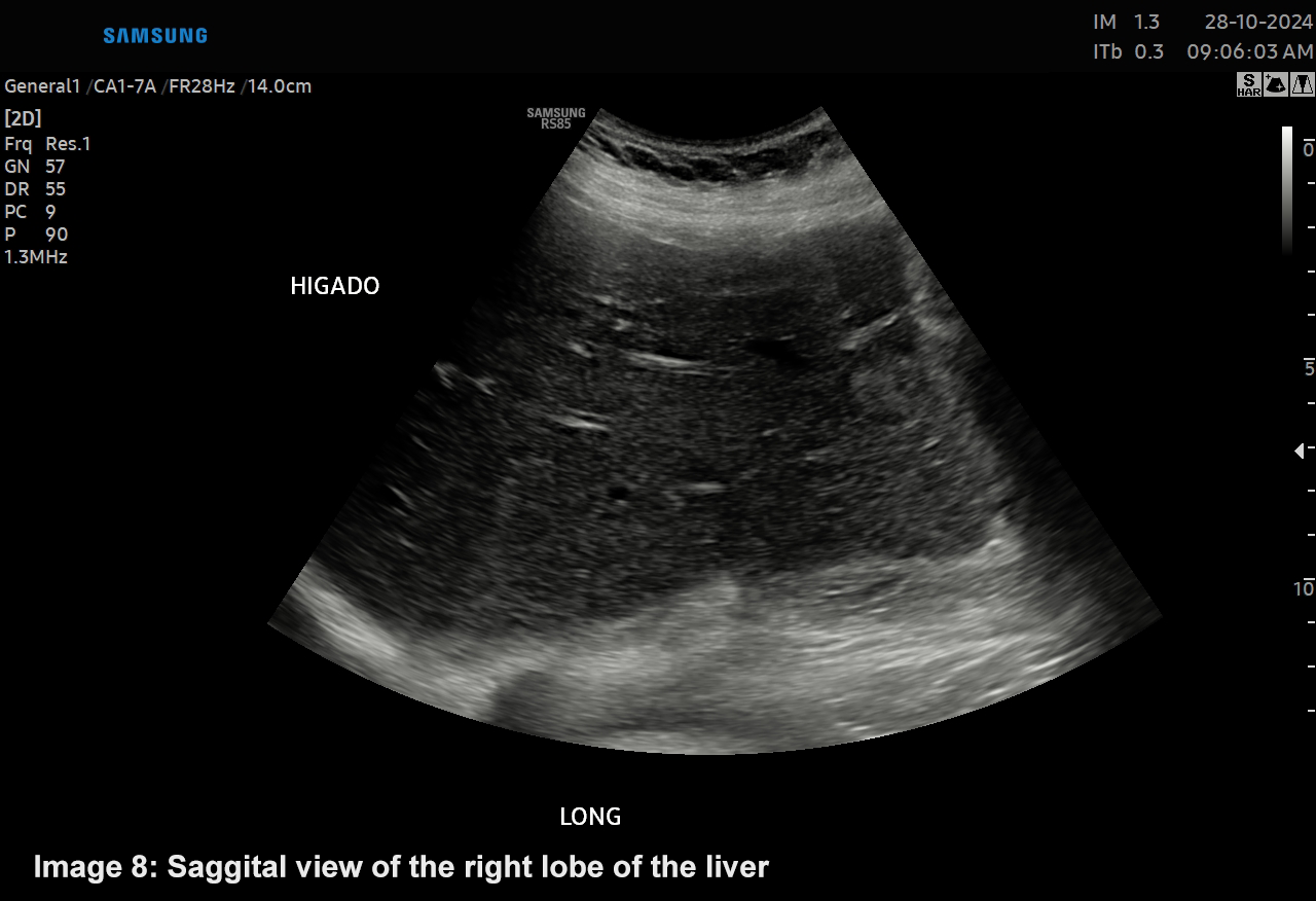
The ultrasound confirmed the clinical suspicion of appendicitis, however during the evaluation the liver is explored obtaining the following images.
Question 3: What is your diagnostic impression?
Correct
CORRECT ANSWER EXPLAINED BELOW Correct answer to Q3 is: None of the above
Incorrect
ANSWER EXPLAINED BELOW That is incorrect.
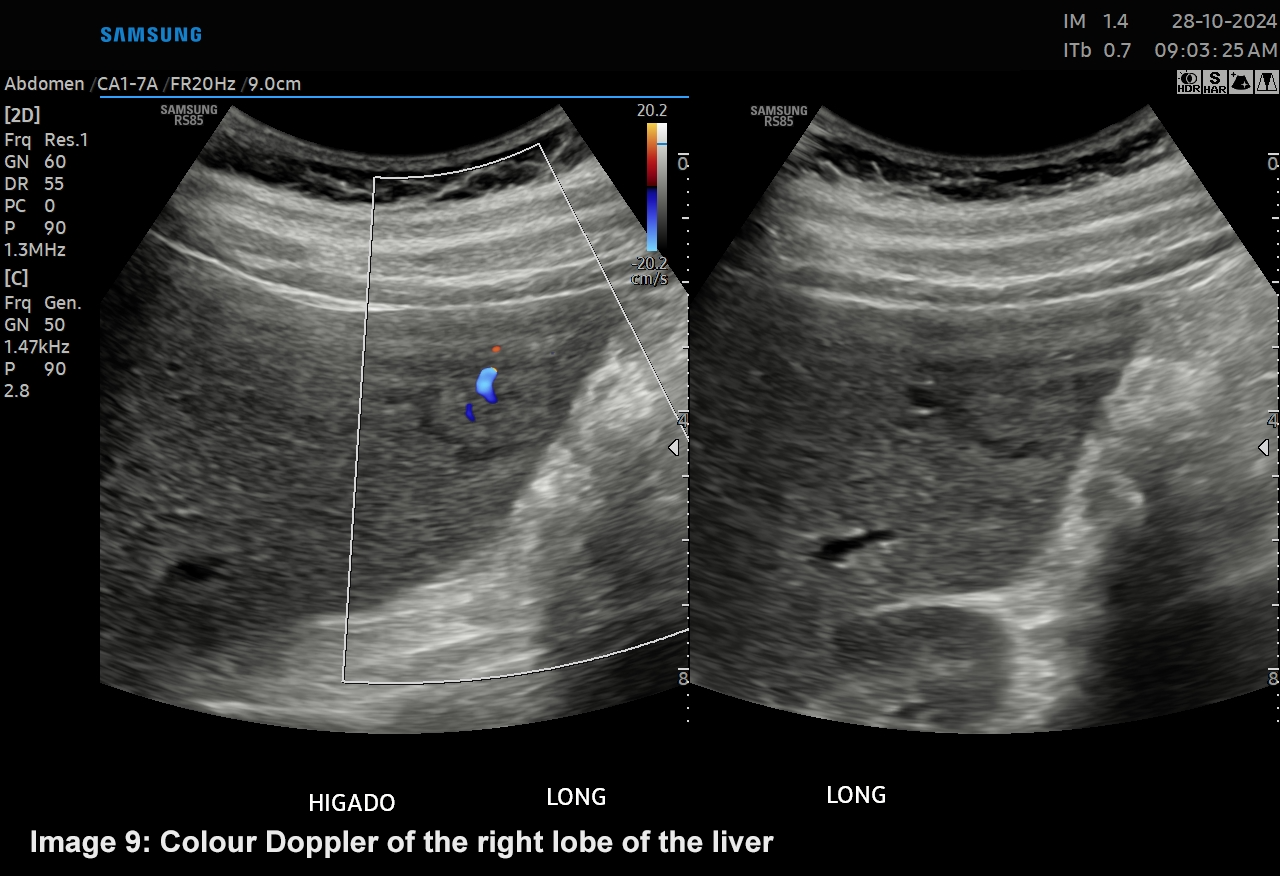
There is a small focal liver lesion, a 1.6 x 1.8 x 1.3 cms slightly hypoechoic with an echogenic center which was demonstrated by colour ultrasound to be a vascular component, central scar, it’s not an injury since the patient had no trauma
-
Question 4 of 4
4. Question
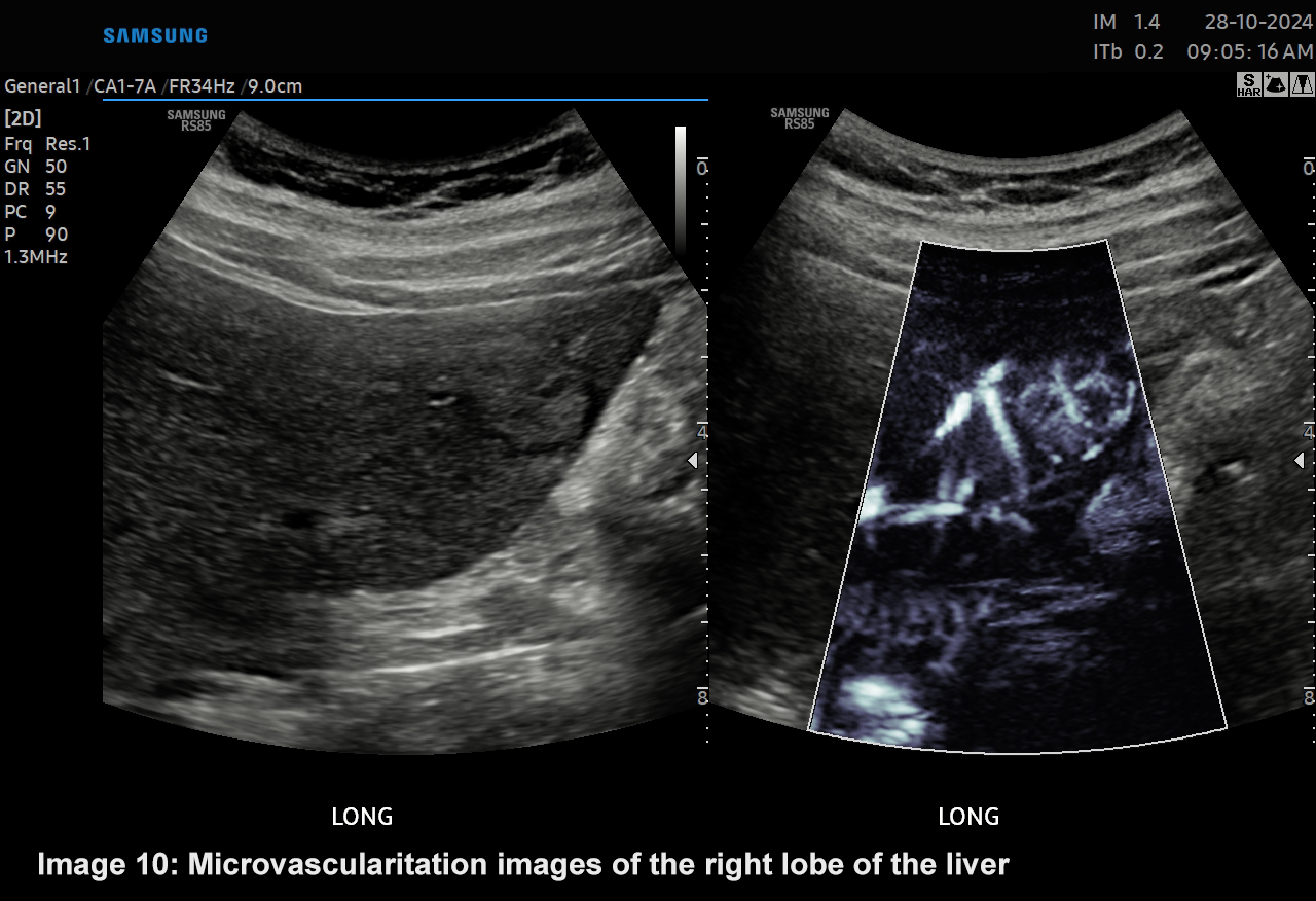
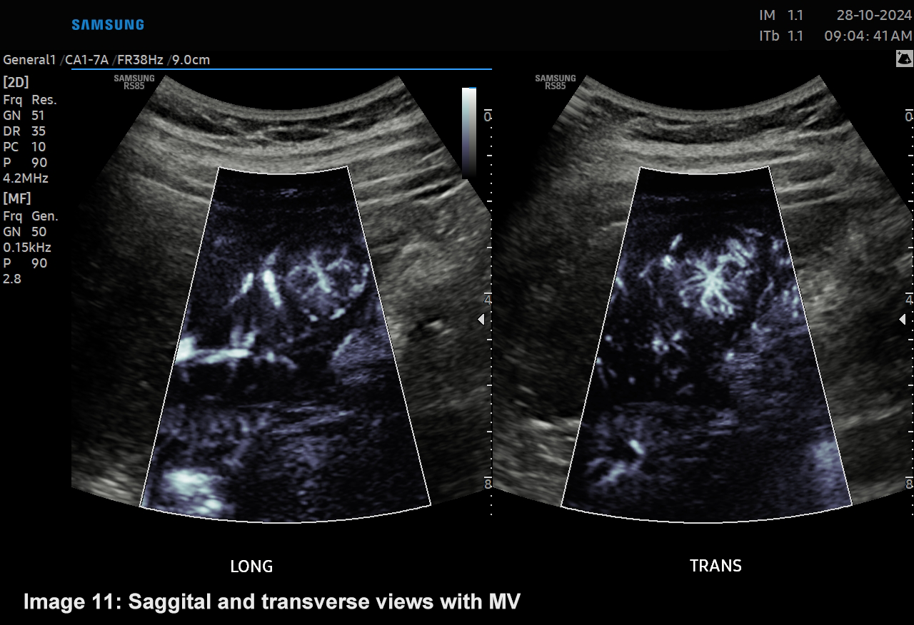
The study was complemented with color Doppler and microvascular flow
Question 4: What is your diagnostic impression:
Correct
CORRECT ANSWER EXPLAINED BELOW Correct answer to Q4 is: Nodular focal hyperplasia
Discussion
The ultrasound study confirmed the clinical suspicion of appendicitis, although initially difficult to assess, this led to a thorough examination of the patient. While looking for another cause of the pain, a focal liver lesion was found. We identified, in the right lobe of the liver, a 1.6 x 1.8 x 1.3 cms a hypoechoic lesion with a central scar and dominant vascular component. This is considered a characteristic image of Focal nodular hyperplasia (FNH). FNH is the most common benign focal liver lesion in women aged between 25 and 50 years, often discovered incidentally in asymptomatic patients. In general, there is no relation between FNH with the intake of exogenous estrogens. These lesions from a histopathological point of view demonstrate proliferation of hyperplastic hepatocytes that surround a central scar, typically it is a solitary lesion, due to damage to a portal path which causes the formation of arteriovenous shunts. The recommendation in asymptomatic patients with lesions such as the case presented, is control with images every 6 months for a period of three years
Conclusion
It is important not to limit ourselves exclusively to the evaluation of FID when we perform a study for suspected appendicitis, since additional examination may provide important findings for patient follow-up and control. In the case presented despite the fact of been younger than the average age of presentation, the finding of an hypoechoic focal liver lesion with a central scar and dominant vascular component, led to the diagnosis of nodular focal hyperplasia. It was decided not to expose the patient to another study such as tomography, especially considering it is a very small lesion in an asymptomatic patient with no abnormal findings in her blood samples. Follow-up studies every 6 month was suggested.
Conflicts of interest
“The authors declare no conflict of interest.”
References
- Ciencia Latina Revista Científica multidisciplinaria, Ciudad de México, México, ISSN 2707-2207/ ISSN 2707-2215, marzo-abril 2024, volumen 8 Número 2. Hiperplasia Nodular Focal. Hallazgo incidental en colecistectomía laparoscopia electiva. Reporte de caso
- Marrero, Jorge A MD1; Ahn, Joseph MD, FACG2; Reddy, Rajender K MD, FACG3 on behalf of the Practice Parameters Committee of the American College of Gastroenterology. ACG Clinical Guideline: The Diagnosis and Management of Focal Liver Lesions. American Journal of Gastroenterology 109(9):p 1328-1347, September 2014.
- Gaillard F, Walizai T, Kogan J, et al. Focal nodular hyperplasia. Reference article, Radiopaedia.org (Accessed on 29 Oct 2024)
- Hamad S, Willyard CE, Mukherjee S. Focal Nodular Hyperplasia. [Updated 2022 Sep 26]. In: StatPearls [Internet]. Treasure Island (FL): StatPearls Publishing; 2025 Jan
Incorrect
CORRECT ANSWER EXPLAINED BELOW Correct answer to Q4 is: Nodular focal hyperplasia
Discussion
The ultrasound study confirmed the clinical suspicion of appendicitis, although initially difficult to assess, this led to a thorough examination of the patient. While looking for another cause of the pain, a focal liver lesion was found. We identified, in the right lobe of the liver, a 1.6 x 1.8 x 1.3 cms a hypoechoic lesion with a central scar and dominant vascular component. This is considered a characteristic image of Focal nodular hyperplasia (FNH). FNH is the most common benign focal liver lesion in women aged between 25 and 50 years, often discovered incidentally in asymptomatic patients. In general, there is no relation between FNH with the intake of exogenous estrogens. These lesions from a histopathological point of view demonstrate proliferation of hyperplastic hepatocytes that surround a central scar, typically it is a solitary lesion, due to damage to a portal path which causes the formation of arteriovenous shunts. The recommendation in asymptomatic patients with lesions such as the case presented, is control with images every 6 months for a period of three years
Conclusion
It is important not to limit ourselves exclusively to the evaluation of FID when we perform a study for suspected appendicitis, since additional examination may provide important findings for patient follow-up and control. In the case presented despite the fact of been younger than the average age of presentation, the finding of an hypoechoic focal liver lesion with a central scar and dominant vascular component, led to the diagnosis of nodular focal hyperplasia. It was decided not to expose the patient to another study such as tomography, especially considering it is a very small lesion in an asymptomatic patient with no abnormal findings in her blood samples. Follow-up studies every 6 month was suggested.
Conflicts of interest
“The authors declare no conflict of interest.”
References
- Ciencia Latina Revista Científica multidisciplinaria, Ciudad de México, México, ISSN 2707-2207/ ISSN 2707-2215, marzo-abril 2024, volumen 8 Número 2. Hiperplasia Nodular Focal. Hallazgo incidental en colecistectomía laparoscopia electiva. Reporte de caso
- Marrero, Jorge A MD1; Ahn, Joseph MD, FACG2; Reddy, Rajender K MD, FACG3 on behalf of the Practice Parameters Committee of the American College of Gastroenterology. ACG Clinical Guideline: The Diagnosis and Management of Focal Liver Lesions. American Journal of Gastroenterology 109(9):p 1328-1347, September 2014.
- Gaillard F, Walizai T, Kogan J, et al. Focal nodular hyperplasia. Reference article, Radiopaedia.org (Accessed on 29 Oct 2024)
- Hamad S, Willyard CE, Mukherjee S. Focal Nodular Hyperplasia. [Updated 2022 Sep 26]. In: StatPearls [Internet]. Treasure Island (FL): StatPearls Publishing; 2025 Jan