WFUMB / EFSUMB Students Webinar Series: 2 December 2021
December 3, 2021
Echoes Issue No. 25 [June 2021]
December 15, 2021Niloofar Sherazi Dreyer* 1, Tobias Todsen 1
1 Department of Otorhinolaryngology, Rigshospitalet, Copenhagen, Denmark; Niloofar.sherazi.dreyer@regionh.dk
* Correspondence: Niloofar.sherazi.dreyer@regionh.dk
Clinical History:
A female in her 30s was referred to the department of otorhinolaryngology, Head and neck surgery by her general practitioner.
She had noticed a lump on the neck and was unsure of the time of debut. She had no family history of cancer, but was raised in a city near Chernobyl, Ukraine. Beside night sweats and a weight loss of 13 kg over a couple of months, she was healthy and had no other symptoms.
The physical examination revealed a big painless mass ventrally on the neck.
Quiz-summary
0 of 1 questions completed
Questions:
- 1
Information
View the May Case below, answer the question and then click check >
You have already completed the quiz before. Hence you can not start it again.
Quiz is loading...
You must sign in or sign up to start the quiz.
You have to finish following quiz, to start this quiz:
Results
0 of 1 questions answered correctly
Your time:
Time has elapsed
You have reached 0 of 0 points, (0)
Categories
- Not categorized 0%
- 1
- Answered
- Review
-
Question 1 of 1
1. Question
Question: What is the most likely diagnosis?
Correct
CORRECT ANSWER EXPLAINED BELOW Correct answer is: Papillary thyroid carcinoma
Discussion
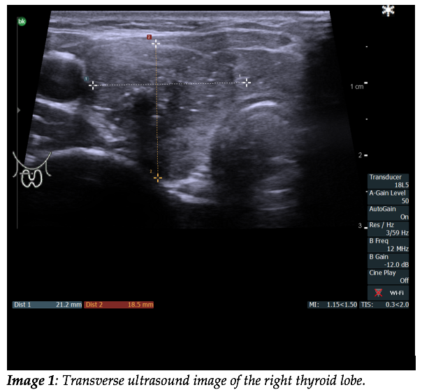
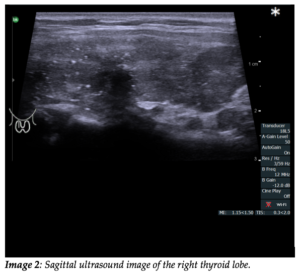
The ultrasound images show the right thyroid lobe with a solitary mass with irregular outline. The nodule has a “taller-than-wide” shape and is hypoechogenic with small punctuate regions of hyperechogenicity representing microcalcifications, which is highly specific for papillary thyroid cancer (1). The image also demonstrates chaotic intranodular vascularity. The video displays multiple enlarged (>10mm) metastatic lymph nodes on the neck with sharp borders. They are predominantly hyperechoic with intranodal necrosis with a cystic appearance. Furthermore the lymph nodes display mixed vascularity (peripheral and hilar) and peripheral microcalcifications.
Nodules in the thyroid are usually benign; fewer than 5% are cancerous. They are mostly asymptomatic, but some patients present with symptoms as difficulty swallowing/breathing, stridor and hoarseness.
Papillary thyroid cancer (PTC) is the most common type of thyroid cancer (80%) followed by follicular (10%), medullary carcinoma (5-10%) and anaplastic carcinoma (1-2%). PTC is more common in the middle-aged (30-50 years of age) and more frequent in women. It has a good prognosis. PTC tends to metastasize early to local lymph nodes, with 40-50% of patients having nodal involvement at presentation (2). Commonly recognized risk factors for thyroid malignancy are family history of thyroid cancer/hereditary syndromes that predispose to thyroid cancer, accidental exposure to ionizing radiation in childhood or adolescence and medical irradiation during childhood. This patient lived in a city near Chernobyl where the “Chernobyl accident” happened in 1986 and she might have been exposed to radioactive iodine-131(I-131, a radioactive isotope). This isotope is believed to be responsible for the significant raise in thyroid cancers that are still occurring among people, who lived in the Chernobyl area and were children and adolescents at the time (3).
Differential diagnoses include anaplastic thyroid carcinoma, which is an aggressive cancer that occurs more commonly in elderly women. Anaplastic thyroid cancers are hypoechoic with increased vascularity and can present with or without microcalcifications(1).
Multinodular goiter is a diffuse enlargement of the thyroid gland with heterogeneous nodules. It is a common, benign thyroid disease, and nodules are often isoechoic and may have cystic components, coarse calcifications and perinodular vascularity(1,4). A benign goiter is not accompanied by metastatic lymph nodes.
Lymphoma of the thyroid is very rare cancer of the thyroid, incidence (1:1000000). Hashimotos thyroiditis is a major risk factor. Its radiologic findings are non-specific and can be found in a goiter with multiple hyper vascularized nodules and posterior acoustic enhancement, but calcifications are uncommon. Some present with lymphadenopathies (5).
When ultrasound or clinical findings raise suspicion of thyroid malignancy, fine needle aspiration biopsy should be performed to confirm the diagnosis(6).
Take Home Message
Thyroid nodules are common and often benign. Ultrasound characteristics associated with malignancy include: hypoechogenicity; infiltrative, irregular, or lobulated margins; intranodular microcalcifications; and a taller-than-wide shape (4,7). Beside thyroid ultrasound it is also important to perform cervical neck ultrasound for suspicious lymph nodes, a finding, which will increase the likelihood of malignant disease.
Conflicts of Interest
The authors declare no conflict of interest
Ethics
The patient gave informed consent to publish this anonymized case.
References
- Ahuja AT. Diagnostic Ultrasound: Head and Neck. 2nd ed. Wang DTC, editor. Elsevier Health Sciences; 2019.
- Mao J, Zhang Q, Zhang H, Zheng K, Wang R, Wang G. Risk Factors for Lymph Node Metastasis in Papillary Thyroid Carcinoma: A Systematic Review and Meta-Analysis. Front Endocrinol (Lausanne). 2020;11:265.
- Samet JM, Berrington de González A, Dauer LT, Hatch M, Kosti O, Mettler FAJ, et al. Gilbert W. Beebe Symposium on 30 Years after the Chernobyl Accident: Current and Future Studies on Radiation Health Effects. Vol. 189, Radiation research. 2018. p. 5–18.
- Alexander LF, Patel NJ, Caserta MP, Robbin ML. Thyroid Ultrasound: Diffuse and Nodular Disease. Radiol Clin North Am. 2020 Nov;58(6):1041–57.
- Xia Y, Wang L, Jiang Y, Dai Q, Li X, Li W. Sonographic appearance of primary thyroid lymphoma-preliminary experience. PLoS One. 2014;9(12):e114080.
- Todsen T, Bennedbaek FN, Kiss K, Hegedüs L. Ultrasound-guided fine-needle aspiration biopsy of thyroid nodules. Head Neck. 2021 Mar;43(3):1009–13.
- Russ G, Bonnema SJ, Erdogan MF, Durante C, Ngu R, Leenhardt L. European Thyroid Association Guidelines for Ultrasound Malignancy Risk Stratification of Thyroid Nodules in Adults: The EU-TIRADS. Eur Thyroid J. 2017 Sep;6(5):225–37.
Incorrect
CORRECT ANSWER EXPLAINED BELOW Correct answer is: Papillary thyroid carcinoma
Discussion
The ultrasound images show the right thyroid lobe with a solitary mass with irregular outline. The nodule has a “taller-than-wide” shape and is hypoechogenic with small punctuate regions of hyperechogenicity representing microcalcifications, which is highly specific for papillary thyroid cancer (1). The image also demonstrates chaotic intranodular vascularity. The video displays multiple enlarged (>10mm) metastatic lymph nodes on the neck with sharp borders. They are predominantly hyperechoic with intranodal necrosis with a cystic appearance. Furthermore the lymph nodes display mixed vascularity (peripheral and hilar) and peripheral microcalcifications.
Nodules in the thyroid are usually benign; fewer than 5% are cancerous. They are mostly asymptomatic, but some patients present with symptoms as difficulty swallowing/breathing, stridor and hoarseness.
Papillary thyroid cancer (PTC) is the most common type of thyroid cancer (80%) followed by follicular (10%), medullary carcinoma (5-10%) and anaplastic carcinoma (1-2%). PTC is more common in the middle-aged (30-50 years of age) and more frequent in women. It has a good prognosis. PTC tends to metastasize early to local lymph nodes, with 40-50% of patients having nodal involvement at presentation (2). Commonly recognized risk factors for thyroid malignancy are family history of thyroid cancer/hereditary syndromes that predispose to thyroid cancer, accidental exposure to ionizing radiation in childhood or adolescence and medical irradiation during childhood. This patient lived in a city near Chernobyl where the “Chernobyl accident” happened in 1986 and she might have been exposed to radioactive iodine-131(I-131, a radioactive isotope). This isotope is believed to be responsible for the significant raise in thyroid cancers that are still occurring among people, who lived in the Chernobyl area and were children and adolescents at the time (3).
Differential diagnoses include anaplastic thyroid carcinoma, which is an aggressive cancer that occurs more commonly in elderly women. Anaplastic thyroid cancers are hypoechoic with increased vascularity and can present with or without microcalcifications(1).
Multinodular goiter is a diffuse enlargement of the thyroid gland with heterogeneous nodules. It is a common, benign thyroid disease, and nodules are often isoechoic and may have cystic components, coarse calcifications and perinodular vascularity(1,4). A benign goiter is not accompanied by metastatic lymph nodes.
Lymphoma of the thyroid is very rare cancer of the thyroid, incidence (1:1000000). Hashimotos thyroiditis is a major risk factor. Its radiologic findings are non-specific and can be found in a goiter with multiple hyper vascularized nodules and posterior acoustic enhancement, but calcifications are uncommon. Some present with lymphadenopathies (5).
When ultrasound or clinical findings raise suspicion of thyroid malignancy, fine needle aspiration biopsy should be performed to confirm the diagnosis(6).
Take Home Message
Thyroid nodules are common and often benign. Ultrasound characteristics associated with malignancy include: hypoechogenicity; infiltrative, irregular, or lobulated margins; intranodular microcalcifications; and a taller-than-wide shape (4,7). Beside thyroid ultrasound it is also important to perform cervical neck ultrasound for suspicious lymph nodes, a finding, which will increase the likelihood of malignant disease.
Conflicts of Interest
The authors declare no conflict of interest
Ethics
The patient gave informed consent to publish this anonymized case.
References
- Ahuja AT. Diagnostic Ultrasound: Head and Neck. 2nd ed. Wang DTC, editor. Elsevier Health Sciences; 2019.
- Mao J, Zhang Q, Zhang H, Zheng K, Wang R, Wang G. Risk Factors for Lymph Node Metastasis in Papillary Thyroid Carcinoma: A Systematic Review and Meta-Analysis. Front Endocrinol (Lausanne). 2020;11:265.
- Samet JM, Berrington de González A, Dauer LT, Hatch M, Kosti O, Mettler FAJ, et al. Gilbert W. Beebe Symposium on 30 Years after the Chernobyl Accident: Current and Future Studies on Radiation Health Effects. Vol. 189, Radiation research. 2018. p. 5–18.
- Alexander LF, Patel NJ, Caserta MP, Robbin ML. Thyroid Ultrasound: Diffuse and Nodular Disease. Radiol Clin North Am. 2020 Nov;58(6):1041–57.
- Xia Y, Wang L, Jiang Y, Dai Q, Li X, Li W. Sonographic appearance of primary thyroid lymphoma-preliminary experience. PLoS One. 2014;9(12):e114080.
- Todsen T, Bennedbaek FN, Kiss K, Hegedüs L. Ultrasound-guided fine-needle aspiration biopsy of thyroid nodules. Head Neck. 2021 Mar;43(3):1009–13.
- Russ G, Bonnema SJ, Erdogan MF, Durante C, Ngu R, Leenhardt L. European Thyroid Association Guidelines for Ultrasound Malignancy Risk Stratification of Thyroid Nodules in Adults: The EU-TIRADS. Eur Thyroid J. 2017 Sep;6(5):225–37.


