Echoes Issue No. 25 [June 2021]
December 15, 2021
Case of the Month February 2022 – Intrauterine pregnancy, or is it?
January 31, 2022Malene Nerstrøm1,*, Jonathan Cohen1
1 Department of Radiology, Copenhagen University Hospital, Rigshospitalet, Copenhagen, Denmark; malene_nerstroem@hotmail.com
* Correspondence: malene_nerstroem@hotmail.com
Clinical History:
A 66-year-old female with no former history of abdominal surgery presented to our clinic with a palpable, superficial tumor superior to umbilicus. The tumor was not tender on palpation and persisted when the patient was lying on her back. The patient had no abdominal pain and normal gastrointestinal function.
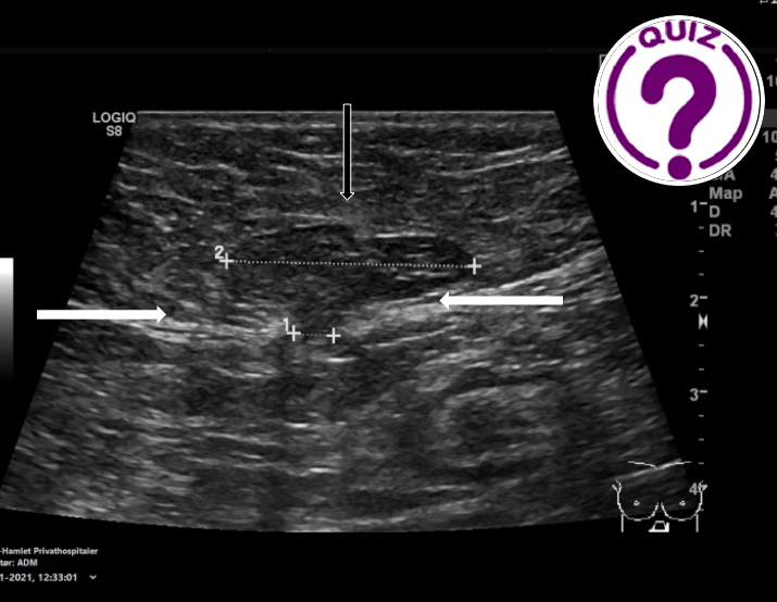
An ultrasound examination was performed revealing the images below.
Quiz-summary
0 of 1 questions completed
Questions:
- 1
Information
View the Case below, answer the question and then click check >
You have already completed the quiz before. Hence you can not start it again.
Quiz is loading...
You must sign in or sign up to start the quiz.
You have to finish following quiz, to start this quiz:
Results
0 of 1 questions answered correctly
Your time:
Time has elapsed
You have reached 0 of 0 points, (0)
Categories
- Not categorized 0%
- 1
- Answered
- Review
-
Question 1 of 1
1. Question
Question: What is the most likely diagnosis due to the ultrasound findings?
Correct
CORRECT ANSWER EXPLAINED BELOW Correct answer is: Epigastric hernia with coexisting rectus abdominis diastasis
Discussion
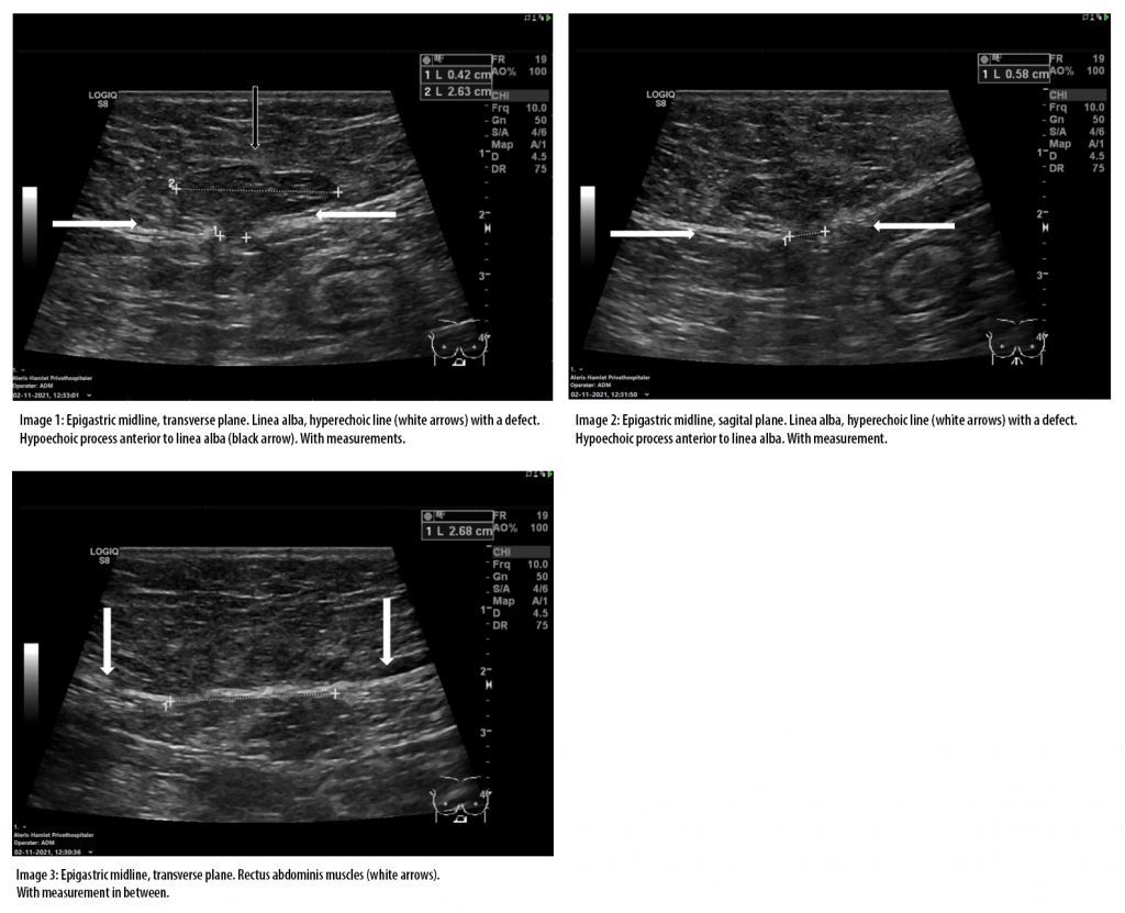
The ultrasound images revealed an epigastric hernia with coexisting rectus abdominis diastasis.
Image 1 and 2 show a small defect in the linea alba measuring 0.4 x 0.6 cm with herniation of preperitoneal fat. On image 3, the gap between the rectus abdominis muscles is seen measuring 2.7 cm. On ultrasound examination, sometimes only a small defect in linea alba is seen, without herniation of preperitoneal/omental fat, and rarely one finds herniation of bowel or stomach (1).
Epigastic hernias are defined as hernias occurring through a defect in the midline from above the umbilicus to the xiphoid process of the sternum. They represent 1.6 to 3.6 % of all abdominal wall hernias and are more common in men. Other risk factors include obesity, pregnancy, rectus diastasis, extensive physical training and connective tissue disorders. Epigastric hernias are often asymptomatic and the patient often presents to the clinic because of a small lump palpated in the upper abdomen. In most cases they are diagnosed by clinical examination alone, but imaging by ultrasound or CT can be considered when the diagnosis is uncertain. Asymptomatic patients can follow a watchful waiting strategy, but symptomatic patients often undergo surgery with repair of the midline defect with sutures or mesh (2-3).
Conflicts of Interest
The authors declare no conflict of interest
References
- Epigastric hernia | Radiology Reference Article | Radiopaedia.org. Available online: https://radiopaedia.org/articles/epigastric-hernia (accessed on 22.12.2021)
- A. Henriksen, A. Montgomery, R. Kaufmann, F. Berrevoet, B. East, J. Fischer, W. Hope, D. Klassen, R. Lorenz, Y. Renard , M. A. Garcia Urena and M. P. Simons on behalf of the European and Americas Hernia Societies (EHS and AHS). Guidelines for treatment of umbilical and epigastric hernias from the European Hernia Society and Americas Hernia Society. Br J Surg 2020 Feb;107(3):171-190.
- Overview of abdominal wall hernias in adults – UpToDate. Available online:
https://www.uptodate.com/contents/overview-of-abdominal-wall-hernias-in-adults?search=epigastric%20hernia&source=search_result&selectedTitle=1~19&usage_type=default&display_rank=1 (accessed on 26.12.2021).
Incorrect
CORRECT ANSWER EXPLAINED BELOW Correct answer is: Epigastric hernia with coexisting rectus abdominis diastasis
Discussion
The ultrasound images revealed an epigastric hernia with coexisting rectus abdominis diastasis.
Image 1 and 2 show a small defect in the linea alba measuring 0.4 x 0.6 cm with herniation of preperitoneal fat. On image 3, the gap between the rectus abdominis muscles is seen measuring 2.7 cm. On ultrasound examination, sometimes only a small defect in linea alba is seen, without herniation of preperitoneal/omental fat, and rarely one finds herniation of bowel or stomach (1).
Epigastic hernias are defined as hernias occurring through a defect in the midline from above the umbilicus to the xiphoid process of the sternum. They represent 1.6 to 3.6 % of all abdominal wall hernias and are more common in men. Other risk factors include obesity, pregnancy, rectus diastasis, extensive physical training and connective tissue disorders. Epigastric hernias are often asymptomatic and the patient often presents to the clinic because of a small lump palpated in the upper abdomen. In most cases they are diagnosed by clinical examination alone, but imaging by ultrasound or CT can be considered when the diagnosis is uncertain. Asymptomatic patients can follow a watchful waiting strategy, but symptomatic patients often undergo surgery with repair of the midline defect with sutures or mesh (2-3).
Conflicts of Interest
The authors declare no conflict of interest
References
- Epigastric hernia | Radiology Reference Article | Radiopaedia.org. Available online: https://radiopaedia.org/articles/epigastric-hernia (accessed on 22.12.2021)
- A. Henriksen, A. Montgomery, R. Kaufmann, F. Berrevoet, B. East, J. Fischer, W. Hope, D. Klassen, R. Lorenz, Y. Renard , M. A. Garcia Urena and M. P. Simons on behalf of the European and Americas Hernia Societies (EHS and AHS). Guidelines for treatment of umbilical and epigastric hernias from the European Hernia Society and Americas Hernia Society. Br J Surg 2020 Feb;107(3):171-190.
- Overview of abdominal wall hernias in adults – UpToDate. Available online:
https://www.uptodate.com/contents/overview-of-abdominal-wall-hernias-in-adults?search=epigastric%20hernia&source=search_result&selectedTitle=1~19&usage_type=default&display_rank=1 (accessed on 26.12.2021).


