WFUMB / EFSUMB Students Webinar Series: 5 February 2022
February 8, 2022
WFUMB / EFSUMB Students Webinar Series: 5 March 2022
March 7, 2022Rania Soussi1, Hassen Gharbi1 and Wiem Douira-Khomsi1*
1 Department of Paediatric Radiology, Béchir Hamza Children’s Hospital, Tunis, Tunisia
* Correspondence: khomsiwiem@yahoo.fr
Clinical History:
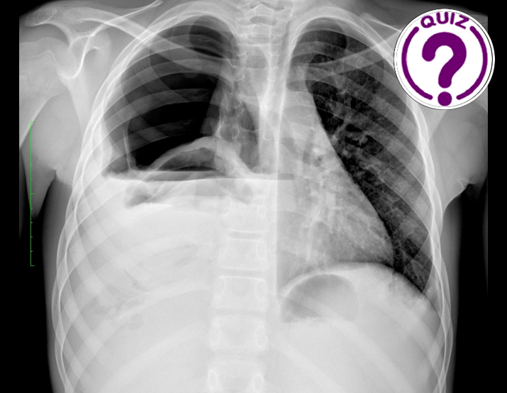
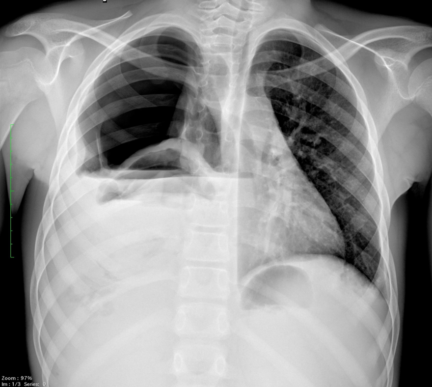
A 14-year-old boy was referred to our department due to coughing, fever, and shortness of breath. Physical examination showed an epigastric mass, and an x-ray of the thorax was requested.
Quiz-summary
0 of 2 questions completed
Questions:
- 1
- 2
Information
View the MARCH Case below, answer the question and then click check >
You have already completed the quiz before. Hence you can not start it again.
Quiz is loading...
You must sign in or sign up to start the quiz.
You have to finish following quiz, to start this quiz:
Results
0 of 2 questions answered correctly
Your time:
Time has elapsed
You have reached 0 of 0 points, (0)
Categories
- Not categorized 0%
- 1
- 2
- Answered
- Review
-
Question 1 of 2
1. Question
Question 1: What are the x-ray findings?
Correct
CORRECT ANSWER EXPLAINED BELOW Correct answer to Q1 is: Right sided opacity, right sided hydropneumothorax and elevation of the right diaphragm
Incorrect
CORRECT ANSWER EXPLAINED BELOW Correct answer to Q1 is: Right sided opacity, right sided hydropneumothorax and elevation of the right diaphragm
-
Question 2 of 2
2. Question

The x-ray revealed a right sided opacity with an air-fluid level and ipsilateral hydropneumothorax.
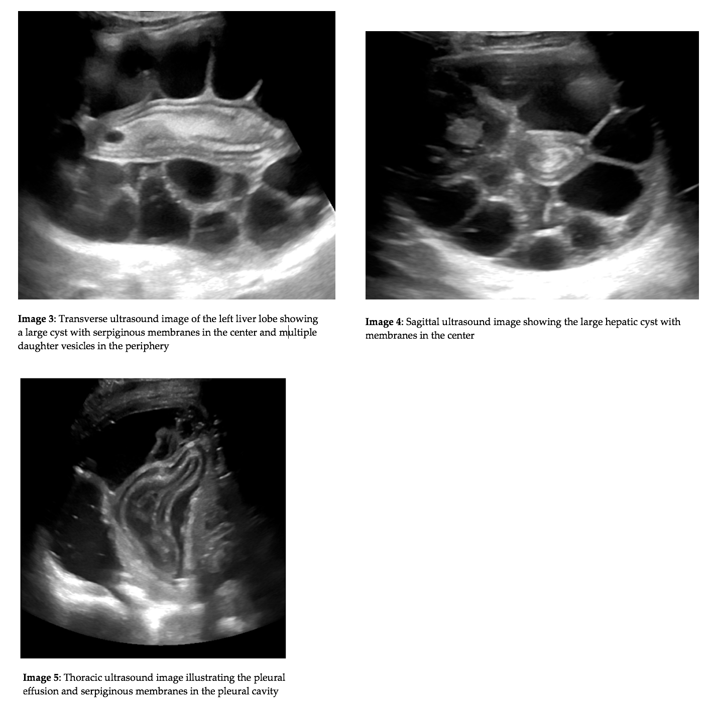
Due to the findings on the x-ray an abdominal and thoracic ultrasound was performed. The abdominal ultrasound examination revealed a large epigastric cyst attached to the left liver lobe. The cyst had serpiginous membranes in the center of the cyst and multiple daughter vesicles in the periphery. Chest ultrasound showed another cyst in the lung filled with serpentine linear structures which was ruptured into the pleural cavity.

Question 2: What is the most likely diagnosis?
Correct
CORRECT ANSWER EXPLAINED BELOW Correct answer to Q2 is: Hydatic cysts.
Discussion:
Firstly, the patient underwent surgical treatment consisting of decortication and cystotomy of the lung cyst. Secondly, surgeons performed a closed total pericystectomy for the liver-attached hydatic cyst. In addition to that, Albendazole was administrated for three months after the intervention.
Ultrasound is the imaging modality of choice in the diagnosis of hydatid cysts. It is used to establish the diagnosis and to describe the location, number and stage of lesions. Both the WHO and Gharbi classifications are based on the ultrasound findings (1-2). The WHO classification divides cysts into active, transitional and inactive stages, which is relevant for treatment and follow up. Last, but not least, ultrasound helps to find complications such as pleural effusion or bile duct rupture. In these cases, treatment is essentially surgical with a controversial approach and a high risk of complications and recurrence (3).
Conflicts of interest
“The authors declare no conflict of interest.”
References
- Gharbi HA, Hassine W et al. Ultrasound examination of the hydaticliver. Radiology. 1981; 139:459–463.
- WHO informal working group. International classification of ultrasound images in cystic echinococcosis for application in clinical and field epidemiological settings. Acta Trop 2003; 85:253-261
- Brunetti E, Kern P. Expert consensus for the diagnosis and treatment of cystic and alveolar echinococcosis in humans. Acta Trop 2010; 114:1-16
Incorrect
CORRECT ANSWER EXPLAINED BELOW Correct answer to Q2 is: Hydatic cysts.
Discussion:
Firstly, the patient underwent surgical treatment consisting of decortication and cystotomy of the lung cyst. Secondly, surgeons performed a closed total pericystectomy for the liver-attached hydatic cyst. In addition to that, Albendazole was administrated for three months after the intervention.
Ultrasound is the imaging modality of choice in the diagnosis of hydatid cysts. It is used to establish the diagnosis and to describe the location, number and stage of lesions. Both the WHO and Gharbi classifications are based on the ultrasound findings (1-2). The WHO classification divides cysts into active, transitional and inactive stages, which is relevant for treatment and follow up. Last, but not least, ultrasound helps to find complications such as pleural effusion or bile duct rupture. In these cases, treatment is essentially surgical with a controversial approach and a high risk of complications and recurrence (3).
Conflicts of interest
“The authors declare no conflict of interest.”
References
- Gharbi HA, Hassine W et al. Ultrasound examination of the hydaticliver. Radiology. 1981; 139:459–463.
- WHO informal working group. International classification of ultrasound images in cystic echinococcosis for application in clinical and field epidemiological settings. Acta Trop 2003; 85:253-261
- Brunetti E, Kern P. Expert consensus for the diagnosis and treatment of cystic and alveolar echinococcosis in humans. Acta Trop 2010; 114:1-16


