Echoes Issue No. 26 [December 2021]
April 7, 2022
WFUMB / EFSUMB Students Webinar Series: 7 May 2022
May 13, 2022Ammy Prada1, Daniel Marquez1, Carlos Villegas1, Isaac Rodriguez2 Edda L. Chaves3
1Unidad de Perinatología, Hospital Universitario de Caracas. Universidad Central de Venezuela
2Servicio de Radiodiganóstico. Hospital Universitario de Caracas. Universidad Central de Venezuela
3 Servicio de Radiología. Hospital Regional Nicolas Solano. La Chorrera. Panamá
Corresponding author: Daniel Marquez danielmarquez33@gmail.com
Clinical History:
A 27-year-old female patient in her second pregnancy, 27 weeks 5 days on. She was referred to our health center for reevaluation because of findings in her routine exam. In our center we performed an anatomical detailed ultrasound with the following findings:
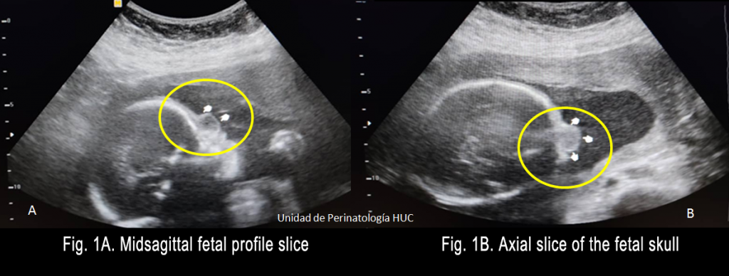
In the midsagittal fetal profile section, at the level of the frontal nasal region an anechoic rounded mass, with well-defined margins, left nasal predominance, is seen. It seems to emerge from the nasal bridge and is without colour Doppler signal (Fig. 1A). The axial section of the fetal skull shows an ovoid structure in the frontal nasal region. It is hyperechoic compared to the brain, and isoechoic with soft tissue.
Quiz-summary
0 of 2 questions completed
Questions:
- 1
- 2
Information
View the May Case below, answer the question and then click check >
You have already completed the quiz before. Hence you can not start it again.
Quiz is loading...
You must sign in or sign up to start the quiz.
You have to finish following quiz, to start this quiz:
Results
0 of 2 questions answered correctly
Your time:
Time has elapsed
You have reached 0 of 0 points, (0)
Categories
- Not categorized 0%
- 1
- 2
- Answered
- Review
-
Question 1 of 2
1. Question
Question 1: Which probable malformation of the central nervous system would you suspect from the ultrasound images?
Correct
CORRECT ANSWER EXPLAINED BELOW Correct answer to Q1 is: Frontonasal encephalocele
Incorrect
CORRECT ANSWER EXPLAINED BELOW Correct answer to Q1 is: Frontonasal encephalocele
-
Question 2 of 2
2. Question
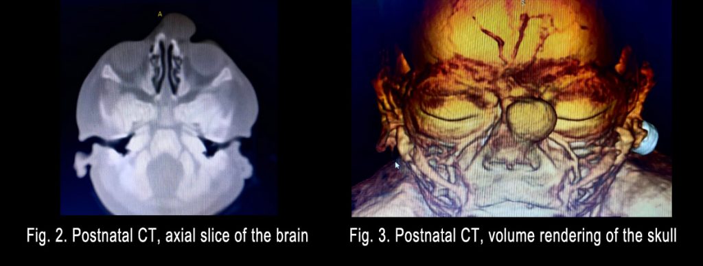
The postnatal CT of the brain in the axial plane and volume rendering (Fig 2-3) shows an ovoid nodular lesion in the left frontal nasal region. It has regular borders, is hypodense compared to soft tissue and measures 1.7 x 1.1 x 1.7 cm. The lesion does not cause erosion, fracture or sclerosis of the proper bones of the nose.
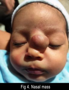
The clinical finding (Fig. 4.) was an asymmetrical nasal pyramid at the expense of the nasal bridge. It was not compressible, was negative for transillumination, not pulsatile, and did not increase in volume with Valsalva’s maneuver (cry).
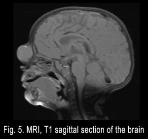
MRI of the brain showed a nodular rounded lesion with well-defined edges, hypointense, with slight enhancement after contrast administration on T1 weighted sequences, located in the nasal pyramid. There are no signs of calcification, no evidence of vascular paths, no adjacent edema, no signs of communication with the brain parenchyma and, no relation with the CNS, and it does not have any fatty tissue.
Question 2: Which diagnosis do you suggest?
Correct
CORRECT ANSWER EXPLAINED BELOW Correct answer to Q2 is: Nasal glioma
Discussion:
To diagnose tumors of the facial middle line, different imaging must be performed pre-natal and post-natal. These include pre-natal ultrasound and post-natal MRI and CT. The final diagnosis requires histology after surgical resection.
A congenital nasal mass, in the midline, is a rare developmental abnormality. Nasal glioma or heterotopic nasal glial is a rare benign congenital tumor. Its origin is in the ectopic glial cells located at the level of the nasal root with no communication with the central nervous system. They represent approximately 5% of all congenital nasal abnormalities and may present as intranasal masses (30%), extranasal masses (60%) or mixed (10%). With a predominance of 3:1 in male sex.
The encephaloceles are herniations of the contents of the cranium that maintain communication with the brain through a bone defect, they are characterized by being pulsatile, increasing in size with weeping with Valsalva or jugular compression (Furstemberg’s sign). Illumination through the tumor is possible.3-4
Dermoid cysts are the most frequent congenital midline nasal masses, with an incidence of 61% with a higher frequency in boys .3,5,6-9
Hemangiomas are vascular lesions. They are frequent in infancy, especially located in the head and neck. We ruled out this diagnosis, since there was no Doppler signal during the ultrasound evaluation
CT is useful to define bone anatomy, and MRI is essential to determine the content of the mass. It will show a fluid signal in an epidermoid cyst, fat in a dermoid and encephaloceles are isointense masses with the gray matter, contiguous with the brain parenchyma that extends through a bone defect. While in the case of gliomas a well circumscribed mass hypointense or isointense with the grey matter is seen. 3,7,10,11 The last features coincide with the findings in the post-natal imaging studies in our case.
Conclusion
Congenital midline nasal tumors are rare entities, but susceptible to prenatal diagnosis. If there is no compromise of the central nervous system, the prognosis is generally favorable. Nasal gliomas should be treated by complete excision to prevent recurrence. Prenatal follow up should be with ultrasound. After birth all imaging findings and clinical findings must be assessed by a multidisciplinary team. The treatment of choice is surgical resection, and the definitive diagnosis is made by histology.
Conflicts of interest
“The authors declare no conflict of interest.”
References
- Hughes GB, Sharpino G, Hunt W, Tucker HM. Management of the congenital midline nasal mass: a review. Head Neck Surg. [Internet]. 1980. [consultado 17 de abril 2022]; 2(3):222-33. doi: 10.1002/hed.2890020308.
- Casas E, Benlloch M, Cerdá N. A propósito de un caso de glioma nasal en un recién nacido. An Esp Pediatr [Internet]. 1998 [citado 17 de abril 2022];49:624-6. Disponible en: efaidnbmnnnibpcajpcglclefindmkaj/viewer.html?pdfurl=https%3a %2F %2Fwww.aeped.es%2Fsites%2Fdefault%2Ffiles%2Fanales%2F49-6 14.pdf& clen=74201&chunk=true.
- Morales G, Fauque L., Gerard A y col. Tumores nasales congénitos de la línea media. Med. infant ; [Internet]. Junio 2018 [citado 17 de abril 2022]; 25(2): 205-12. Disponible en: https://pesquisa.bvsalud.org/portal/resource/pt/biblio-909962.
- Zhang, W., Tang, L. Intranasal glial heterotopia in a male infant: A case report. Medicine, [Internet]. 2017. [consultado 17 de abril 2022]; 99(29). doi: 10.1097/MD.0000000000021200.
- Van Wyhe RD, Chamata ES, Hollier LH. Midline Craniofacial Masses in Children. Semin Plast Surg. [Internet]. [consultado abril 2022] 30(4):176-80. doi: 10.1055/s-0036-1593482.
- Marrugo G, Torres J masa nasal congénita: quiste epidermoide.. Rev. Fac. Med. [Internet]. 2010 [consultado 17 de abril 2022]; 58 no.4. Disponible en: http://www.scielo.org.co/scielo.php?script=sci_arttext&pid=S0120-00112010000400011.
- Clarós P, Ribeiro I, Clarós A. Nasal Glioma: Prenatal Imaging Diagnosis. J Radiol Clin Imaging [Internet]. 2019; [citado 17 de abril 2022]; 2 (1): 033-41 doi: 10.26502/jrci.2644-2809008.
- James D, Pierre M, Philip Cogen Congenital Nasal Masses: CT and MR Imaging Features in 16 Cases. AJNR:12, January/February 1991.
- Chtira K, Elallouchi Y, Zahrou F, Assamadi M, Ait El Qadi A, Ghannane H, Laghmari M. Nasofrontal surgical reconstruction by external table flap of frontal bone following removal of a dermoid cyst revealed by a fistula: A case report and review of the literature. GMS Interdiscip Plast Reconstr Surg DGPW. [Internet]. [citado 17 de abril de 2022]; 8: Doc01. doi: 10.3205/iprs000127.
- Ni K, Li X, Zhao L, Wu J, Liu X, Shi H. Diagnosis and treatment of congenital nasal dermoid and sinus cysts in 11 infants: A consort compliant study. Medicine (Baltimore). [Internet]. 2020 May [consultado 17 de abril 2022]; 22;99(21):e19435. doi: 10.1097/MD.0000000000019435.
- Choudhary G, Udayasankar U, Saade C, Winegar B, Maroun G, Chokr J. A systematic approach in the diagnosis of paediatric skull lesions: what radiologists need to know. Pol J Radiol. [Internet]. [citado 17 de abril de 2022]; 84:e92-e111. doi: 10.5114/pjr.2019.83101.
Incorrect
CORRECT ANSWER EXPLAINED BELOW Correct answer to Q2 is: Nasal glioma
Discussion:
To diagnose tumors of the facial middle line, different imaging must be performed pre-natal and post-natal. These include pre-natal ultrasound and post-natal MRI and CT. The final diagnosis requires histology after surgical resection.
A congenital nasal mass, in the midline, is a rare developmental abnormality. Nasal glioma or heterotopic nasal glial is a rare benign congenital tumor. Its origin is in the ectopic glial cells located at the level of the nasal root with no communication with the central nervous system. They represent approximately 5% of all congenital nasal abnormalities and may present as intranasal masses (30%), extranasal masses (60%) or mixed (10%). With a predominance of 3:1 in male sex.
The encephaloceles are herniations of the contents of the cranium that maintain communication with the brain through a bone defect, they are characterized by being pulsatile, increasing in size with weeping with Valsalva or jugular compression (Furstemberg’s sign). Illumination through the tumor is possible.3-4
Dermoid cysts are the most frequent congenital midline nasal masses, with an incidence of 61% with a higher frequency in boys .3,5,6-9
Hemangiomas are vascular lesions. They are frequent in infancy, especially located in the head and neck. We ruled out this diagnosis, since there was no Doppler signal during the ultrasound evaluation
CT is useful to define bone anatomy, and MRI is essential to determine the content of the mass. It will show a fluid signal in an epidermoid cyst, fat in a dermoid and encephaloceles are isointense masses with the gray matter, contiguous with the brain parenchyma that extends through a bone defect. While in the case of gliomas a well circumscribed mass hypointense or isointense with the grey matter is seen. 3,7,10,11 The last features coincide with the findings in the post-natal imaging studies in our case.
Conclusion
Congenital midline nasal tumors are rare entities, but susceptible to prenatal diagnosis. If there is no compromise of the central nervous system, the prognosis is generally favorable. Nasal gliomas should be treated by complete excision to prevent recurrence. Prenatal follow up should be with ultrasound. After birth all imaging findings and clinical findings must be assessed by a multidisciplinary team. The treatment of choice is surgical resection, and the definitive diagnosis is made by histology.
Conflicts of interest
“The authors declare no conflict of interest.”
References
- Hughes GB, Sharpino G, Hunt W, Tucker HM. Management of the congenital midline nasal mass: a review. Head Neck Surg. [Internet]. 1980. [consultado 17 de abril 2022]; 2(3):222-33. doi: 10.1002/hed.2890020308.
- Casas E, Benlloch M, Cerdá N. A propósito de un caso de glioma nasal en un recién nacido. An Esp Pediatr [Internet]. 1998 [citado 17 de abril 2022];49:624-6. Disponible en: efaidnbmnnnibpcajpcglclefindmkaj/viewer.html?pdfurl=https%3a %2F %2Fwww.aeped.es%2Fsites%2Fdefault%2Ffiles%2Fanales%2F49-6 14.pdf& clen=74201&chunk=true.
- Morales G, Fauque L., Gerard A y col. Tumores nasales congénitos de la línea media. Med. infant ; [Internet]. Junio 2018 [citado 17 de abril 2022]; 25(2): 205-12. Disponible en: https://pesquisa.bvsalud.org/portal/resource/pt/biblio-909962.
- Zhang, W., Tang, L. Intranasal glial heterotopia in a male infant: A case report. Medicine, [Internet]. 2017. [consultado 17 de abril 2022]; 99(29). doi: 10.1097/MD.0000000000021200.
- Van Wyhe RD, Chamata ES, Hollier LH. Midline Craniofacial Masses in Children. Semin Plast Surg. [Internet]. [consultado abril 2022] 30(4):176-80. doi: 10.1055/s-0036-1593482.
- Marrugo G, Torres J masa nasal congénita: quiste epidermoide.. Rev. Fac. Med. [Internet]. 2010 [consultado 17 de abril 2022]; 58 no.4. Disponible en: http://www.scielo.org.co/scielo.php?script=sci_arttext&pid=S0120-00112010000400011.
- Clarós P, Ribeiro I, Clarós A. Nasal Glioma: Prenatal Imaging Diagnosis. J Radiol Clin Imaging [Internet]. 2019; [citado 17 de abril 2022]; 2 (1): 033-41 doi: 10.26502/jrci.2644-2809008.
- James D, Pierre M, Philip Cogen Congenital Nasal Masses: CT and MR Imaging Features in 16 Cases. AJNR:12, January/February 1991.
- Chtira K, Elallouchi Y, Zahrou F, Assamadi M, Ait El Qadi A, Ghannane H, Laghmari M. Nasofrontal surgical reconstruction by external table flap of frontal bone following removal of a dermoid cyst revealed by a fistula: A case report and review of the literature. GMS Interdiscip Plast Reconstr Surg DGPW. [Internet]. [citado 17 de abril de 2022]; 8: Doc01. doi: 10.3205/iprs000127.
- Ni K, Li X, Zhao L, Wu J, Liu X, Shi H. Diagnosis and treatment of congenital nasal dermoid and sinus cysts in 11 infants: A consort compliant study. Medicine (Baltimore). [Internet]. 2020 May [consultado 17 de abril 2022]; 22;99(21):e19435. doi: 10.1097/MD.0000000000019435.
- Choudhary G, Udayasankar U, Saade C, Winegar B, Maroun G, Chokr J. A systematic approach in the diagnosis of paediatric skull lesions: what radiologists need to know. Pol J Radiol. [Internet]. [citado 17 de abril de 2022]; 84:e92-e111. doi: 10.5114/pjr.2019.83101.


