Case of the Month February 2023 – Importance of diagnostic imaging in the management of a tumor lesion
February 14, 2023
Echoes Issue No. 30 [December 2022]
March 31, 2023Marta Duczkowska 1,*, Thomas Skaarup Kristensen 1
1 Department of Radiology, Copenhagen University Hospital, Rigshospitalet, Copenhagen, Denmark
* Correspondence: marta.duczkowska@regionh.dk
Clinical history
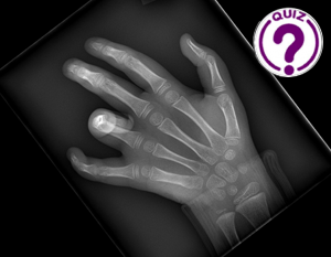
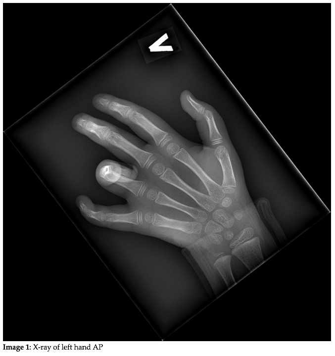
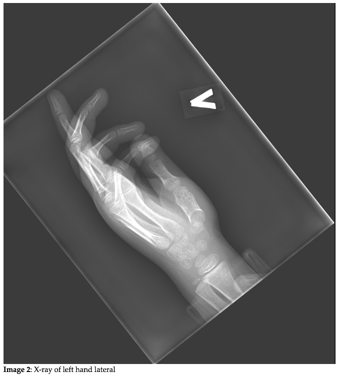
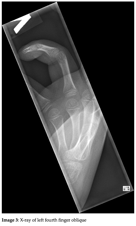
A 5-year-old girl was referred to our hospital 7 days after a fall with unknown mechanism. She had persistent inability to extend her fourth finger. Physical examination revealed her left fourth finger in flexion at the PIP joint with indirect pain of the finger as well as direct pain of the proximal phalanx. The finger could be extended using force, causing pain in both the finger and the volar aspect of the hand at the level of 4th ray. A left hand X-ray series were obtained to look for fractures.
Quiz-summary
0 of 2 questions completed
Questions:
- 1
- 2
Information
View the May Case below, answer the question and then click check >
You have already completed the quiz before. Hence you can not start it again.
Quiz is loading...
You must sign in or sign up to start the quiz.
You have to finish following quiz, to start this quiz:
Results
0 of 2 questions answered correctly
Your time:
Time has elapsed
You have reached 0 of 0 points, (0)
Categories
- Not categorized 0%
- 1
- 2
- Answered
- Review
-
Question 1 of 2
1. Question
Question 1: What are the X-ray findings?
Correct
CORRECT ANSWER EXPLAINED BELOW Correct answer to Q1 is: No visible bony lesions
Incorrect
CORRECT ANSWER EXPLAINED BELOW Correct answer to Q1 is: No visible bony lesions
-
Question 2 of 2
2. Question
Discussion
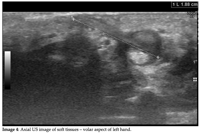
Left hand X-ray series confirmed volar flexion of the left fourth finger at the PIP joint and showed no dislocations or fractures. No radiopaque foreign bodies in the soft tissues were visible. On closer examination edema and a small wound could be seen on volar aspect of the hand at the level of 4th ray. Supplementary ultrasound of the palmar surface of the left hand was performed to look for a foreign body.
Additional Images
Question 2: What is the correct diagnosis?
Correct
CORRECT ANSWER EXPLAINED BELOW Correct answer to Q2 is: Foreign body penetrating into the flexor tendon
Additional discussion
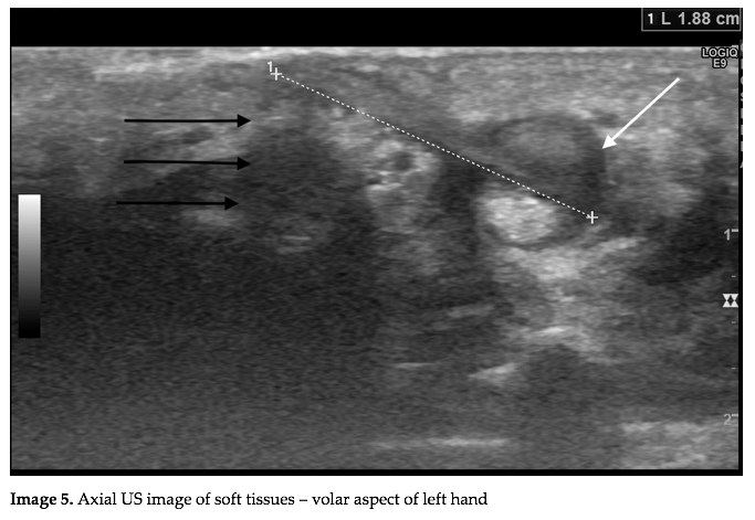
US examination using a 6-15 MHz linear probe showed a 19-mm-long echogenic structure extending into the flexor tendon of the left fourth finger at the level of the distal metacarpal bone, locking the tendon in place. There is a hypoechoic rim around the tendon associated with thickened synovium (white arrow). Slight posterior shadowing of the foreign body is noted (black arrows).
Surgical exploration was performed without complications with successful retrieval of a 2 cm long wood splinter. The procedure yielded a good result with immediate return to full range of motion of the finger.
Hand injuries are among the most common causes of emergency room visits in clinical practice and they are frequently associated with the presence of a retained foreign body. Foreign bodies are often overlooked at initial examination and can lead to a wide range of complications including infection, neurological deficits or tendon lacerations (1,2,3).
While wood slivers are among the most commonly reported foreign bodies, their detection remains exceptionally challenging, because plain radiography fails to visualize them in the majority of cases (1,4). The correct diagnosis becomes even more difficult when patients present without a reported history of penetrating injury.
Ultrasound is the modality of choice for evaluation of soft tissues, aiding not only in the detection of foreign materials, but also in providing their more precise localization and evaluating concomitant complications. It is especially useful in cases of radiolucent materials, which can otherwise remain undetected (2,3). Wood splinters typically appear as linear echogenic structures with pronounced acoustic shadowing (3).
Conclusion
Retained wooden foreign bodies can be easily missed on clinical evaluation and radiography. US allows accurate detection of radiolucent soft-tissue foreign bodies and aids in assessment of their associated complications.
Conflicts of Interest:
No conflict of interest.
References
- Anderson MA, Newmeyer WL 3rd, Kilgore ES. Diagnosis and treatment of retained foreign bodies in the hand. Am J Surg 1982; 144:63– 67
- Boyse TD, Fessell DP, Jacobson JA, Lin J, van Holsbeeck MT, Hayes CW. US of soft-tissue foreign bodies and associated complications with surgical correlation. Radiographics 2001; 21:1251–1256
- Jacobson JA, Powell A, Craig JG, Bouffard JA, van Holsbeeck MT. Wooden foreign bodies in soft tissue: detection at US. Radiology 1998; 206:45– 48.
- Peterson JJ, Bancroft LW, Kransdorf MJ. Wooden foreign bodies: imaging appearance. AJR Am J Roentgenol 2002; 178(3):557–562
Incorrect
CORRECT ANSWER EXPLAINED BELOW Correct answer to Q2 is: Foreign body penetrating into the flexor tendon
Additional discussion
US examination using a 6-15 MHz linear probe showed a 19-mm-long echogenic structure extending into the flexor tendon of the left fourth finger at the level of the distal metacarpal bone, locking the tendon in place. There is a hypoechoic rim around the tendon associated with thickened synovium (white arrow). Slight posterior shadowing of the foreign body is noted (black arrows).
Surgical exploration was performed without complications with successful retrieval of a 2 cm long wood splinter. The procedure yielded a good result with immediate return to full range of motion of the finger.
Hand injuries are among the most common causes of emergency room visits in clinical practice and they are frequently associated with the presence of a retained foreign body. Foreign bodies are often overlooked at initial examination and can lead to a wide range of complications including infection, neurological deficits or tendon lacerations (1,2,3).
While wood slivers are among the most commonly reported foreign bodies, their detection remains exceptionally challenging, because plain radiography fails to visualize them in the majority of cases (1,4). The correct diagnosis becomes even more difficult when patients present without a reported history of penetrating injury.
Ultrasound is the modality of choice for evaluation of soft tissues, aiding not only in the detection of foreign materials, but also in providing their more precise localization and evaluating concomitant complications. It is especially useful in cases of radiolucent materials, which can otherwise remain undetected (2,3). Wood splinters typically appear as linear echogenic structures with pronounced acoustic shadowing (3).
Conclusion
Retained wooden foreign bodies can be easily missed on clinical evaluation and radiography. US allows accurate detection of radiolucent soft-tissue foreign bodies and aids in assessment of their associated complications.
Conflicts of Interest:
No conflict of interest.
References
- Anderson MA, Newmeyer WL 3rd, Kilgore ES. Diagnosis and treatment of retained foreign bodies in the hand. Am J Surg 1982; 144:63– 67
- Boyse TD, Fessell DP, Jacobson JA, Lin J, van Holsbeeck MT, Hayes CW. US of soft-tissue foreign bodies and associated complications with surgical correlation. Radiographics 2001; 21:1251–1256
- Jacobson JA, Powell A, Craig JG, Bouffard JA, van Holsbeeck MT. Wooden foreign bodies in soft tissue: detection at US. Radiology 1998; 206:45– 48.
- Peterson JJ, Bancroft LW, Kransdorf MJ. Wooden foreign bodies: imaging appearance. AJR Am J Roentgenol 2002; 178(3):557–562

