Publications Committee EFSUMBOdd Helge Gilja
June 1, 2023
PRESIDENT ELECTGeorge Condous – ASUM
June 2, 2023Odd Helge Gilja
National Centre for Ultrasound in Gastroenterology, Haukeland University Hospital, Bergen, and Department of Clinical Medicine, University of Bergen, Norway.
Tel: +47 55972133
e-mail: odd.helge.gilja@helse-bergen.no
Clinical history
A 22-year-old man was admitted to hospital due to abdominal pain, fever, and minor diarrhoea for one week. The pain was mostly periumbilical but was also localized in the right lower and right upper part of the abdomen.
Images
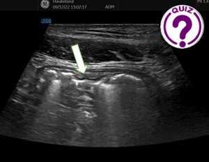
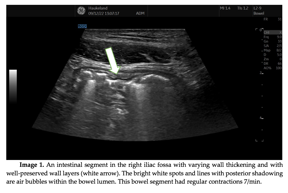
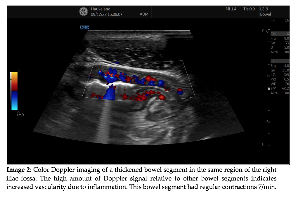
First, cholecystitis was excluded by ultrasound scanning the upper right quadrant. Subsequently, the probe was moved to the lower right part of the abdomen.
Quiz-summary
0 of 2 questions completed
Questions:
- 1
- 2
Information
View the June Case below, answer the question and then click check >
You have already completed the quiz before. Hence you can not start it again.
Quiz is loading...
You must sign in or sign up to start the quiz.
You have to finish following quiz, to start this quiz:
Results
0 of 2 questions answered correctly
Your time:
Time has elapsed
You have reached 0 of 0 points, (0)
Categories
- Not categorized 0%
- 1
- 2
- Answered
- Review
-
Question 1 of 2
1. Question
Question 1: What is the most likely diagnosis?
Correct
CORRECT ANSWER EXPLAINED BELOW Correct answer to Q1 is: Crohn’s disease.
Incorrect
CORRECT ANSWER EXPLAINED BELOW Correct answer to Q1 is: Crohn’s disease.
-
Question 2 of 2
2. Question
Discussion
Pseudomembranous colitis may display varying bowel wall thickness and increased vascularity. However, the right colon usually has infrequent contractions. Furthermore, no haustration can be seen in these images.
Yersinia enteritis often affects the distal part of the ileum and gives rise to increased Doppler signal. The symptoms of the patient and the relatively short duration (one week) supports this diagnosis. To determine the diagnosis of Yersinia enteritis, serum antibodies and biopsies of this particular bowel segment during ileocolonoscopy should be taken.
Appendicitis is very frequent and could very well present with the symptoms reported by this patient. However, the images do not support this diagnosis as the inflamed appendix is usually less wide, has a different shape, and does not have such wall thickening. On the other hand, increased Doppler signal in the wall is also frequently seen in acute appendicitis.
Crohn’s disease frequently presents in this age group, but often with a longer history of abdominal symptoms. The most frequent intestinal affection of Mb. Crohn is the terminal ileum, which is seen in image 1 and 2. The ultrasound images are typical of Crohn’s disease with the variation in wall thickness over a short segment with abrupt air interfaces on the luminal surface indicating ulcerations. The increase in color Doppler signal is usually seen in the acute phase of this disease. To prove this diagnosis ileocolonoscopy with biopsies was performed. The diagnosis of Mb. Crohn was confirmed and anti-inflammatory drugs were prescribed.
Additional Images
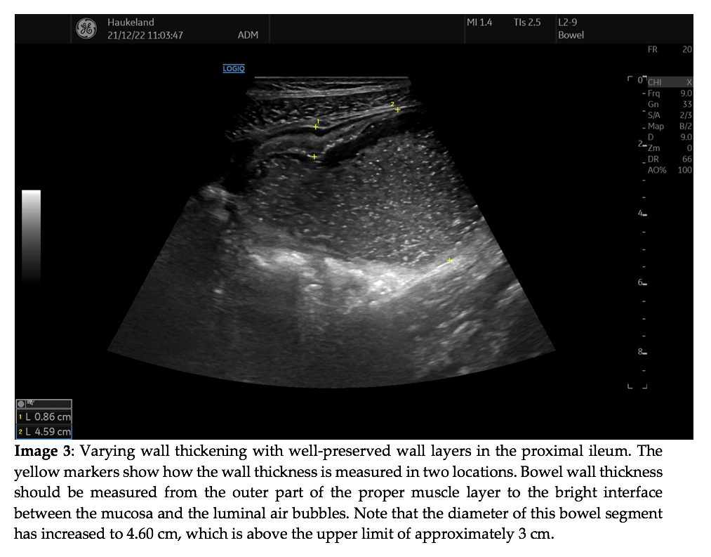
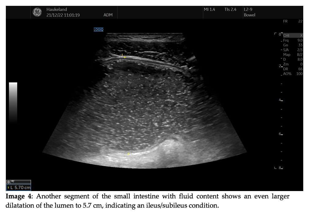
Video 1: This video demonstrates luminal content of the small intestine flowing through a narrow stenosis with markedly thickened bowel walls. The stenosis is short, only 2.5 cm in length.
Question 2: What would you do now while the patient is lying at your scanning facility?
Correct
CORRECT ANSWER EXPLAINED BELOW Correct answer to Q2 is: Consult the GI/IBD surgeon ASAP.
Additional discussion
There is only one of these alternatives which is definitely wrong, and that is number 4, as this patient needs urgent treatment and intensive surveillance. In our facility we would recommend calling the GI-surgeon on duty and arrange for an evaluation regarding hospital admittance, surgery etc. Surgeons at our facility will often drop by to observe the ultrasound examination in order to gain a better understanding of the findings.
A CT scan of the abdomen is often possible to perform urgently and is also often warranted by the operating surgeon to get an overview of the intestines. MRI is another possibility but may be difficult to obtain in the acute setting.
Conclusion
The patient underwent preoperative CT of the abdomen. It confirmed the bowel dilatations and stenoses shown by ultrasound. Subsequently adequate resections and stricturoplastics were performed.
Conflicts of Interest:
The author declares no conflict of interest with respect to this case.
References
- Atkinson NSS, Bryant RV, Dong Y, Maaser C, Kucharzik T, Maconi G, Asthana AK, Blaivas M, Goudie A, Gilja OH, Nuernberg D, Schreiber-Dietrich D, Dietrich CF. How to perform gastrointestinal ultrasound: Anatomy and normal findings. World J Gastroenterol. 2017 Oct 14;23(38):6931-694
- Maconi G, Nylund K, Ripolles T, Calabrese E, Dirks K, Dietrich CF, Hollerweger A, Sporea I, Saftoiu A, Maaser C, Hausken T, Higginson AP, Nürnberg D, Pallotta N, Romanini L, Serra C, Gilja OH. EFSUMB Recommendations and Clinical Guidelines for Intestinal Ultrasound (GIUS) in Inflammatory Bowel Diseases. Ultraschall Med. 2018 Jun;39(3):304-317. doi: 10.1055/s-0043-125329. Epub 2018 Mar 22. PubMed
- Hollerweger A, Maconi G, Ripolles T, Nylund K, Higginson A, Serra C, Dietrich CF, Dirks K, Gilja OH. Gastrointestinal Ultrasound (GIUS) in Intestinal Emergencies – An EFSUMB Position Paper. Ultraschall Med. 2020 Apr 20. doi: 10.1055/a-1147-1295.
- Sævik F, Gilja OH, Nylund K. Gastrointestinal Ultrasound Can Predict Endoscopic Activity in Crohn’s Disease. Ultraschall Med. 2020 Apr 24. doi: 10.1055/a-1149-9092. [Epub ahead of print] PubMed PMID: 32330994.
- Nylund K, Gilja OH, Dietrich CF. Gastrointestinal ultrasound. In Book: WFUMB ultrasound Book. Editors: Nurnberg D, Chammas C, Gilja OH, Sporea I, Sirli, R. ISBN: 978-1-8384990-0-6. WFUMB Publisher 2021. 251-264.
Incorrect
CORRECT ANSWER EXPLAINED BELOW Correct answer to Q2 is: Consult the GI/IBD surgeon ASAP.
Additional discussion
There is only one of these alternatives which is definitely wrong, and that is number 4, as this patient needs urgent treatment and intensive surveillance. In our facility we would recommend calling the GI-surgeon on duty and arrange for an evaluation regarding hospital admittance, surgery etc. Surgeons at our facility will often drop by to observe the ultrasound examination in order to gain a better understanding of the findings.
A CT scan of the abdomen is often possible to perform urgently and is also often warranted by the operating surgeon to get an overview of the intestines. MRI is another possibility but may be difficult to obtain in the acute setting.
Conclusion
The patient underwent preoperative CT of the abdomen. It confirmed the bowel dilatations and stenoses shown by ultrasound. Subsequently adequate resections and stricturoplastics were performed.
Conflicts of Interest:
The author declares no conflict of interest with respect to this case.
References
- Atkinson NSS, Bryant RV, Dong Y, Maaser C, Kucharzik T, Maconi G, Asthana AK, Blaivas M, Goudie A, Gilja OH, Nuernberg D, Schreiber-Dietrich D, Dietrich CF. How to perform gastrointestinal ultrasound: Anatomy and normal findings. World J Gastroenterol. 2017 Oct 14;23(38):6931-694
- Maconi G, Nylund K, Ripolles T, Calabrese E, Dirks K, Dietrich CF, Hollerweger A, Sporea I, Saftoiu A, Maaser C, Hausken T, Higginson AP, Nürnberg D, Pallotta N, Romanini L, Serra C, Gilja OH. EFSUMB Recommendations and Clinical Guidelines for Intestinal Ultrasound (GIUS) in Inflammatory Bowel Diseases. Ultraschall Med. 2018 Jun;39(3):304-317. doi: 10.1055/s-0043-125329. Epub 2018 Mar 22. PubMed
- Hollerweger A, Maconi G, Ripolles T, Nylund K, Higginson A, Serra C, Dietrich CF, Dirks K, Gilja OH. Gastrointestinal Ultrasound (GIUS) in Intestinal Emergencies – An EFSUMB Position Paper. Ultraschall Med. 2020 Apr 20. doi: 10.1055/a-1147-1295.
- Sævik F, Gilja OH, Nylund K. Gastrointestinal Ultrasound Can Predict Endoscopic Activity in Crohn’s Disease. Ultraschall Med. 2020 Apr 24. doi: 10.1055/a-1149-9092. [Epub ahead of print] PubMed PMID: 32330994.
- Nylund K, Gilja OH, Dietrich CF. Gastrointestinal ultrasound. In Book: WFUMB ultrasound Book. Editors: Nurnberg D, Chammas C, Gilja OH, Sporea I, Sirli, R. ISBN: 978-1-8384990-0-6. WFUMB Publisher 2021. 251-264.

