WFUMB AFSUMB Student Webinar on Ultrasound safety, technology and development
October 20, 2023
Beijing, China WFUMB COE
November 15, 2023Yuko Kono
University of California, San Diego. Department of Medicine, Department of Radiology
Correspondence: ykono@health.ucsd.edu
Clinical history
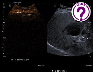
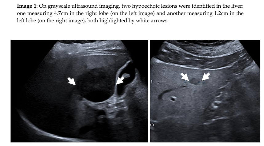
A 69-year-old female with a medical history of hypertension, hyperlipidemia, and NASH (non-alcoholic steatohepatitis) – without known cirrhosis – presents with liver lesions noted on images obtained at an outside hospital.
Images
Quiz-summary
0 of 3 questions completed
Questions:
- 1
- 2
- 3
Information
View the November Case below, answer the question and then click check >
You have already completed the quiz before. Hence you can not start it again.
Quiz is loading...
You must sign in or sign up to start the quiz.
You have to finish following quiz, to start this quiz:
Results
0 of 3 questions answered correctly
Your time:
Time has elapsed
You have reached 0 of 0 points, (0)
Categories
- Not categorized 0%
- 1
- 2
- 3
- Answered
- Review
-
Question 1 of 3
1. Question
Question 1: What is the likely diagnosis of this liver lesion?
Correct
CORRECT ANSWER EXPLAINED BELOW Correct answer to Q1 is: Cholangiocarcinoma
Incorrect
CORRECT ANSWER EXPLAINED BELOW Correct answer to Q1 is: Cholangiocarcinoma
-
Question 2 of 3
2. Question
Discussion
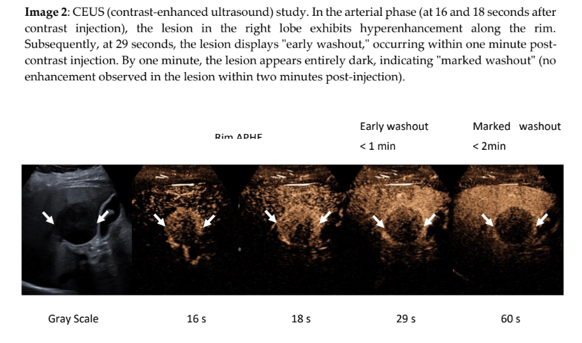
Rim arterial phase hyperenhancement (APHE), early washout (becomes darker compared to surrounding liver in less than 60 seconds after contrast injection), and marked washout (turning notably dark within 2 minutes after contrast injection) are distinctive CEUS imaging traits associated with intrahepatic cholangiocarcinoma or metastasis (1). A biopsy of this lesion confirmed the diagnosis of intrahepatic cholangiocarcinoma.
Question 2: What is the CEUS LI-RADS category for this hepatic lesion.
Correct
CORRECT ANSWER EXPLAINED BELOW Correct answer to Q2 is: Cannot Apply CEUS LI-RADS
Incorrect
CORRECT ANSWER EXPLAINED BELOW Correct answer to Q2 is: Cannot Apply CEUS LI-RADS
-
Question 3 of 3
3. Question
Discussion
LI-RADS (liver imaging Reporting and Data System) is a standardized system for the diagnosis of liver lesion on patients at high risk for HCC (hepatocellular carcinoma) (1, 2).
The imaging characteristics of this case align with those typically seen in cholangiocarcinoma, meeting the major imaging features for CEUS LI-RADS M. However, it is crucial to note that this patient has non alcoholic fatty liver without cirrhosis, making the application of CEUS LI-RADS inappropriate in this context. Its imperative to reserve LI-RADS for high-risk patients in order to maintain its high specificity of HCC for LI-RADS 5.
Additional images & videos
Question 3: What is the diagnosis of this lesion?
Correct
CORRECT ANSWER EXPLAINED BELOW Correct answer to Q3 is: Hemangioma
Additional Discussion:
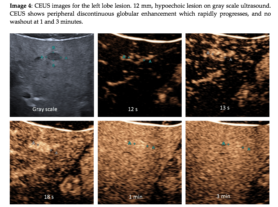
Although MRI is usually a good imaging modality to diagnose liver lesions, including hemangiomas, occasionally, it can misdiagnose with cholangiocarcinoma or vice versa as both lesions have rim enhancement and do not show washout on MRI or CT. The reason cholangiocarcinoma does not show “washout”, the hallmark for malignancy, is due to the leakage of the small molecule contrast agents for CT and MRI into the interstitium of this schirrous tumor. On CEUS, which utilizes pure intravascular microbubble contrast agent, this leakage does not happen, and thus cholangiocarcinoma would show washout. This discrepancy between CT/MRI and CEUS is an important and very useful feature for cholangiocarcinoma diagnosis (3).
CEUS is a great imaging modality for the diagnosis of hemangiomas (4). Hemangioma is typically hyperechoic on gray scale ultrasound, however, with increasing incidence of fatty liver disease, more hemangiomas and other lesions are hypoechoic on gray scale imaging.
Conclusion
CEUS is a useful tool for the diagnosis of focal liver lesions, especially for lesions indeterminate on CT or MRI as highlighted in this case. CEUS LI-RADS is a standardized system for HCC diagnosis, providing non invasive diagnosis for HCC, however, it cannot be applied on patients without high risk for HCC.
Conflicts of interest
Research support from Bracco Diagnostics Inc, Lantheus, and Cannon Medical Systems co.
References
- Wilson SR, Lyshchik A, Piscaglia F, Cosgrove D, Jang HJ, Sirlin C, et al. CEUS LI-RADS: algorithm, implementation, and key differences from CT/MRI. Abdom Radiol (NY). 2018;43(1):127-42.
- Chernyak V, Fowler KJ, Do RKG, Kamaya A, Kono Y, Tang A, et al. LI-RADS: Looking Back, Looking Forward. Radiology. 2023;307(1):e222801.
- Wilson SR, Kim TK, Jang HJ, Burns PN. Enhancement patterns of focal liver masses: discordance between contrast-enhanced sonography and contrast-enhanced CT and MRI. AJR Am J Roentgenol. 2007;189(1):W7-W12.
- Liu LP, Dong BW, Yu XL, Liang P, Zhang DK, An LC. Focal hypoechoic tumors of Fatty liver: characterization of conventional and contrast-enhanced ultrasonography. Journal of ultrasound in medicine : official journal of the American Institute of Ultrasound in Medicine. 2009;28(9):1133-42.
Incorrect
CORRECT ANSWER EXPLAINED BELOW Correct answer to Q3 is: Hemangioma
Additional Discussion:
Although MRI is usually a good imaging modality to diagnose liver lesions, including hemangiomas, occasionally, it can misdiagnose with cholangiocarcinoma or vice versa as both lesions have rim enhancement and do not show washout on MRI or CT. The reason cholangiocarcinoma does not show “washout”, the hallmark for malignancy, is due to the leakage of the small molecule contrast agents for CT and MRI into the interstitium of this schirrous tumor. On CEUS, which utilizes pure intravascular microbubble contrast agent, this leakage does not happen, and thus cholangiocarcinoma would show washout. This discrepancy between CT/MRI and CEUS is an important and very useful feature for cholangiocarcinoma diagnosis (3).
CEUS is a great imaging modality for the diagnosis of hemangiomas (4). Hemangioma is typically hyperechoic on gray scale ultrasound, however, with increasing incidence of fatty liver disease, more hemangiomas and other lesions are hypoechoic on gray scale imaging.
Conclusion
CEUS is a useful tool for the diagnosis of focal liver lesions, especially for lesions indeterminate on CT or MRI as highlighted in this case. CEUS LI-RADS is a standardized system for HCC diagnosis, providing non invasive diagnosis for HCC, however, it cannot be applied on patients without high risk for HCC.
Conflicts of interest
Research support from Bracco Diagnostics Inc, Lantheus, and Cannon Medical Systems co.
References
- Wilson SR, Lyshchik A, Piscaglia F, Cosgrove D, Jang HJ, Sirlin C, et al. CEUS LI-RADS: algorithm, implementation, and key differences from CT/MRI. Abdom Radiol (NY). 2018;43(1):127-42.
- Chernyak V, Fowler KJ, Do RKG, Kamaya A, Kono Y, Tang A, et al. LI-RADS: Looking Back, Looking Forward. Radiology. 2023;307(1):e222801.
- Wilson SR, Kim TK, Jang HJ, Burns PN. Enhancement patterns of focal liver masses: discordance between contrast-enhanced sonography and contrast-enhanced CT and MRI. AJR Am J Roentgenol. 2007;189(1):W7-W12.
- Liu LP, Dong BW, Yu XL, Liang P, Zhang DK, An LC. Focal hypoechoic tumors of Fatty liver: characterization of conventional and contrast-enhanced ultrasonography. Journal of ultrasound in medicine : official journal of the American Institute of Ultrasound in Medicine. 2009;28(9):1133-42.


