Case of the Month March 2024 – Ultrasound as an auxiliary tool in the diagnosis of dermato-rheumatological conditions

WFUMB 2027 in Lima, Peru
February 20, 2024
Webinar: Update to WFUMB 2018 Guidelines on Liver Ultrasound Elastography – 15-04-2024
March 4, 2024Eliza Ducati 1*and Isabella Wender 2
1 Radiology Department, Hospital das Clínicas São Paulo – São Paulo, Brazil.
2 Dermatology Department, Hospital São Lucas da PUCRS – Porto Alegre, Brazil.
* Correspondence to: lilajusto@gmail.com
Clinical history
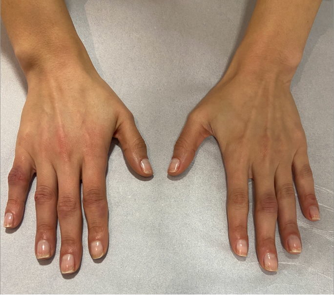
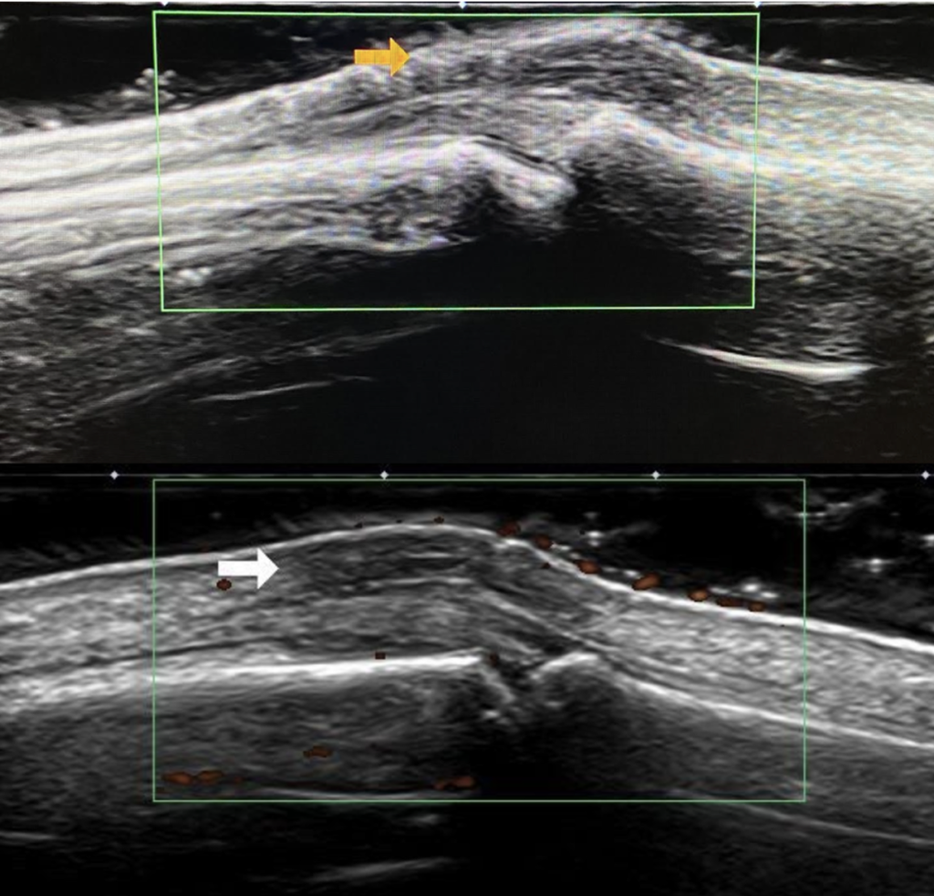
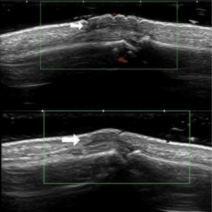
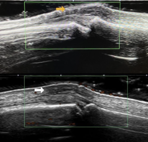
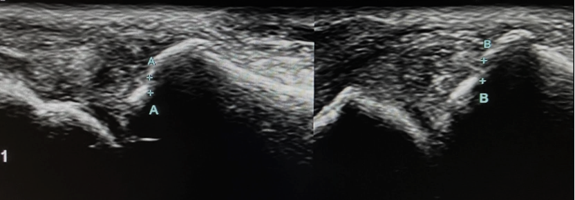
A 35-year-old healthy woman was evaluated in the primary care clinic of south Brazil for edema, pain and reddish macules in the proximal interphalangeal joints of the second and fifth finger of her right hand and the third finger of her left hand for several weeks. The symptons started during the winter months. The patient also had morning stiffnes and positive rheumatoid factor and CCP (cyclic citrullinated peptide) antibodies. No associated systemic symptoms were found and there was no family history of rheumatological diseases. The differential diagnosis included rheumatoid arthritis, musculoskeletal injury and a vasospastic process. The doctor chose to order a dermatological and rheumatological ultrasonography of the hands and wrists.
Images
Quiz-summary
0 of 2 questions completed
Questions:
- 1
- 2
Information
View the March Case below, answer the question and then click check >
You have already completed the quiz before. Hence you can not start it again.
Quiz is loading...
You must sign in or sign up to start the quiz.
You have to finish following quiz, to start this quiz:
Results
0 of 2 questions answered correctly
Your time:
Time has elapsed
You have reached 0 of 0 points, (0)
Categories
- Not categorized 0%
- 1
- 2
- Answered
- Review
-
Question 1 of 2
1. Question
Question 1: What are the Ultrasound findings?
Correct
CORRECT ANSWER EXPLAINED BELOW Correct answer to Q1 is: No signs of synovitis in the joints
Discussion
No increased synovial thickness, joint effusion or power Doppler signals were detected, which ruled out rheumatoid arthritis. Subcutaneous thickening was predominant, and the patient was not aware of any recent trauma or injury. The image findings, in addition to the clinic of worsening during winter days and the physical exam showing multiple erythrocyanotic lesions on the fingers, lead to a suspicion of erythema pernio.
Incorrect
CORRECT ANSWER EXPLAINED BELOW Correct answer to Q1 is: No signs of synovitis in the joints
Discussion
No increased synovial thickness, joint effusion or power Doppler signals were detected, which ruled out rheumatoid arthritis. Subcutaneous thickening was predominant, and the patient was not aware of any recent trauma or injury. The image findings, in addition to the clinic of worsening during winter days and the physical exam showing multiple erythrocyanotic lesions on the fingers, lead to a suspicion of erythema pernio.
-
Question 2 of 2
2. Question
Additional Images
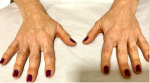
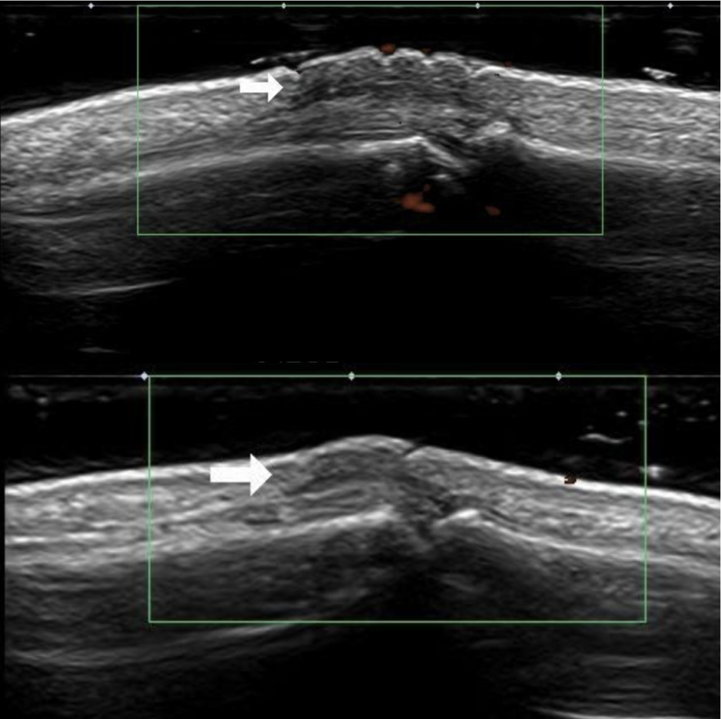
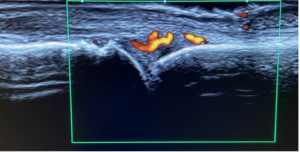
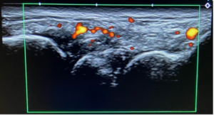
Image 4: Another healthy middle-aged woman with a clinical suspicion of erythema pernio. No personal or family history of rheumatological disease was present. The patient complained of joint pain for two months and had worsening of the symptoms in the last 15 days, during the winter period in southern Brazil. An ultrasound was performed to help elucidate the diagnosis.
Question 2: About the second case, what can we conclude?
Correct
CORRECT ANSWER EXPLAINED BELOW Correct answer to Q2 is: Considering the involvement of deeper structures as synovitis and cortical erosion, rheumatoid arthritis must be the diagnosis.
Additional discussion
Erythema pernio, also known as chilblains, is a rare inflammatory skin condition characterized by painful, red or purple lesions on the extremities, typically in response to exposure to cold conditions. It primarily affects the fingers and toes. In contrast to conditions like rheumatoid arthritis, which affects joints and soft tissues, erythema pernio is essentially a skin-related disease.
In the context of erythema pernio, the use of ultrasound is not a standard diagnostic approach. However, in this case it was essential to exclude the involvment of deeper structures like joints and tendons rather than the superficial layers of the skin.
The pathophysiology is unknown, but it is associated with vasospasm, particularly when the patient is exposed to cold, damp conditions for a prolonged period. A comprehensive history and physical examination is essential for the assessment and diagnosis of pernio. Inquiries about the timing of the appearance of the lesions, recent cold exposures, and symptom improvement upon removing cold exposure are instrumental in narrowing down the potential causes to pernio or other vasospastic diseases. Skin biopsies shows nonspecific findings and are not helpful for the diagnosis.
The disease is largely considered idiopathic, although there are instances of secondary pernio. One notable form of secondary pernio is linked to systemic lupus erythematosus, leading to the development of chilblain lupus erythematosus, and also Raynaud phenomenon, acrocyanosis, cryoglobulinemia and cold panniculitis should be considered.
Conclusion
Dermatological conditions can often be diagnosed by clinical history and physical exam, however the use of ultrasound as an auxiliary tool is growing. In the context of our case with a suspicion of erythema pernio, ultrasound was essential to rule out involvement of the deeper structures, as the condition primarily involves the superficial layers of the skin. This is particularly important when trying to differentiate erythema pernio from conditions like rheumatoid arthritis, which affect these deeper structures.
Conflicts of Interest:
The author declares no conflict of interest.
References
- Almuhanna N, Wortsman X, Wohlmuth-Wieser I, Kinoshita-Ise M, Alhusayen R. Overview of Ultrasound Imaging Applications in Dermatology [Formula: see text]. J Cutan Med Surg. 2021 Sep;25(5):521-529. doi: 10.1177/1203475421999326. Epub 2021 Mar 7. PMID: 33682489; PMCID: PMC8474315.
- Middleton HT, Boswell CL, Houwink EJ, Allen-Rhoades WA, Kuhn AK, Wright JA. Vincristine-Induced Acrocyanosis and Erythema Pernio. Journal of Primary Care & Community Health. 2023;14. doi:1177/21501319231181879
- Di Matteo, A., Mankia, K., Azukizawa, M. et al.The Role of Musculoskeletal Ultrasound in the Rheumatoid Arthritis Continuum. Curr Rheumatol Rep 22, 41 (2020). https://doi.org/10.1007/s11926-020-00911-w
Incorrect
CORRECT ANSWER EXPLAINED BELOW Correct answer to Q2 is: Considering the involvement of deeper structures as synovitis and cortical erosion, rheumatoid arthritis must be the diagnosis.
Additional discussion
Erythema pernio, also known as chilblains, is a rare inflammatory skin condition characterized by painful, red or purple lesions on the extremities, typically in response to exposure to cold conditions. It primarily affects the fingers and toes. In contrast to conditions like rheumatoid arthritis, which affects joints and soft tissues, erythema pernio is essentially a skin-related disease.
In the context of erythema pernio, the use of ultrasound is not a standard diagnostic approach. However, in this case it was essential to exclude the involvment of deeper structures like joints and tendons rather than the superficial layers of the skin.
The pathophysiology is unknown, but it is associated with vasospasm, particularly when the patient is exposed to cold, damp conditions for a prolonged period. A comprehensive history and physical examination is essential for the assessment and diagnosis of pernio. Inquiries about the timing of the appearance of the lesions, recent cold exposures, and symptom improvement upon removing cold exposure are instrumental in narrowing down the potential causes to pernio or other vasospastic diseases. Skin biopsies shows nonspecific findings and are not helpful for the diagnosis.
The disease is largely considered idiopathic, although there are instances of secondary pernio. One notable form of secondary pernio is linked to systemic lupus erythematosus, leading to the development of chilblain lupus erythematosus, and also Raynaud phenomenon, acrocyanosis, cryoglobulinemia and cold panniculitis should be considered.
Conclusion
Dermatological conditions can often be diagnosed by clinical history and physical exam, however the use of ultrasound as an auxiliary tool is growing. In the context of our case with a suspicion of erythema pernio, ultrasound was essential to rule out involvement of the deeper structures, as the condition primarily involves the superficial layers of the skin. This is particularly important when trying to differentiate erythema pernio from conditions like rheumatoid arthritis, which affect these deeper structures.
Conflicts of Interest:
The author declares no conflict of interest.
References
- Almuhanna N, Wortsman X, Wohlmuth-Wieser I, Kinoshita-Ise M, Alhusayen R. Overview of Ultrasound Imaging Applications in Dermatology [Formula: see text]. J Cutan Med Surg. 2021 Sep;25(5):521-529. doi: 10.1177/1203475421999326. Epub 2021 Mar 7. PMID: 33682489; PMCID: PMC8474315.
- Middleton HT, Boswell CL, Houwink EJ, Allen-Rhoades WA, Kuhn AK, Wright JA. Vincristine-Induced Acrocyanosis and Erythema Pernio. Journal of Primary Care & Community Health. 2023;14. doi:1177/21501319231181879
- Di Matteo, A., Mankia, K., Azukizawa, M. et al.The Role of Musculoskeletal Ultrasound in the Rheumatoid Arthritis Continuum. Curr Rheumatol Rep 22, 41 (2020). https://doi.org/10.1007/s11926-020-00911-w

