Case of the month November 2024: Sudden abdominal pain
November 19, 2024
Case of the month January 2025: Girl with abdominal pain
January 13, 2025Edda Chaves 1,*
1 Servicio de Radiología. Hospital Regional Nicolas Solano La Chorrera. Panamá; echavess@minsa.gob.pa
* Correspondence: echavess@minsa.gob.pa
Clinical history
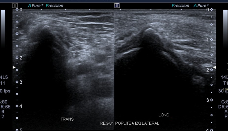
A 45-year-old female was evaluated in the primary care clinic for pain in her right knee. The pain worsened during the day, causing walking difficulties. The clinical exam did not show any important signs of swelling, only tenderness in the posterior lateral region of the knee. There was no history of trauma or any other important event that could cause the pain. The patient was sent to the orthopedic surgeon who suggested an ultrasound to exclude pathology of tendons or bursae.
Images
Quiz-summary
0 of 2 questions completed
Questions:
- 1
- 2
Information
View the December Case below, answer the question and then click check >
You have already completed the quiz before. Hence you can not start it again.
Quiz is loading...
You must sign in or sign up to start the quiz.
You have to finish following quiz, to start this quiz:
Results
0 of 2 questions answered correctly
Your time:
Time has elapsed
You have reached 0 of 0 points, (0)
Categories
- Not categorized 0%
- 1
- 2
- Answered
- Review
-
Question 1 of 2
1. Question
Question 1: What do you see on the ultrasound image?
Correct
CORRECT ANSWER EXPLAINED BELOW Correct answer to Q1 is: No abnormality
Incorrect
CORRECT ANSWER EXPLAINED BELOW Correct answer to Q1 is: No abnormality
-
Question 2 of 2
2. Question
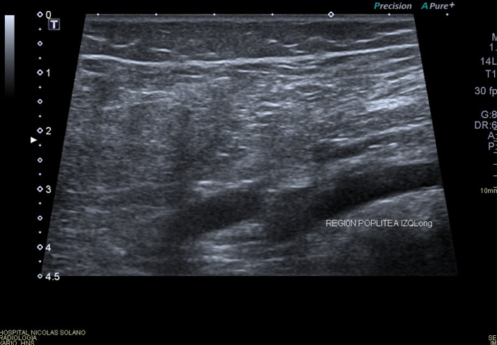
Image 2: Ultrasound image of the popliteal region, lateral view, transverse and longitudinal view.
Question 2: What do you see on the ultrasound image?
Correct
CORRECT ANSWER EXPLAINED BELOW Correct answer to Q2 is: Accessory bone – sesamoid bone
Discussion
There are no changes in the superficial tissues. No change in the cartilage. The imaging findings correspond with an echogenic structure, with increased sound attenuation, indicating bone. In this location it corresponds to the fabella.
The fabella is a sesamoid bone located in the posterolateral part of the knee (1), associated with the lateral head of the gastrocnemius muscle articulating with the lateral femoral condyle. It is an articular sesamoid bone occurring in 20 to 30% of the population (2). Rarely it can be found in the medial head of the gastrocnemius.
The patient said she occasionally had similar symptoms in her right knee, and the ultrasound examination revealed similar images.
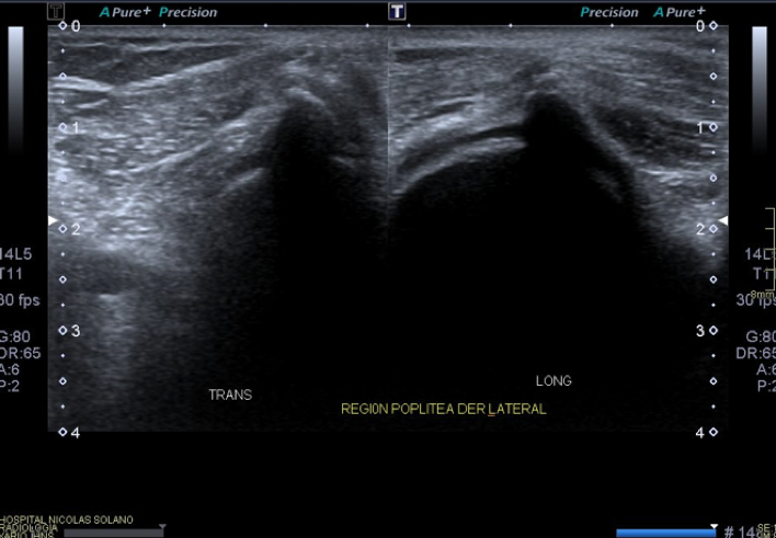
Image 3: Right popliteal region, transverse and longitudinal view.In view of the findings, and to confirm the diagnosis of fabella, an x-ray of both knees was done.
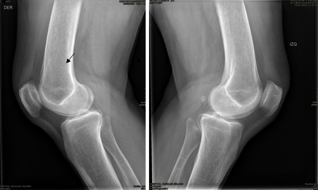
Image 4: x-ray of both knees, lateral view. The fabella is marked with an arrow.The fabella pain syndrome (3) should be considered as a differential diagnosis when a patient presents with persistent posterolateral knee pain, which could also be due to meniscal tears, lateral ligament instability, Baker’s cyst, and proximal tibiofibular joint hypomobility. Patients with fabella pain syndrome usually complain that the posterolateral knee pain is worse on fully extending the the knee joint, which is due to compression of the common peroneal nerve.
Conclusion
The fabella can be mistaken for loose bodies or osteophytes which are usually asymptomatic in patients. Ultrasound in a useful tool to see the relation to anatomical structures when an incidental finding occurs.
References
- Dalip D, Iwanaga J, Oskouian RJ, Tubbs RS. A Comprehensive Review of the Fabella Bone. Cureus. 2018 Jun 5;10(6):e2736. doi: 10.7759/cureus.2736. PMID: 30087812; PMCID: PMC6075638
- Kawashima T, Takeishi H, Yoshitomi S et-al. Anatomical study of the fabella, fabellar complex and its clinical implications. Surg Radiol Anat. 2007;29 (8): 611-6. doi:10.1007/s00276-007-0259-4
- Driessen, Arne; Balke, Maurice; Offerhaus, Christoph; White, William; Shafizadeh, Sven; Becher, Christoph; Bouillon, Bertil; Höher, Jürgen (2014). The fabella syndrome – a rare cause of posterolateral knee pain: a review of the literature and two case reports. BMC Musculoskeletal Disorders, 15(1), 100–.doi:10.1186/1471-2474-15-100
Incorrect
CORRECT ANSWER EXPLAINED BELOW Correct answer to Q2 is: Accessory bone – sesamoid bone
Discussion
There are no changes in the superficial tissues. No change in the cartilage. The imaging findings correspond with an echogenic structure, with increased sound attenuation, indicating bone. In this location it corresponds to the fabella.
The fabella is a sesamoid bone located in the posterolateral part of the knee (1), associated with the lateral head of the gastrocnemius muscle articulating with the lateral femoral condyle. It is an articular sesamoid bone occurring in 20 to 30% of the population (2). Rarely it can be found in the medial head of the gastrocnemius.
The patient said she occasionally had similar symptoms in her right knee, and the ultrasound examination revealed similar images.

Image 3: Right popliteal region, transverse and longitudinal view.In view of the findings, and to confirm the diagnosis of fabella, an x-ray of both knees was done.

Image 4: x-ray of both knees, lateral view. The fabella is marked with an arrow.The fabella pain syndrome (3) should be considered as a differential diagnosis when a patient presents with persistent posterolateral knee pain, which could also be due to meniscal tears, lateral ligament instability, Baker’s cyst, and proximal tibiofibular joint hypomobility. Patients with fabella pain syndrome usually complain that the posterolateral knee pain is worse on fully extending the the knee joint, which is due to compression of the common peroneal nerve.
Conclusion
The fabella can be mistaken for loose bodies or osteophytes which are usually asymptomatic in patients. Ultrasound in a useful tool to see the relation to anatomical structures when an incidental finding occurs.
References
- Dalip D, Iwanaga J, Oskouian RJ, Tubbs RS. A Comprehensive Review of the Fabella Bone. Cureus. 2018 Jun 5;10(6):e2736. doi: 10.7759/cureus.2736. PMID: 30087812; PMCID: PMC6075638
- Kawashima T, Takeishi H, Yoshitomi S et-al. Anatomical study of the fabella, fabellar complex and its clinical implications. Surg Radiol Anat. 2007;29 (8): 611-6. doi:10.1007/s00276-007-0259-4
- Driessen, Arne; Balke, Maurice; Offerhaus, Christoph; White, William; Shafizadeh, Sven; Becher, Christoph; Bouillon, Bertil; Höher, Jürgen (2014). The fabella syndrome – a rare cause of posterolateral knee pain: a review of the literature and two case reports. BMC Musculoskeletal Disorders, 15(1), 100–.doi:10.1186/1471-2474-15-100

